DERLEME MAKALE
GERIATRI YAŞ GRUBUNDA GASTROINTESTINAL
KANAMA VE TEDAVISI
Approximately 35%-40% of geriatric patients seek medical care for gastrointestinal symptoms at least once a year. At least 1% of the population aged over 80 years is hospitalized each year due to gastrointestinal hemorrhage. Gastrointestinal hemorrhage is frequently observed in this population and associated with high mortality and morbidity. Also, gastrointestinal hemorrhage is one of the main causes of hospitalization among geriatric patients. Geriatric patients with GIH constitute a subgroup of patients requiring special care in hospitals. The bleeding may occur in the upper or lower gastrointestinal tract and manifest itself with a variety of symptoms depending on its location. The incidence and natural course of hemorrhage are affected by antiplatelet and anticoagulant medications. The outcome of gastrointestinal hemorrhage in geriatric patients depends largely on the characteristics of bleeding lesion and comorbidities.
Keywords: Geriatrics; Gastrointestinal hemorrhage; Aged
A
BSTRACTGeriatri yaş grubundaki hastaların yaklaşık %35-40’lık bir kısmı, yılda en az 1 kez gastrointestinal semptomlar nedeni ile tıbbi yardıma ihtiyaç duymaktadır. Seksen yaş üstü nüfusun en az %1’i, her yıl gastrointestinal kanama nedeni ile hastaneye yatmak zorunda kalmaktadır. Gastrointestinal kanaması olan geriatric olgular, hastanelerde özel bakım gerektiren hasta grubunu oluşturmaktadır. Geriatrik çağda gastrointestinal kanama sık gözlenmektedir ve beraberinde yüksek morbidite ve mortalite riski taşımaktadır. Kanama üst veya alt gastrointestinal sistemden köken alabilir ve kanama lokalizasyonuna göre semptom çeşitliliği gösterebilmektedir. Kanamanın insidansı ve klinik seyri, hastanın kullandığı antiplatellet veya antikoagülan tedaviden etkilenmektedir. Geriatrik olgulardaki kanamanın seyri çoğunlukla kanayan lezyonun karakteristiğine ve eşlik eden hastalık olup olmamasına bağlıdır.
Anahtar sözcükler: Geriatri; Gastrointestinal hemoraji; Yaşlı
Ö
ZTurkish Journal of Geriatrics DOI: 10.31086/tjgeri.2019.85 2019;22 (2):122-131
CORRESPONDANCE Dilek OĞUZ
Kırıkkale University, Faculty of Medicine, Department of Gastroenterology, Kırıkkale, Turkey.
Phone: +903182251000 e-mail: [email protected] Received: 08/02/2019 Accepted: 18/03/2019
1 TOBB University of Economics and
Technology, Faculty of Medicine, Department of Gastroenterology, Ankara, Turkey.
2 Kırıkkale University, Faculty of Medicine,
Department of Gastroenterology, Kırıkkale, Turkey.
MANAGEMENT IN GERIATRIC AGE GROUP
Cem CENGİZ1 Dilek OĞUZ2
INTRODUCTION
At least 1% of the population aged over 80 years is hospitalized each year due to gastrointestinal hemorrhage (GIH). Gastrointestinal hemorrhage can be caused by age-specific lesions or lesions that are observed in all age groups. Mortality or morbidity depends on the characteristics of the bleeding lesion, the presence of comorbidities, and medication use. GIH-causing lesions can develop because of comorbidities or can be initiated by medications used to relieve the effects of aging. Gastrointestinal hemorrhage incidence increases with age, and elderly patients constitute a subgroup of patients requiring special care in hospitals (1).
UPPER GASTROINTESTINAL HEMORRHAGE
Despite the decline in the total incidence of non-variceal upper GIHs since the 1990s, there has been an increase in the number of patients aged over 60 years (2). Approximately 70% of patients seeking medical attention for GIH are aged over 60 years with the incidence of hemorrhage increasing with age. Advanced age is a significant risk factor for mortality following GIH because of the increased prevalence of pulmonary and cardiovascular diseases in this population (3).
Etiology
Despite the advancements in the diagnostic and treatment techniques for GIH, mortality rates remain unchanged, the primary reason being the steadily increasing elderly population. The elderly (at least 60 years of age) are prone to GIH, and excessive use of NSAIDs in elderly patients is one of the primary causes of GIH (4). In addition, elderly patients comprise 44.5% of the GIH patients and 75% of GIH-originated mortality (5).
Many studies have been conducted on the etiology of upper gastrointestinal bleeding among elderly (1-10). The causes of upper gastrointestinal hemorrhage are the same across all age groups. Peptic ulcer is the primary cause in both elderly
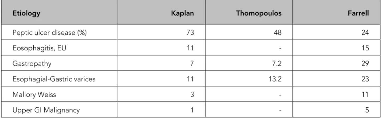
and young patients, and the other most frequent causes of GIH are esophagitis, esophageal ulcer, gastropathy, gastric and esophageal varices, Mallory-Weiss tear, and malignancy (Table 1).
Clinic and laboratory
For elderly patients admitted with upper GIH, a detailed medical history should be obtained, including information regarding prior surgeries, prior GIH, medications used, comorbidities, NSAIDs use, and the use of drugs for neurological disorders (e.g., Parkinson’s and Alzheimer’s diseases). The presence of a chronic liver disorder should be evaluated and detailed by physical examination. Laboratory results should be urgently and consistently assessed, and the determination and replacement of acute blood loss should be the primary goal. Moreover, a treatment plan should be developed according to the clinical results that are obtained following admission. Total blood count, coagulation parameters, and evaluation of electrolyte imbalance are all critical tests following which additional tests, such as a liver function test, can be performed to enable physicians to detect underlying disease. Hypotension is an important indicator of a higher risk of mortality and blood loss in elderly patients with peptic ulcer hemorrhage (6).
Notably, 40%–50% of patients with GIH have hematemesis, 75%–80% have melena, and 15%– 20% have hematochezia. Medical history and physical examination results have prognostic value. Indeed, advanced age, a positive tilt test, and shock symptoms indicate a poor prognosis, and cases with these symptoms have high mortality. An increase in comorbidities related to the cardiovascular system, central nervous system, gastrointestinal system (GIS), hepatic system, pulmonary system, renal system, and physiological stress increase mortality rates (7).
The color of feces depends on the transit time and the amount of blood in the GIS. Hematochezia sometimes reflects ulcer hemorrhage and fast transit time which are good indicators of hemodynamic
instability. Interestingly, 71% of patients with upper GIH have melena, but mortality rates are lower compared to those having hematochezia (9.4% vs. 13.6%). In cases with negative upper GIS on endoscopy, colonoscopy remains the most appropriate tool for evaluating melena. Melena is observed in 18% of patients with lower GIS hemorrhage (8).
Treatment of upper gastrointestinal hemorrhage
The patient’s hemodynamic condition should be the primary parameter to be evaluated. Additionally, the presence of tachycardia, a pulse rate over 100–120 beats/min, hypotension (blood pressure lower than 90 mm Hg), incidences of syncope, findings of shock, and blackout require immediate resuscitation of the patient.
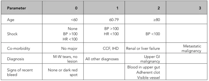
The severity of the hemorrhage must be evaluated, and the clinical status of the patient must be determined (e.g., using the Glasgow and Rockall coma scores, the APACHE II score, and the Child-Pugh score). Specifically, the Rockall coma score combines general and endoscopic symptoms (Table 2) (7) and predicts mortality and rehemorrhage using a risk scoring system.
The patient’s blood volume loss should be detected according to the methods mentioned above, and the patient should be hemodynamically
resuscitated. Compared with young patients, elderly patients are less tolerant to anemia and hypoxia. Therefore, in the presence of cardiac comorbidity, mortality would significantly increase in elderly patients if they are not promptly treated. Blood volume deficiency must be rapidly replaced using plasma, erythrocyte suspension, or other plasma volume expanders.
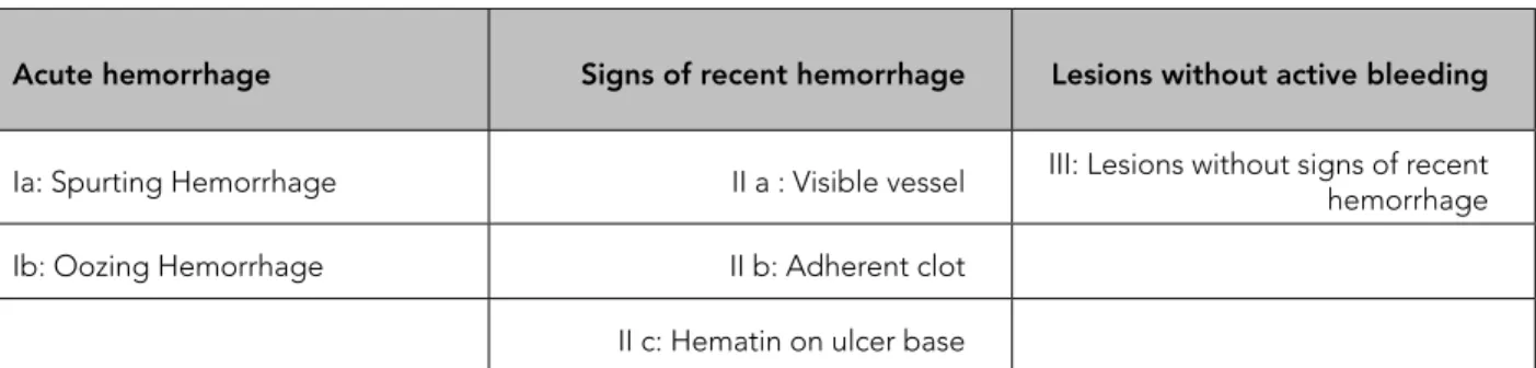
Continuous infusion of 8 mg/h proton pump inhibitor (PPI) therapy following 80 mg i.v. bolus of PPI (esomeprazole, omeprazole, or pantoprazole) can improve endoscopic stigmata and can accelerate platelet aggregation in the stomach by increasing intragastric pH. Esophagogastroduodenoscopy (EGD) should be performed when the patient is hemodynamically stable to detect hemorrhage and bleeding lesion, and should be endoscopically treated. An endoscopic examination should be performed imminently because the observed symptoms influence the treatment required. The significant complication rate because of endoscopy is 0.5%, whereas the mortality rate is 0.13%. However, an EGD can provide information on the localization of bleeding up to the ligament of Treitz and information on the activity and rebleeding risk of peptic ulcers according to the Forrest classification (Table 3) (8).
Table 1. Etiology of upper gastrointestinal bleeding among elderly (1, 10).
Etiology Kaplan Thomopoulos Farrell
Peptic ulcer disease (%) 73 48 24
Eosophagitis, EU 11 - 15
Gastropathy 7 7.2 29
Esophagial-Gastric varices 11 13.2 23
Mallory Weiss 3 - 11
In hemodynamically-stabilized patients, endoscopic procedures can be performed for Forrest Ia, Ib, IIa, and IIb ulcers. Currently, endoscopic treatments for peptic ulcer bleeding involve the use of a sclerosing agent, adrenaline injection, thermal coagulation, endoscopic clipping, or a combination of these procedures. However, in several rare hemorrhages (e.g., Dieulafoy’s lesion and gastric antral vascular ectasia hemorrhages) and in esophagus/stomach variceal bleeding, endoscopic treatments also involve tissue adhesives, band ligation, and argon plasma coagulation.
If the endoscopic treatment is unsuccessful or if the hemorrhage cannot be adequately controlled during subsequent endoscopy, radiological or surgical treatments should be considered, and surgical consultation should be performed for these patients. Additionally, multidisciplinary evaluation of the patient should be conducted with radiology and other departments as appropriate.
Once the hemorrhage is controlled, the underlying illnesses should be appropriately treated. Treatments may include Helicobacter
pylori (Hp) treatment, eradication of esophageal
and fundal varices, specific treatment (if possible) for hepatic diseases, treatment of esophagitis, and argon plasma coagulation in telangiectasia. Notably, long-term treatment programs for elderly patients appear to be crucial in preventing rebleeding.
LOWER GASTROINTESTINAL HEMORRHAGE
Despite the incidence of lower GIH being lesser than upper GIH, it certainly does increase with age. Lower GIH refers to hemorrhages below the ligament of Treitz. Several studies have shown a 200-fold increase in the incidence of lower GIH from the third to the ninth decade of life. The average age of patients with lower GIH is 63-77 years. The reason for an increased incidence of lower GIH with age is because of increased risk of diverticular hemorrhage, which is the most common cause of lower GIHs in elderly individuals. Compared with younger individuals, elderly individuals have to endure extended hospital stay and higher treatment expenditure (9). There is no difference between the diagnostic approach for lower GIH
Table 2. Rockall score (7).
Parameter 0 1 2 3 Age <60 60-79 ≥80 Shock None BP >100 HR <100 BP >100 HR <100 BP <100
Co-morbidity No major CCF, IHD Renal or liver failure malignancyMetastatic Diagnosis M-W tears, no lesion All other diagnoses malignancyUpper GI
Signs of recent
bleed None or dark red spot
Blood in upper gut Adherent clot Visible vessel
CCF:Congestive Cardiac Failure, IHD:Ischemic heart disease, M-W:Mallory-Weiss BP:Blood-Pressure, HR: Heart rate
and that for other GIHs (i.e., evaluating the status of the patient and identifying the hemorrhage). Once the patient is stabilized, the procedure for the determination of the etiology and treatment is the same as that for upper GIH.
Etiology
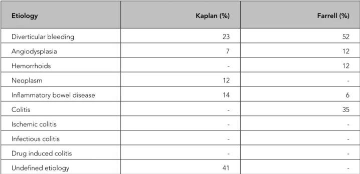
Concerning factors for lower GIH are vascular ectasia, diverticular disease, malignancy, ischemic intestinal disorders, and anorectal diseases in patients aged over 50 years. Vascular ectasia and diverticular disease constitute 60% of major GIHs, with vascular ectasia being the most common cause. Postpolypectomy bleeding, radiation proctitis, and solitary ulcer are also rare causes of lower GIH in geriatric population. (Table 4) (1,10).
Clinical symptoms and diagnosis
Obtaining a detailed medical history is critical in lower GIH patients to evaluate possible etiologies. Blood color, presence of hematochezia or melena, and volume of blood loss may help to locate the hemorrhage. Blood in the feces usually indicates anorectal hemorrhages. In addition to upper GIH, melena may also indicate hemorrhage in the proximal colon. Abdominal pain with cramps and rectal bleeding can indicate inflammatory bowel disease. If the patient is hemodynamically stable, a sudden onset of painless bleeding is a typical indicator of diverticular hemorrhage. Vascular dysplasia hemorrhages can involve a broad spectrum of bleeding, from occult to
severe. Depending on the type of bleeding, flexible sigmoidoscopy may be performed after colonoscopy.
Treatment of lower gastrointestinal bleeding
Diverticulosis coli
Despite studies showing right colon diverticula to be the primary source of diverticular hemorrhage, it can originate from any segment of the colon. Interestingly, 80% of cases occur in elderly individuals. In most studies, diverticulosis is reported to be the most common source of colonic and in the majority of cases bleeding stops spontaneously although it can sometimes be severe (11). Treatment plan includes flexible sigmoidoscopy/colonoscopy and endoscopic treatment (i.e., adrenaline injection, thermal coagulation, and band ligation). Radiological diagnosis and treatment should be considered when the endoscopic approach fails. Multidetector CT/CT angiography and Tc-99m sulfur colloid scintigraphy can assist in locating the bleeding. Once the location of the hemorrhage is identified, selective mesenteric angiography with embolisation can be used to occlude the bleeding vein. Nevertheless, intermittent hemorrhage prevents an easy diagnosis. Surgery is required only in a small proportion of patients, and is typically an elective surgery. Emergent surgery has a high rate of mortality in elderly patients with a study reporting mortality of 37% in patients aged over 70 years (12).
Table 3. Forrest classification (8).
Acute hemorrhage Signs of recent hemorrhage Lesions without active bleeding Ia: Spurting Hemorrhage II a : Visible vessel III: Lesions without signs of recent hemorrhage
Ib: Oozing Hemorrhage II b: Adherent clot
Ischemic colitis
Typically, ischemic colitis results from a reduction in colonic blood circulation or dehydration. The risk for ischemic colitis increases with age, especially in patients with comorbidities, such as vasculitis, or in those who use diuretics or vasoactive agents.
The clinical symptoms typically include an abdominal pain with cramps and the appearance of thickened colon wall on abdominal CT. Colonoscopy shows fragile, inflammatory, mucosal, and submucosal bleeding structures in the affected region, and ischemic colitis must be distinguished from mesenteric and chronic ischemia by the presence of acute emboli. Hemorrhage rarely causes hemodynamic problems in patients with ischemic colitis; however, stenosis can occur between attacks, and the patients typically present with obstructive symptoms. Additionally, in ischemic colitis secondary stenosis should be considered in patients who have obstructive symptoms.
Vascular dysplasia
Vascular dysplasia is a frequent source of apparent or occult hemorrhage in elderly patients. They are generally located in the small intestine but have also been reported in the colon; however, the involvement of the right colon is rare . The clinical spectrum varies from chronic occult to acute massive bleedings (13). It is often difficult to reach the lesions in the small intestine and balloon enteroscopy can be used for this purpose. Endoscopic, angiographic, and surgical treatment is possible for a single, massively bleeding vascular dysplasia located in the cecum. The primary endoscopic treatment is argon plasma coagulation; however, the treatment choice depends on the technical capabilities and the expertise level of each center. Heat application by using a heater probe was a popular procedure in the past; however, argon plasma coagulation is the first choice of treatment due to its higher success rates.
Table 4. Etiology of lower gastrointestinal bleeding among elderly (1,10).
Etiology Kaplan (%) Farrell (%)
Diverticular bleeding 23 52
Angiodysplasia 7 12
Hemorrhoids - 12
Neoplasm 12
-Inflammatory bowel disease 14 6
Colitis - 35
Ischemic colitis -
-Infectious colitis -
-Drug induced colitis -
-Hemorrhoids
The prevalence of hemorrhoids decreases after the age of 65. However, patients who had chronic constipation during their youth continue to experience hemorrhoids as they advance in age (14). Hemorrhoids can cause a variety of hemorrhages, from a small number of fresh rectal bleeds to massive bleeding. They are best evaluated by performing a retroflexion technique in the rectum. Typically, hemorrhoids cause low-volume bleeding, and treatment (e.g., medical or surgical) depends on the complication.
Stercoral ulcer and solitary rectal ulcer
Both stercoral and solitary rectal ulcers can cause massive rectal bleedings (15). Notably, a large percentage of patients with hemorrhagic ulcers are aged over 60 years. Stercoral ulcer originates from mucosal trauma in the rectum caused by petrified feces, whereas solitary rectal ulcer evolves following prolapse of the rectal mucosa because of constipation and rupture. If active bleeding from the ulcer is detected, the endoscopic treatments used for peptic ulcer can be used to treat stercoral and solitary rectal ulcers.
Colon tumors and postpolypectomy hemorrhages
Typically, colon tumors cause occult bleeding, but they can also cause overt hemorrhages. Bleeding can occur following polypectomy in the early or late phase. Several studies documented that different techniques were available to prevent post-polypectomy bleeding. These techniques include injectable solutions, placement of endoclips, deployment of a detachable loop, and application of thermal energy (with coagulation forceps or argon plasma coagulation) as well as a combination of these techniques Additionally, aspirin and anticoagulant use should be regulated (16).
Radiation proctitis
Radiation proctitis occurs in patients with
prostatic, genitourinary, and gynecologic malignancies who underwent radiotherapy. Symptoms may occur years after the therapy and it can cause overt bleedings that require chronic transfusion. Endoscopic measures especially argon plasma coagulation (APC) are effective and safe. Treatment includes formalin application, sucralfate enema, and hyperbaric oxygen application are other treatment alternatives; however, data regarding the success of these various treatments other than APC is scant. Surgery is considered for refractory or severe cases. (17).
OCCULT/OBSCURE HEMORRHAGES
A diagnosis of occult or obscure hemorrhage should be made when the location of the hemorrhage cannot be detected on either upper endoscopy or colonoscopy. Occult hemorrhages occur mainly because of an inability to locate lesions during the initial endoscopy or intermittent bleeding that make diagnosing hemorrhage challenging (18). Endoscopy should be repeated, and angiography and scintigraphy results should be evaluated in case of occult hemorrhages. The location of the hemorrhage can also be the small bowel, which was considered to be a blind spot until recently. In such cases, push jejunoscopy, balloon enteroscopy and capsule endoscopy can help physicians locate the source of the hemorrhage.
The diagnostic capability of capsule endoscopy in occult hemorrhages has been reported to be 58%–80%. Vascular dysplasia, bleeding tumors of the small bowel, and ulcers because of NSAIDs use that have been frequently detected in recent years can be diagnosed using capsule endoscopy. Comparative studies have shown the superiority of capsule endoscopy over push enteroscopy, small bowel enteroclysis, and mesenteric angiography (19). Swallowing disorders and delays in gastric emptying in elderly patients can create problems for the passage of capsule to the small bowel
ultimately requiring endoscopic aid. Capsule use is inappropriate in patients with a suspicion of intestinal obstruction. In addition to recent developments in capsule technology, ongoing research is focusing on improving battery life and image quality.
Both single and double balloon enteroscopy have enabled visualization and treatment of small bowel illnesses by using endoscopic procedures. The diagnostic efficiency of balloon enteroscopy in occult hemorrhage cases is noted to be 60%–67% (20). Despite its advantages over other techniques (e.g., the possibility of argon application to treat vascular dysplasia and the suitability to use endoscopic applications), it is an invasive technique, and the targeted area cannot always be reached.
NSAIDS AND GASTROINTESTINAL HEMORRHAGE
Aspirin use increases the risk of upper GIH (21), and the addition of a nonaspirin antiplatelet agent or NSAID also increases the risk of hemorrhage. Similarly, concurrent use of aspirin and NSAIDs increases the risk of lower GIH, particularly of diverticular hemorrhage (22). Interestingly, the use of both aspirin and NSAIDs in elderly patients with cardiovascular and rheumatologic illnesses increase the risk of both lower and upper GIH. Therefore, it is crucial to determine the GIH risk in patients who are required to use aspirin. The hemorrhage history of the patient and illnesses and drugs that predispose patients to hemorrhages should be delineated, and minimal cardiac protection should be maintained by using the smallest possible dose of aspirin. The long-term hemorrhage risk because of aspirin use can be reduced using a PPI. NSAIDs are significant risk factors of GIH during hospitalization. Aspirin or selective COX-2 inhibitor use and peptic ulcer history were found to be predictors of upper GIH in patients hospitalized with hip fractures. Prophylactic H2 receptor blocker or PPI usage
reduced the risk of GIH during hospitalization (23).
RELIABILITY OF ENDOSCOPY
The reliability of endoscopy has been observed throughout the general population. The mortality rate of elective EGD has been determined to be 0.0004%, whereas the mortality rate of endoscopies performed during hemorrhage is 0.01% (24).
Lower and upper endoscopic procedures have similar success and mortality rates for the entire population. Most studies dealt with elective endoscopy performed for surveying. Mortality and morbidity of elderly patients during endoscopy depend on the severity and magnitude of the bleeding and comorbidities. Procedure-related perforation and complications due to premedication are the most significant causes of mortality and morbidity. Zenker’s diverticulum and anatomical deformities (cervical eminentia) are among the most critical causes of perforation during endoscopy in elderly patients. Perforations during a colonoscopy performed to detect lower GIHs occur because of excessive looping, a relaxed venter, hernia, previous operations, colon shrinkage, and adhesions. These perforations are significant predictors of mortality and morbidity during endoscopy for lower GIH.
Sedation during all endoscopic procedures is a crucial problem for elderly patients. Typically, in elderly patients, benzodiazepines and other anesthetic substances are used in smaller amounts compared to that in young patients of similar height and weight. Also propofol has a longer recovery time in elderly patients when compared with sedation with a combiation of midazolam and fentanyl (25). Additionally, patients should be adequately monitored during sedation to detect changes in oxygen saturation, blood pressure, pulse rate, and carbon dioxide levels.
CONCLUSION
Acute hemorrhages require evaluation and stabilization of general health condition before endoscopy in elderly. The factors affecting the course of hemorrhage, comorbidities, and
medication should be promptly evaluated. Endoscopy can be performed in geriatric patients as safely and efficiently as in younger patients; however, each case should be individually assessed for risk factors.
REFERENCES
1. Kaplan RC, Heckbert SR, Koepsell TD, et al. Risk factors for hospitalized gastrointestinal bleeding among older persons. J Am Geriatr Soc 2001;49:126-33. (PMID:11207865).
2. Thomopoulos KC, Vagenas KA, Vagianos CE, et al. Changes in aetiology and clinical outcome of acute upper gastrointestinal bleeding during the last 15 years. Eur J Gastroenterol Hepatol 2004;16:177-82. (PMID:15075991).
3. Katschinski B, Logan R, Davies J, et al. Prognostic factors in upper gastrointestinal bleeding. Dig Dis Sci 1994;39(4):706-12. (PMID:7908623).
4. Solomon DH, Gurwitz JH. Toxicity of nonsteroidal anti-inflammatory drugs in the elderly: is advanced age a risk factor? Am J Med 1997;102(2):208-15. (PMID:9217572).
5. Lingenfelser T, Ell C. Gastrointestinal bleeding in the elderly. Best Pract Res Clin Gastroenterol 2001;15(6):963-82. (PMID:11355905).
6. Yachimski PS, Friedman LS. Gastrointestinal bleeding in the elderly. Nat Clin Pract Gastroenterol Hepatol 2008;5(2):80-93. (PMID:18253137).
7. Robertson M, Majumdar A, Boyapati R, et al. Risk stratification in acute upper GI bleeding: comparison of the AIMS65 score with the Glasgow-Blatchford and Rockall scoring systems. Gastrointest Endosc 2016 June;83(6):1151-60. (PMID:26515955).
8. Forrest JA, Finlayson ND, Shearman DJ. Endoscopy in gastrointestinal bleeding. Lancet 1974;2:394-97. (PMID:4136718).
9. Speir EJ, Ermentrout RM, Martin JG. Management of acute lower gastrointestinal bleeding. Tech Vasc Interv Radiol 2017 Dec;20(4):258-62. (PMID:29224658). 10. Farrell JJ, Friedman LS. Gastrointestinal bleeding
in older people. Gastroenterol Clin North Am 2000;29(1):1-36. (PMID:10752016).
11. Olafsson GD, Hreinsson JP, Björnsson ES. Incidence of diverticular bleeding: a population-based study.
Scand J Gastroenterol 2019;10:1-5. (PMID:30739520). 12. Bender JS, Wiencek RG, Bouwman DL. Morbidity
and mortality following total abdominal colectomy for massive lower gastrointestinal bleeding. Am Surg 1991;57(8):536-41. (PMID:1928995).
13. Baum S, Athanasoulis CA, Waltman AC, et al. Angiodysplasia of the right colon: a cause of gastrointestinal bleeding. AJR Am J Roentgenol 1977;129(5):789-94. (PMID:410241).
14. Stewart RB, Moore MT, Marks RG, Hale WE. Correlates of constipation in an ambulatory elderly population. Am J Gastroenterol 1992;87(7):859-64. (PMID:1615939).
15. Tseng CA, Chen LT, Tsai Kbet al. Acute hemorrhagic rectal ulcer syndrome: a new clinical entity? Report of 19 cases and review of the literature. Dis Colon Rectum 2004;47(6):895-905. (PMID:15129312).
16. Lee JM, Kim WS, Kwak MS, et al. Clinical outcome of endoscopic management in delayed postpolypectomy bleeding. Intest Res 2017;15(2):221-27. (PMID:28522953).
17. Vanneste BG, Van De Voorde L, de Ridder RJ, et al. Chronic radiation proctitis: tricks to prevent and treat. Int J Colorectal Dis 2015;30(10):1293-303. (PMID:26198994).
18. Descamps C, Schmit A, Van Gossum A. “Missed” upper gastrointestinal tract lesions may explain “occult” bleeding. Endoscopy 1999;31(6):452-55. (PMID:10494684).
19. Leighton JA, Sharma VK, Srivathsan K, et al. Safety of capsule endoscopy in patients with pacemakers. Gastrointest Endosc 2004;59(4):567-69. (PMID:15044901).
20. Sun B, Rajan E, Cheng S, et al. Diagnostic yield and therapeutic impact of double-balloon enteroscopy in a large cohort of patients with obscure gastrointestinal bleeding. Am J Gastroenterol 2006;101(9):2011-15. (PMID:16848814).
21. Diener HC. Preventing major gastrointestinal bleeding in elderly patients. Lancet 2017 Jul 29;390(10093):435-37. (PMID:28622952).
22. Pilotto A, Franceschi M, Leandro G, et al. The risk of upper gastrointestinal bleeding in elderly users of aspirin and other non-steroidal anti-inflammatory drugs: the role of gastroprotective drugs. Aging Clin Exp Res 2003;15(6):494-99. (PMID:14959953).
23. Fisher L, Fisher A, Pavli P, Davis M. Perioperative acute upper gastrointestinal haemorrhage in older patients with hip fracture: incidence, risk factors and
prevention. Aliment Pharmacol Ther 2007;25(3):297-308. (PMID:17217452).
24. Silvis SE, Nebel O, Rogers G, Sugawa C, Mandelstam P. Endoscopic complications. Results of the 1974 American Society for Gastrointestinal Endoscopy Survey. JAMA 1976;235(9):928-30. (PMID:128642). 25. Lovett P, Gómez V, Hodge DO, Ladlie B. Propofol
versus midazolam/fentanyl sedation for colonoscopy in the elderly patient population. J Perianesth Nurs 2017;32(3):210-14.