RESEARCH ARTICLE
Importance of fasting serum glucose, haemoglobin A1c and fructosamine in the
diag-nosis of diabetes
Bahadır Ersan, Mahmut Ok* Özet
Erşan B, Ok M. Diyabetin teşhisinde serum açlık glikoz, he-moglobin A1c ve fruktozaminin önemi. Eurasian J Vet Sci, 2011, 27, 1, 13-18
Amaç: Tavşanlarda iki farklı doz (100 ve 175 mg/kg) allok-san ile oluşturulan deneysel diabetes mellitus’un teşhisin-de, klinik, hematolojik ve biyokimyasal parametrelerdeki değişimlerin değerlendirilmesidir. İkinci amacı ise hastalı-ğın teşhisinde glikoz, glikozile hemoglobin ve fruktozami-nin önemini ortaya koymaktır.
Gereç ve Yöntem: Tavşanlar rastlantısal olarak 2 gruba ay-rıldı. Birinci grupta 12, ikinci grupta 13 tavşan vardı. Birin-ci gruptaki tavşanlara 100 mg/kg dozunda, 2. gruptaki tav-şanlara 175 mg/kg dozunda alloksan intravenöz yolla veril-di. Eritrosit, lökosit, hemotokrit, hemoglobin, trombosit, or-talama hücre volümü ve oror-talama hücre hemoglobin kon-santrasyonları belirlendi. Serum açlık glikoz, fruktozamin, insülin, hemoglobin HbA1c, kolesterol, trigliserid, üre, as-partat aminotransferaz ve alanin aminotransferaz düzeyle-ri ölçüldü.
Bulgular: İstatistiksel olarak, her iki grupta serum açlık gli-koz, fruktozamin, HbA1c, total kolesterol ve trigliserid dü-zeylerinde önemli artış, insülin, eritrosit, hemotokrit ve he-moglobin düzeylerinde ise azalma (p<0.05) belirlendi.
Öneri: Diabetus mellitusun teşhisinde serum açlık glikoz, fruktozamin ve hemoglobin A1c önemli diagnostik indika-tör olabilir.
Abstract
Ersan B, Ok M. Importance of fasting serum glucose, hae-moglobin A1c and fructosamine in the diagnosis of diabe-tes. Eurasian J Vet Sci, 2011, 27, 1, 13-18
Aim: The primary aim of this study was to evaluate the changes in hematological and certain biochemical param-eters in the diagnosis of rabbits with experimental diabetes mellitus induced with two different doses of alloxan mono-hydrate (100 and 175 mg/kg). The secondary aim of this study was to determine the importance of glucose, haemo-globin A1c and fructosamine in the diagnosis of the disease. Materials and Methods: Rabbits were randomly allocated to two groups. The first group included 12 rabbits and the second group included 13 rabbits. Hundred and 175 mg/ kg dose of the alloxan solutions were given intravenously to the first group and second group, respectively. White blood cell, red blood cell, haemoglobin, platelet counts, paced cell volume, mean corpuscular haemoglobin concentration and mean corpuscular volume values were determined. Fasting serum glucose, fructosamine, insulin, haemoglobin HbA1c, total cholesterol, triglyceride, urea, aspartate amino trans-ferase and alanin amino transtrans-ferase levels were measured. Results: Statistically significantly increases of serum fast-ing glucose, fructosamine, haemoglobin A1c, total cho-lesterol and triglyceride concentrations and decreases of serum insulin levels and red blood cell, paced cell volume and haemoglobin values were determined (p<0.05) in both groups.
Conclusion: Fasting serum glucose, fructosamine and hae-moglobin A1c could be more important indicators for the diagnosis of diabetes mellitus.
Department of Internal Medicine, Faculty of Veterinary Medicine, University of Selcuk, Campus, 42075, Konya, Turkey
Received: 10.05.2010, Accepted: 29.08.2010 *[email protected]
Anahtar kelimeler: Alloksan, diabetus mellitus, hemoglobin A1c, fruktozamin
Keywords: Alloxan, diabetes mellitus, haemoglobin A1c, fructos-amine
Eurasian
Journal of Veterinary Sciences
www.ejvs.selcuk.edu.trIntroduction
Diabetes mellitus (DM) is a complex disorder caused by a multitude of factors; it is characterized by insulin deficiency or dysfunction, resulting in hyperglycemia, glycosuria, and abnormal lipid and protein metabo-lism (Remillard 1999, Başoğlu and Sevinç 2000, Kim et al 2006, Bakırel et al 2008). The disease occurs in two forms based on the ability of pancreatic beta cells to continue to produce and secrete insulin: (I) insulin-dependent diabetes mellitus (IDDM), in which the loss or destruction of beta cells leads to complete lack of insulin and (II) non-insulin-dependent diabe-tes mellitus (NIDDM), in which beta cells retain some functional ability (Garcia and Bruyette 1998, Burski et al 2004). Worldwide projections suggest that more than 300 million people will have diabetes by the year 2025 and the global cost of treating diabetes and its complications could annually reach up to trillion US$ (Somani et al 2006).
DM is well documented in dogs, cats, rats and mice and probably occurs in most mammals (Kimmel et al 2002). The cause of DM is probably multifactorial. Factors such as genetics, obesity, diet, inflammatory/ infectious conditions, exposure to toxic chemicals or drugs causing insulin resistance, immune-mediated disease destruction of islet cells, and destruction of islet cells secondary to pancreatitis may play a role (Feldman and Nelson 2004, Parsons et al 2007). There have been changes in the human diabetic population in last decade especially; an increase in the incidence of type 1 diabetes has been noted world wide (Onka-ma et al 1999). Spontaneous DM is reported in most dog breeds (Feldman and Nelson 2004). Female dogs had an increased risk of DM when compared to males (Guptill et al 2003). DM is characterized by glycosuria, osmotic polyuria, polyphagia and polydipsia (Tiftik et al 1992, Feldman and Nelson 2004, Kim et al 2006). It is reported that serum alanine amino transferase (Smith et al 2002), glucose (Kimmel 2000), fructos-amine (Davison et al 2005, Chansaisakorn et al 2009), haemoglobin A1c (HbA1c) (Lost and Marca 2001, Chansaisakorn et al 2009), triglyceride (Goldberg 2001), and total cholesterol (Nelson 2000) concentra-tions increase in DM. Endogenous insulin level usu-ally decreases in DM (Khan 2005, Martin et al 2006). Parsons et al (2007) also reported that endogenous insulin level in diabetic dogs with ketoacidosis signifi-cantly decreased.
The primary aims of this study were to evaluate the changes in hematological and biochemical param-eters in the diagnosis of rabbits with experimental in-duced diabetes mellitus by injection of two different doses of alloxan monohydrate (100 and 175 mg/kg) and to determine the importance of glucose, HbA1c and fructosamine in the diagnosis of the disease.
Materials and Methods
Animal material
The study involved 25 healthy white male rabbits (7-9 months, 2.5-3.7 kg). Rabbits were kept in a cage for 20 days for adaptation and standardization at 21 0C
in a well ventilated air-conditioned room during the study. Rabbits were fed commercially prepared pellet feed (CP® 5701), containing 12% water, 18% crude protein, 12% crude cellulose, 8% crude ash, 1-2% Ca, 0.5% P, 0.1-0.2% Na, 0.6% NaCl, vitamins and some micro elements. The rabbits were given free access to sufficient tap water. This experimental study was con-ducted in accordance with Veterinary Faculty Ethic Committee of Selcuk University.
Induction of the diabetes
Rabbits were randomly allocated to two groups. The first group included 12 rabbits and the second group included 13 rabbits. The rabbits were fasted for 24 h prior to the induction of diabetes mellitus. Blood sam-ples were collected from the Arteria auricularis of all the rabbits, before alloxan monohydrate administra-tion for inducadministra-tion diabetes mellitus. Fresh soluadministra-tion of alloxan monohydrate (Sigma, Aldirch, 6125, Switzer-land) was prepared by dissolving it in normal saline at a concentration of 100 mg/mL just prior to injec-tion. This solution was given via auricular vena at a dose of 100 mg/kg in the first group (Kim et al 2006), and given via at a dose of 175 mg/kg in the second group (Waters 1950) immediately after preparation. Following the injection, 100 mL glucose solution (5% dextrose) was added to the water of the rabbits for three days in order to prevent hypoglycemic shock. Diabetes was confirmed on the 4th day of the ex-periment after the alloxan injection by the presence of fasting hyperglycemia (fasting blood glucose level >300 mg/dL).
Collection of blood samples
Blood samples were obtained at 0 minute (just before alloxan injection) and 4, 8, 12 and 16 days after the alloxan injection. Blood samples were taken from the Arteria auricularis of all the rabbits. The rabbits were fasted for 16 h prior to blood sampling. Blood sam-ples were taken at 09.00 a.m. each time, an aliquot of blood was placed into EDTA–containing a plastic tube for haematocytometry. Blood samples for serum bio-chemical analyses were collected into plain tubes, al-lowed to clot at room temperature, and centrifuged and the serum was harvested and stored at -20 0C
un-til analyzed.
Blood and serum biochemical parameters
A complete blood count (CBC) was performed by Me-donic CA 530 Thor within two hours of collection. The serum fasting glucose, total cholesterol (TCh), tri-glyceride (TG), urea (BUN), aspartate aminotransfer-ase (AST) and alanine aminotransferaminotransfer-ase (ALT) levels were measured by using an autoanalyser (Vitros-350, Johnson &Johnson Firm). Blood HbA1c level was mea-sured with HPLC method in three hours of collection
by using the autoanalyser (Drew D5-5) (Davidson et al 2005). Serum insulin level was detected using che-miluminesence method on an autoanalyser (Boeh-ringer-Mainheim Elecsyse 1010, Roche diagnostic), Serum fructosamine was determined by colorimetric method on an autoanalyser (Cobas integra 400, Roche diagnostic).
Statistical analysis
Statistical analyses were performed using package programme (SPSS 12.0), as reported by Akgül (2003). One way analysis of variance (ANOVA) and Duncan test were used for comparing the data (p<0.05).
Results
Clinical findings
Rabbits were clinically observed throughout the ex-periment. The rabbits exhibited polyuria, polydipsia, polyphagia and weight loss. These clinical signs ob-served in the rabbits were more severe in the second group when compared to the first group. Five rabbits in the first group and 7 rabbits in the second group died within three days of alloxan injection.
Biochemical findings
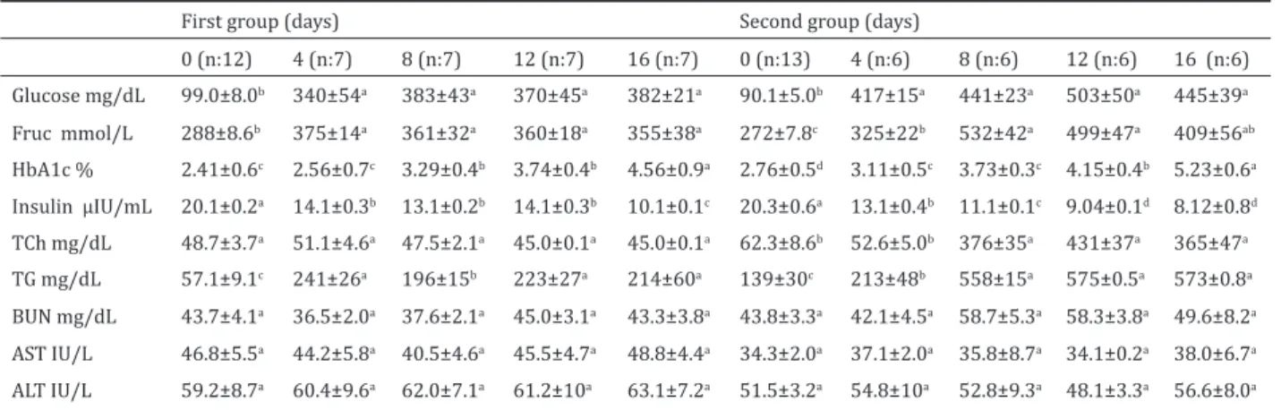
Biochemical parameters are presented in Table 1. Fasting serum glucose, HbA1c, fructosamine, and tri-glyceride concentrations of the rabbits in the first and the second group significantly increased (p<0.001) throughout the experiment. Changes in serum cho-lesterol levels of rabbits in the first group were not significant while it significantly increased in the sec-ond group (p<0.001) during the experiment. Serum insulin concentration determined in both of groups significantly decreased (p<0.001) during the experi-ment. Alterations in serum BUN, ALT and AST concen-trations were insignificant in both groups.
Hematologic findings
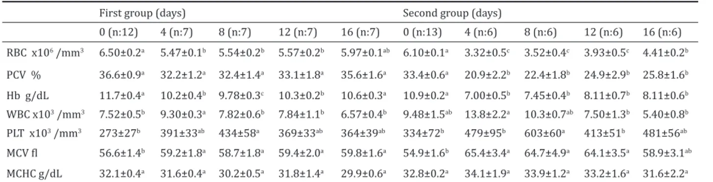
Hematologic results are presented in Table 2. Blood RBC, PCV and Hb concentrations significantly
de-creased in both groups throughout the experiment. Blood WBC counts on the 4th day (p<0.05) and thrombocyte counts throughout the study (p<0.001) increased in both groups. MCV concentrations also slightly increased (p<0.05) in both groups during the experiment. MCHC levels did not significantly change between the groups throughout the experiment.
Discussion
Alloxan monohydrate induces diabetes mellitus through causing permanent damage to the beta cells of the pancreas (Arai et al 2002, Burski et al 2004). DM is characterized by glycosuria, osmotic polyuria, polyphagia and polydipsia (Tiftik et al 1992, Kim et al 2006). Kim et al (2006) reported that diabetes melli-tus was induced in dogs by injection of alloxan mono-hydrate at the dose of 100 mg/kg, While Zalesk and Bryla (1978) reported induction of diabetes mellitus in rabbits by injecting alloxan monohydrate at the dose of 200 mg/kg through the Auricular vein. Waters (1950) also reported alloxan monohydrate injection via auricular vein at the dose of 175 mg/kg induced diabetes mellitus in rabbits. The results of this in-vestigation demonstrated that the administration of a single dose of alloxan to rabbits produced a repro-ducible model of diabetes mellitus. Three days after alloxan injection, rabbits exhibited polyuria, polypha-gia and polydipsia. The clinical and laboratory find-ings were more severe in rabbits in the second group when compared to the rabbits in the first group. Se-vere polydipsia was observed in many of the rabbits. Moreover, 5 rabbits in the first group and 7 rabbits in the second group died within three days of alloxan injection. The cause of high mortality among rabbits might have been related to hypoglycemia or renal tox-icity induced by alloxan (Kim et al 2006).
Fasting blood glucose levels in healthy cats and dogs were reported to be 75-120 mg/dL (Nelson 1985, Başoğlu ve Sevinç 2004, Fracassi et al 2007), while
15
Table 1. Some serum biochemical parameters of the first and second groups (mean±SE).
First group (days) Second group (days)
0 (n:12) 4 (n:7) 8 (n:7) 12 (n:7) 16 (n:7) 0 (n:13) 4 (n:6) 8 (n:6) 12 (n:6) 16 (n:6) Glucose mg/dL 99.0±8.0b 340±54a 383±43a 370±45a 382±21a 90.1±5.0b 417±15a 441±23a 503±50a 445±39a Fruc mmol/L 288±8.6b 375±14a 361±32a 360±18a 355±38a 272±7.8c 325±22b 532±42a 499±47a 409±56ab HbA1c % 2.41±0.6c 2.56±0.7c 3.29±0.4b 3.74±0.4b 4.56±0.9a 2.76±0.5d 3.11±0.5c 3.73±0.3c 4.15±0.4b 5.23±0.6a Insulin µIU/mL 20.1±0.2a 14.1±0.3b 13.1±0.2b 14.1±0.3b 10.1±0.1c 20.3±0.6a 13.1±0.4b 11.1±0.1c 9.04±0.1d 8.12±0.8d TCh mg/dL 48.7±3.7a 51.1±4.6a 47.5±2.1a 45.0±0.1a 45.0±0.1a 62.3±8.6b 52.6±5.0b 376±35a 431±37a 365±47a TG mg/dL 57.1±9.1c 241±26a 196±15b 223±27a 214±60a 139±30c 213±48b 558±15a 575±0.5a 573±0.8a BUN mg/dL 43.7±4.1a 36.5±2.0a 37.6±2.1a 45.0±3.1a 43.3±3.8a 43.8±3.3a 42.1±4.5a 58.7±5.3a 58.3±3.8a 49.6±8.2a AST IU/L 46.8±5.5a 44.2±5.8a 40.5±4.6a 45.5±4.7a 48.8±4.4a 34.3±2.0a 37.1±2.0a 35.8±8.7a 34.1±0.2a 38.0±6.7a ALT IU/L 59.2±8.7a 60.4±9.6a 62.0±7.1a 61.2±10a 63.1±7.2a 51.5±3.2a 54.8±10a 52.8±9.3a 48.1±3.3a 56.6±8.0a Fruc; fructosamine, HbA1c; hamoglobin A1c, TCh; total cholesterol, TG; triglyceride. a, b, c, d; different letters in the same line are statistically
fasting blood glucose levels in healthy rabbits were 75-155 mg/dL (Friend ve ark 1981). The most signif-icant laboratory findings in the diagnosis of diabetes mellitus are persistent fasting hyperglycemia (>200 mg/dL) and glycosuria (Nelson 1985, Turgut 2000, Kimmel 2002, McGuire et al 2002, Khan 2005). It was reported that fasting plasma glucose concentrations significantly increased in alloxane-induced diabetic dogs (Tiftik et al 1992, Arai et al 2002, Kim et al 2006). Similarly, Friend et al (1981) and Burski et al (2004) reported that a significant increase in fasting plasma glucose level in alloxane-induced diabetic rabbits. In the present study, it was found that the fasting serum glucose concentrations of the rabbits in the first and the second groups significantly increased (p<0.001) throughout the experiment. However, the increase observed in the serum fasting glucose concentrations of rabbits in the second group was higher than that of rabbits in the first group. The cause of high serum glucose levels observed in this group may be related to the administration of high-dose (175 mg/kg) of alloxan, as administration of high-dose (175 mg/kg) alloxan causes marked damage to pancreatic beta cells, reducing insulin secretion from pancreatic beta cells, and thus resulting in hyperglycemia. These find-ing are consistent with the results obtained by many other researchers (Friend et al 1981, Arai et al 2002, Burski et al 2004, Kim et al 2006, Fracassi et al 2007). Serum fructosamine measurements should be per-formed in the diagnosis and monitoring of diabetes mellitus (Jensen 1993, Loste and Marca 2001, Davi-son et al 2005). Fructosamines are stable ketamine compounds formed when glucose in the open chain form reacts non-enzymatically with amino groups of proteins. Serum fructosamine increases with pro-longed hyperglycemia or propro-longed hyperprotein-emia (Jensen 1995). Kimmel et al (2000) and Davison et al (2005) reported that median serum fructosamine concentrations in diabetic dogs were high and Martin et al (2006) also reported that serum fructosamine concentrations (>340 μmol/L) significantly increased in obese dogs. High serum fructosamine concentra-tions were also determined in hypothroid dogs (Re-usch et al 2002). Lost and Marca (2001) reported that
the determination of blood glycosylated hemoglobin and fructosamine levels was a significant and reliable criterion in the diagnosis of diabetes. In this study, serum fructosamine concentrations of the rabbits in the first and the second groups significantly increased (p<0.001) during the experiment. However, serum fructosamine concentrations of the rabbits in the sec-ond group were higher than that of the rabbits in the first group. It was reported that fructosamine could be used as the indicator of hyperglysemia, either due to stress or diabetes mellitus in some animals, espe-cially in cats (Crenshaw 1996, Loste and Marca 2001). The results of this study indicated that fructosamine could also possibly be used in the diagnosis of diabe-tes mellitus in rabbits. These results were consistent with the results reported by Kimmel et al (2000), Lost and Marca (2001) and Davison et al (2005).
Blood glycosylated hemoglobin measurement may be useful in the diagnosis and monitoring of diabe-tes mellitus in human as it reflect the plasma glucose concentration for the previous months (Cerami et al 1978). In normal human beings, HbA1c constitutes approximately 3% of the total hemoglobin concentra-tion, while in human diabetics, HbA1c values range from 6% to 10% of the total hemoglobin concentra-tion (Trivelli et al 1971, Wong et al 2006). Wood and Smith (1980) found that HbA1c concentration in healthy dogs was 3% of the total hemoglobin con-centration and it was between 3% and 7% in diabetic dogs. The period of hyperglycemia required to elevate HbA1c in dogs is unknown, but it would be expected to be approximately the same as that of human beings, because dogs have a similar RBC life span (approx 120 days). If times are similar, measured HbA1c would re-flect plasma glucose status for preceding 3 to 4 weeks, which would be more useful to monitor insulin ther-apy (Wood and smith 1980). On the other hand, Lost and Marca (2001) reported that the determination of glycosylated hemoglobin and fruktosamin levels could provide useful information in the diagnosis and control of diabetes. Glycosylated hemoglobin is an in-dicator of the blood glucose concentration over the preceding two to three months (Nelson 2000). In this study, it was observed that blood HbA1c concentra-Table 2. Hematologic parameters of the first and second groups (mean±SE).
First group (days) Second group (days)
0 (n:12) 4 (n:7) 8 (n:7) 12 (n:7) 16 (n:7) 0 (n:13) 4 (n:6) 8 (n:6) 12 (n:6) 16 (n:6) RBC x106 /mm3 6.50±0.2a 5.47±0.1b 5.54±0.2b 5.57±0.2b 5.97±0.1ab 6.10±0.1a 3.32±0.5c 3.52±0.4c 3.93±0.5c 4.41±0.2b PCV % 36.6±0.9a 32.2±1.2a 32.4±1.4a 33.1±1.8a 35.6±1.6a 33.4±0.6a 20.9±2.2b 22.4±1.8b 24.9±2.9b 25.8±1.6b Hb g/dL 11.7±0.4a 10.2±0.4b 9.78±0.3c 10.3±0.2b 10.6±0.3a 10.9±0.2a 7.00±0.5b 7.45±0.4b 8.11±0.7b 8.11±0.6b WBC x103 /mm3 7.52±0.5b 9.30±0.3a 7.82±0.6b 7.84±1.1b 6.57±0.4b 9.48±1.5ab 13.8±2.2a 10.3±0.7ab 7.50±1.3b 5.40±0.8b PLT x103 /mm3 273±27b 391±33ab 434±58a 369±33ab 364±39ab 334±72b 479±95b 603±60a 413±51b 481±56ab MCV fl 56.6±1.4b 59.2±1.8a 58.7±1.8a 59.4±2.0a 59.8±1.6a 54.9±1.6b 65.4±3.4a 64.7±4.9a 64.1±3.5a 58.9±3.1ab MCHC g/dL 32.1±0.4a 31.6±0.4a 30.2±0.5a 31.8±1.4a 29.9±0.6a 32.8±0.2a 34.1±1.9a 33.9±1.2a 33.2±1.6a 31.6±2.2a
17
tions of the rabbits in the first and the second group progressively increased (p<0.001) during the experi-ment. However, in our study, the cause of the 2-3 fold increase observed in the HbA1c levels may be related to the short time of the experimental period. Serum fasting glucose, fructosamine and HbA1c parameters in rabbits with diabetes mellitus may be used as diag-nostic indicators. These findings are consistent with the results of Wood and Smith (1980) and Nelson (2000).
Type-I or insulin-dependent DM is caused by the lack of insulin and results from the progressive destruc-tion of the insulin-producing beta cells in the pancre-as. A decrease is observed in the blood endogenous insulin level while an increase in blood glucose level in DM are observed (Nelson 2000, Khan 2005, Kim et al 2006, Martin et al 2006, Parsons et al 2007). Allox-an is the selective agent that destroys the beta cells of pancreas and causes type-1 diabetes mellitus (Pow-ers 2001, Arai et al 2002, Burski et al 2004, Feldman and Nelson 2004). Kirk et al (1993) determined that the insulin levels in cats with type-1 diabetes mellitus were significantly lower when compared to the insu-lin levels in cats with type-2 diabetes mellitus. Par-sons et al (2007) reported that endogenous insulin levels in dogs with diabetic ketoacidosis had a signifi-cant decrease. In this study, it was determined that the serum insulin concentrations of the rabbits in the first and the second groups significantly decreased during the experiment and decrease was more marked in the second group. These results were consistent with the results of Arai et al (2002) and Burski et al (2004). One of the most important problems in patients with diabetes is dyslipidemia. Dyslipidemia is defined as the increase of triglyceride and cholesterol levels in the blood. Insulin regulates the biosynthesis and metabolism of triglycerides and the turnover of tri-glycerides cholesterol in lipoproteins at several ma-jor points in both the metabolic pathway and at the cellular receptor and absorption pathway (Goldberg 2001); in addition, it is a major regulator of adipose tissues lipid metabolism. Insulin resistance develops in patients with diabetes mellitus. Therefore, blood triglyceride and cholesterol levels increase in diabetes mellitus (Khan and Flier 2000). In diabetic patients, high-density lipoprotein (HDL) levels decrease, but low-density lipoprotein (LDL) and triglyceride levels increase (Kreisberg 1998, Nelson et al 2000). Hyper-cholesterolemia and hypertriglyceridemia usually occur in dogs with hypothyroidism and diabetes mel-litus (Ford et al 1993). Nelson et al (2000) reported that serum cholesterol levels significantly increased in a cat with diabetes mellitus. In this study, serum triglyceride concentration of the rabbits in the first and the second group significantly increased and un-like first group, serum total cholesterol levels of rab-bits in the first group during the experiment, serum
total cholesterol concentrations of the rabbits in the second group significantly increased during the ex-periment.
It is reported that normocytic normochromic ane-mia can rarely be observed in the diabetics (Ford et al 1993). Normal values in healthy rabbits are; WBC: 5-12.5 x10³ µL, RBC:5-8 x10⁶ µL, Hb: 10-17 g/dL, PCV: 35-50, thrombocyte 250-650 x103 µL, MCV: 58-67 fl and MCHC: 29-37 g/dL (Khan 2005). In the present study, RBC, PCV and Hb concentrations of the rabbits in the second group were much below the ref-erence values but these were within normal refref-erence limits in first group (Khan 2005), MCV concentrations of the rabbits in the both group slightly increased but MCHC level did not change so it could be stated that mild normocytic normochromic anemia developed in rabbits in the second group. There may be a close relationship between the formation of anemia and the alloxan dose. Nelson (1985) has reported that leucocytosis may occur in diabetic patients. In this study, blood WBC counts of the rabbits in the both groups increased on the 4th day of the experiment. The increase in leucocytes count might be related to the inflammation caused by the drug (Turgut 2000). Blood trombocyte counts of the rabbits in the both groups significantly increase during the experiment, but these values were within normal reference limits reported for rabbits (Khan 2005).
Conclusion
Diabetes mellitus in the rabbits was successfully in-duced by a single dose of alloxan monohydrate via intra-venouse route. The clinical and laboratory find-ings of the rabbits in the second group were more severe. Serum fasting glucose, fructosamine, HbA1c and triglyceride concentrations increased and insulin level decreased in all rabbits with diabetes mellitus. Moreover, normocytic normochromic anemia oc-curred in rabbits in the second group. Serum fasting glucose, fructosamine and HbA1c may be reliable in-dicators for diagnosing DM.
Acknowledgments
This article is the summarized from PhD thesis and supported by Selcuk University Scientific Research Coordinator (No.6202012).
References
Akgül A, 2003. Tıbbi Araştırmalarda istatistiki analiz tek-nikleri “SPSS uygulamaları”. Emek Ofset, Second edition, Ankara, Turkey, pp: 23-26.
Arai T, Nakamura M, Magori E, Fukuda H, Mizutani H, Kawakami E, Sako T, 2002. Changes in activities of en-zymes related to energy metabolism in peripheral leu-kocytes of diabetic dogs with glycemic control by inten-sive insulin treatment. Res Vet Sci, 73, 183-186.
Bakırel T, Bakırel U, Üstüner O, Güneş S, Yardibi H, 2008. In vivo assesment of antidiyabetic and antioxidant activi-ties rosemary (Rosmarinus officinalis) in alloxan-diya-betic rabbits. J Ethnopharmaco, 116, 64-73.
Başoğlu A, Sevinç M, 2004. Evcil hayvanlarda metabolik ve endokrin hastalıklar. S. Ü. Vakfı Yayınları, Konya, Türki-ye, pp: 158-160.
Burski K, Ueland T, Maciejewski R, 2004. Serum amylase ac-tivity disorders in the course of experimental diyabetes in rabbits. Vet Med Czech, 49, 197-200.
Cerami A, Koening R, Peterson, CM, 1978. Hamoglobin A1c and diabetes mellitus. Br J Haematol, 38, 1-4.
Chansaisakorn W, Sprishavatsarakorn P, Sapokdittapong P, Trisiriroj M, Pondeenana S, Buranakarl C, 2009. Oxida-tive stress and intraerythrocytic concentrations of so-dium and potassium in diabetic dogs. Vet Res Commun, 33, 67-75.
Crenshaw KL, Peterson ME, Heeb LA, Moroff SD, Nichols R, 1996. Serum fructosamine concentration as an index of glycemia in cats with diabetes mellitus and stress hy-perglycemia. J Vet Inter Med, 10, 360-364.
Davison LJ, Herrtage ME, Catchpole B, 2005. Study of 253 dogs in the United Kingdom with diabetes mellitus. Vet Rec, 156, 467-471.
Feldman EC, Nelson RW, 2004. Canine and Feline Endocri-nology and Reproduction, 3rd edition, W. B. Saunders, Philadelphia, USA.
Ford SL, Nelson RW, Feldman EC, Niwa D, 1993. Insulin re-sistance in three dogs with hypothyroidism and diabe-tes mellitus. JAVMA, 202, 1478-1480.
Fracassi F, Mandrioli L, Diana A, Hilbe M, Grinwis G, Gandini G, 2007. Pituitary macroadenoma in a cat with diabetes mellitus, hypercortisolism and neuroligical signs. J Vet Med A, 54, 359-363.
Friend J, Kiorpes TC, Thoft RA, 1981. Diabetes mellitus and the rabbit corneal epithelium. Inves Ophthalmol Vis Sci, 21, 317-321.
Garcia JL, Bruyette DS, 1998. Using oral hypoglycemic agents to treat diabetes mellitus in cats. Vet Med, 93, 736-742. Guptill L, Glickman L, Glickman N, 2003. Time trends and
risk factors for diabetes mellitus in dogs: Analysis of vet-erinary medical data base records (1970-1999). Vet J, 165, 240-247.
Goldberg IJ, 2001. Clinical review 124: Diabetic dyslipid-emia: causes and consequences. J Clin Endocrinol Me-tabol, 86, 965-971.
Jensen AL, 1993. Various protein and albumin corrections of the serum fructosamine concentrations in the diag-nosis of canine diabetes mellitus. Vet Res Commun, 17, 13-23.
Jensen AL, 1995. Glycated blood proteins in canine diabetes mellitus. Vet Rec, 137, 401-405.
Khan CM, 2005. Merck Veterinary Manual, Ninth edition, National Publishing Inc., Philadelphia, USA, pp: 2584-2587.
Khan BB, Flier JS, 2000. Obesity and insulin resistance. J Clin Invest Med, 106, 473-481.
Kim JM, Chung JY, Lee SY, Choi EW, Kim MK, Hwang CY, Young HY, 2006. Hipoglycemic effects of vanadium on alloxan monohydrate induced diabetic dogs. J Vet Sci, 7, 391-395.
Kimmel SE, Ward CR, Henthorn PS, Hess RS, 2002. Familial insulin-dependent diabetes mellitus in samoyed dogs. J Am Anim Hospital Assoc, 38, 235-238.
Kimmel SE, Michel KE, Hess, RS, Ward CR, 2000. Effects of insoluble and soluble dietary fiber on glycemic control in dogs with naturally occurring insulin-dependent dia-betes mellitus. JAVMA, 216, 38-42.
Kirk CA, Feldman EC, Nelson RW, 1993. Diagnosis of Natu-rally acquired type-1 and type-2 diabetes mellitus in cats. Am J Vet Res, 54, 463-467.
Kreisberg RA, 1998. Diyabetic dyslipidemia. Am J Cardiol, 82, 89-90.
Loste A, Marca MC, 2001. Fructosamine and glycated hemo-globin in the assessment of glycaemic control in dogs. Vet Res, 32, 55-62.
Martin LJM, Siliart B, Dumon HJV, Nguyen PG, 2006. Hor-monal disturbances associated with obesity in dogs. J Anim Physiol Animal Nutri, 90, 355-360.
McGuire NC, Schulman R, Ridgway MD, Bolero G, 2002. De-tection of occult urinary tract infections in dogs with di-abetes mellitus. J Am Anim Hospital Assoc, 38, 541-544. Nelson RW, 1985. Disorders of glucose metabolism in the
dog-1: diabetes mellitus. Vet Med, 80, 27-36.
Nelson RW, 2000. Diabetes mellitus. In: Textbook of Vet-erinary Internal Medicine, Eds: Ettinger SJ, Feldman FC,5th edition, Philadelphia, USA, pp: 1438-1460. Nelson RW, Scott-Moncrieff JC, Feldman EC,
DeVries-Con-cannon SE, Kass PH, Davenport DJ, Kiernan CT, Neal LA, 2000. Effect of dietary insoluble fiber on control of gly-cemia in cats with naturally acquired diabetes mellitus. JAVMA, 216, 1082-1088.
Onkamo P, Vaanenen S, Karvonem M, Tuomiletho J, 1999. Worldwide increase in incidence of type I diabets-the analysis of the data on published incidence trends. Dia-betol, 42, 1395-1403.
Parsons SE, Drobatz KJ, Lamb SV, Ward CR, Hess RS, 2007. Endogenous serum insulin concentration in dogs with diyabetic ketoacidosis. J Vet Emerg Crit Care, 12, 147-152.
Powers AC, 2001. Diabetes mellitus. In: Harrison’s princi-ples of internal medicine, Ed; Braunwald E, New York, USA, pp: 2109-2137.
Remillard RL, 1999. Nutritional management of diabetic dogs. Compen Cont Edu Pract Vet, 21, 699-700. Reusch CE, Gerber B, Boretti FS, 2002. Serum fruktosamin
concentrations in dogs with hypothyroidism. Vet Res Commun, 26, 531-536.
Smith SA, Freeman LC, Swanson MB, 2002. Hypercalcemia due to iatrogenic secondary hypoadrenocorticism and diabetes mellitus in a cat. J Am Anim Hospital Assoc, 38, 41-44.
Somani R, Kasture S, Singhai AK, 2006. Antidiabetic poten-tial of Bueta monosperma in rats. Fitotherapia, 77, 86-90.
Tiftik AM, Turgut K, Gürbilek M, Sevinç M, 1992. Köpeklerde alloksan ile oluşturulan experimental diyabetes üzeri-nde araştırmalar. S Ü Vet Fak Derg, 8, 18-21.
Trivelli LA, Ranney HM, Lai HT, 1971. Hamoglobin compe-nents in patients with diabetes mellitus. North Eng J Med, 284, 354-357.
Turgut K, 2000. Veteriner Klinik Laboratuvar. Bahcıvanlar Basım Sanayi, Konya, s: 321-326.
Waters JW, 1950. Biochemical and clinical changes in the rabbit lens during alloxan diabetes. Biochem J, 46, 575-578.
Wood PA, Smith JE, 1980. Glycosylated hemoglobin and ca-nine diabetes mellitus. JAVMA, 176, 1267-1268. Wong J, Molyneaux L, Constantio MI, Twing SM, Yue DK,
2006. Metabolic syndrome in type 2 diyabetes: When does it matter? Diabet Obes Metabol, 8, 690-697. Zaleski J, Bryla J, 1978. Effect of alloxan-diyabetes on
gluco-neogenesis and ureogenesis in isolated rabbit liver cells. Biochem J, 176, 563-568.