Effects of BCRP and P-gp Modulators on the Penetration of Aflatoxin
B
1into the Mouse Brain
[1]Bunyamin TRAS
1Gul CETIN
2Kamil UNEY
1
Burak DIK
1Orhan CORUM
3Sema ATALAY
4[1] This study was presented in abstract form at 32nd World Veterinary Congress, Istanbul, Turkey, 13-17 September, 2015 1 Department of Pharmacology and Toxicology, Faculty of Veterinary Medicine, University of Selcuk, TR-42031 Konya - TURKEY 2 Department of Pharmacology and Toxicology, Faculty of Veterinary Medicine, University of Mehmet Akif Ersoy, TR-15030 Burdur - TURKEY
3 Deptartment of Pharmacology and Toxicology, Faculty of Veterinary Med., University of Dicle, TR-21280 Diyarbakır - TURKEY 4 Konya Laboratory and Warehousing, Agricultural, Food, Energy Inc.,TR-42050 Konya - TURKEY
Article Code: KVFD-2016-15904 Received: 20.04.2016 Accepted: 19.06.2016 Published Online: 22.06.2016 Citation of This Article
Tras B, Cetin G, Uney K, Dik B, Corum O, Atalay S: Effects of BCRP and P-gp modulators on the penetration of aflatoxin B1 into the mouse brain.
Kafkas Univ Vet Fak Derg, 23, 95-100, 2017. DOI: 10.9775/kvfd.2016.15904
Abstract
This study was conducted to determine whether the plasma and brain concentrations of AFB1 are affected by the modulation of P-gp and BCRP using zosuquidar (ZQR) and prazosin (PRZ), respectively. In this study, a total of 40 healthy adult male BALB/c mice (32±3.7 g) were used. The animals were randomly divided into 5 groups, with 8 animals per group. Group 1 was used for method validation. Group 2 (AF) received intraperitoneal AFB1 at a dose of 20 mg/kg of body weight. Groups 3 (AF+PRZ), 4 (AF+ZQR), and 5 (AF+PRZ+ZQR) received 20 mg/kg of AFB1 intraperitoneally 30 min after the intraperitoneal administration of prazosin (0.3 mg/kg), zosuquidar (25 mg/kg), and prazosin+zosuquidar (0.3 mg/kg prazosin + 25 mg/kg zosuquidar), respectively. Six hours after the administration of AFB1, blood and brain samples were collected from the animals in Groups 2 to 5. AFB1 concentrations were determined using an HPLC system with fluorescence detection. Individual and simultaneous administration of prazosin and zosuquidar significantly reduced the brain concentrations of AFB1 in comparison to a single administration of AFB1 (P<0.05). The brain/plasma ratio of the AF group was higher than that of the other groups (AF+PRZ, AF+ZQR, and AF+PRZ+ZQR) (P<0.05). Inducers of transmembrane proteins, especially BCRP, can be life saving during acute AFB1 poisoning.
Keywords: Aflatoxin B1, Brain, Drug transporter proteins, Modulation, Mice
Aflatoksin B
1’in Fare Beynine Geçişi Üzerine BCRP ve P-gp Modülatörlerinin
Etkisi
Özet
Çalışma, zosuquidar ve prazosin tarafından sırasıyla P-gp ve BCRP modülasyonunun AFB1’in plazma ve beyin konsantrasyonlarının etkileyip etkilemediğini belirlemek için gerçekleştirildi. Bu çalışmada, 40 adet sağlıklı, erkek BALB/c ırkı fare (32±3.7 g) kullanıldı. Hayvanlar her grupta 8 fare olacak şekilde rastgele 5 gruba ayrıldı. Grup 1, metod validasyon çalışmalarında kullanıldı. Grup 2 (AF)’ye AFB1 20 mg/kg dozda intraperitoneal yolla verildi. Grup 3 (AF+PRZ), 4 (AF+ZQR) ve 5 (AF+PRZ+ZQR)’e ise sırasıyla intraperitoneal yolla prazosin (0.3 mg/kg), zosuquidar (25 mg/kg) ve prazosin+zosuquidar (0.3 mg/kg prazosin + 25 mg/kg zosuquidar) uygulamalarından 30 dk. sonra AFB1 20 mg/kg dozda intraperitoneal yolla uygulandı. Grup 2-5’de bulunan hayvanlardan AFB1 uygulamasından sonraki 6. saatte kan ve beyin örnekleri alındı. AFB1 düzeyleri fluoresans dedektör içeren HPLC sisteminde tayin edildi. Prazosin ve zosuquidarın tek ve eşzamanlı uygulanması AFB1’in beyin konsantrasyonlarında sadece AFB1 uygulamasına göre önemli oranda azalmaya neden oldu (P<0.05). AFB1 grubunda AFB1’in beyin/plazma oranı diğer gruplardan (AF+PRZ, AF+ZQR, and AF+PRZ+ZQR) önemli oranda daha yüksekti (P<0.05). Akut AFB1 zehirlenmelerinde özellikle BCRP gibi transmembran proteinlerin indüklenmesi hayatta kalma oranını artırabilir.
Anahtar sözcükler: Aflatoksin B1, Beyin, İlaç taşıyıcı proteinler, Modülasyon, Fare
INTRODUCTION
Aflatoxin B1 (AFB1), which is an environmental dietary
carcinogen, is one of the most toxic mycotoxins and causes significant losses of livestock [1-3]. While the clinical
signs of acute aflatoxicosis include epistaxis, blood stained faeces, and convulsions, sudden death is observed as the clinical sign of severe acute aflatoxicosis [4,5]. The
elimination half-life of AFB1 was determined to be 77 min
after intraperitoneal administration in mice [6]. There is a
İletişim (Correspondence)
+90 332 2232673high interaction potential to occur of AFB1 with food/feed
because AFB1 is a common food/feed contaminant. Also,
the ingredients found in the food/feed compositions can cause to change the efficiency and disposition of drug and toxic substances through enzyme-and transporter [7-9].
Aflatoxin B1 is a substrate of BCRP, and BCRP function
may affect systemic exposure to this mycotoxin [10,11].
Additionally, AFB1 and its metabolite aflatoxin B1
-epoksit-glutathione are substrates of multidrug resistance protein 1 (MRP1), although the affinity of MRP1 for AFB1 is low [12-14].
BCRP substrates tend to overlap with P-gp substrates [13,15],
but no information is available concerning whether AFB1
is a substrate of P-gp. Van Herwaarden et al.[10]noted that
BCRP plays an important role in the renal excretion of AFB1.
If AFB1 is a substrate of both BCRP and P-gp, the tissue
penetration of AFB1 may be changed by the modulation
of these transmembrane proteins.
The blood-brain barrier (BBB) protects the brain from a variety of endogenous and exogenous substances. The BBB not only limits substance flow from the blood to the brain tissue via the paracellular and transcellular routes but also permits the efflux of substances via several trans- membrane proteins, such as P-glycoprotein (P-gp) and the breast cancer resistance protein (BCRP). P-gp (ABCB1) and BCRP (ABCG2) are members of the ATP-binding cassette transporter superfamily. Both P-gp and BCRP are expressed in mammalian capillary endothelial cells at BBB sites, and these transporters work in tandem to limit the accumulation of substances in tissues [11,16-19]. P-gp and BCRP are inducible
and inhibitable in vivo and in vitro by various substances. The efflux activity of these transmembrane proteins has been described as saturable [19].Because P-gp and BCRP
have a broad substrate specificity and tissue distribution, the modulation of these transmembrane proteins may result in significant alterations in the pharmacokinetics, pharmacodynamics, and toxicity of their substrates. For example, Van Herwaarden et al.[10]found that the brain
concentration of [14C] 2-amino-3-methylimidazo[4,5-f ]
quinoline, which is a BCRP substrate, was higher in bcrp-/- mice than wild-type mice.
The aim of this study was 1) to determine the passage of AFB1 into the brain and 2) to evaluate whether the
plasma and brain concentrations of AFB1 are affected by
the modulation of P-gp and BCRP using prazosin and zosuquidar, respectively. This study is the first in vivo experimental study intended to evaluate the modulation of AFB1 passage into the brain.
MATERIAL and METHODS
Chemicals and Reagents
All reagents were of recognised analytical grade. AFB1
was obtained from Biopure Chemical Co. (Romer Labs
Inc., 1301 Stylemaster Drive Union) and dissolved in corn oil for injection. Zosuquidar hydrochloride (99%) was obtained from MedKoo Biosciences (Canada, USA) and dissolved in a solution that included glycine (15 mg) and mannitol (200 mg) in 1 mL of distilled water for injection. Prazosin hydrochloride (≥99%) was purchased from Sigma Chemical Co. (Saint Louis, MO, USA) and dissolved in 1 mL of distilled water for injection. Immunoaffinity columns (Aflatest® WB) were purchased from Vicam (Watertown, MA, USA). Monobasic potassium phosphate (KH2PO4), sodium
phosphate dibasic (Na2HPO4), sodium chloride (NaCl),
potassium bromide (KBr), nitric acid (4 mol/L), and HPLC-grade acetonitrile and methanol were purchased from Merck (Darmstadt, Germany). All water used in this study was deionised and distilled. For the high performance liquid chromatograpgy (HPLC), water was passed through a water purification system (aqua Max-Ultra System, Younglin Instrument Co. Ltd., South Korea). Phosphate buffer solution (PBS) was prepared by dissolving 0.2 g of KCl, 0.2 g of KH2PO4,
1.16 g of anhydrous Na2HPO4, and 8 g of NaCl in 1,000 mL
of water, and the pH of the solution was adjusted to 7.4 with 0.1 N NaOH. A Tween-20 (Amresco, USA) solution was prepared by adding 15 mL of Tween to 85 mL of PBS and mixing.
Experimental Design
In this study, a total of 40 healthy adult male BALB/c mice (32±3.7 g) were used. Mice were supplied from Experimental Animal Production and Research Laboratory, Faculty of Veterinary Medicine, University of Mehmet Akif Ersoy, Burdur, Turkey. All experimental administrations on animals were carried out in here. The animals were housed individually in plastic cages with grated stainless steel floors in a controlled environment (temperature 25±1°C, relative humidity 60±10%, and artificial lighting sequenced to generate a 12-h light/dark cycle). The animals had ad
libitum access to water and a commercial diet (Optima
Feeds, Kırklareli, Turkey) that included the following: 89% dry matter, 21% crude protein, 2850 kcal/kg metabolic energy, 5% crude fibre, 0.75% methionine and cysteine, 1.0-2.0% calcium, 0.5-1.0% phosphorus, and 0.5% sodium.
All animal protocols in this study were approved by the Ethical Committee of the Faculty of Veterinary Medicine, University of Mehmet Akif Ersoy (No: 2014-71).
The animals were randomly divided into 5 groups of 8 animals each. Group 1 was used for method validation without any treatment of the animals. AFB1
was administered intraperitoneally at a dose of 20 mg/ kg of body weight to mice in Group 2-5. In the present study, the dose of AFB1 was determined by taking into
consideration concerns about AFB1 analysis, the natural
poisoning state and the dose previously reported Bastaki et al.[6] because no experimental research is available
concerning the passage of AFB1 into the brain. Group 2
kg of body weight 30 min after the IP administration of 1 mL of the glycine/mannitol solution. Groups 3 (AF+PRZ), 4 (AF+ZQR), and 5 (AF+PRZ+ZQR) were administered IP AFB1 at a dose of 20 mg/kg of body weight 30 min after the
IP administration of prazosin (0.3 mg/kg of body weight) plus the glycine/mannitol solution (1 mL), zosuquidar (25 mg/kg of body weight), and prazosin (0.3 mg/kg of body weight) plus zosuquidar (25 mg/kg of body weight), respectively. The animals were observed for general behavioural changes, signs of toxicity, and mortality continuously for 6 h following the administration of AFB1.
Six hours after the administration of AFB1 in Groups 2 to
5, blood samples from the hearts of animals under ether anaesthesia were collected into tubes containing heparin. All animals were euthanized using the cervical dislocation method. Plasma and brain tissues were collected from the animals and carried to Department of Pharmacology and Toxicology, Faculty of Veterinary Medicine, University of Selcuk, Konya, Turkey for HPLC analysis. All samples were stored at -70°C until the time of analysis.
HPLC and Chromatographic Conditions
HPLC analyses were performed using an Agilent 1100 series HPLC system, which consisted of a G1379A degasser, a G1310A isocratic pump, a G1313A autosampler, a G1316A column oven, and a fluorescence detector (model G1321A, Agilent Technologies, Palo Alto, California, USA). Data acquisition was performed using the Chemstation 3D software (Agilent Technologies, Palo Alto, California, USA). For HPLC analysis, a reverse-phase ACE C18 analytical column (5 mm particle size, 4.6 x 250 mm) was employed. The column temperature was maintained at 30°C. Postcolumn derivatisation resulting in enhanced fluorescence was achieved with electrochemically-generated bromine in a Cobra cell (Coring System Diagnostics GmbH, Gernsheim, Germany), using a reaction tube that consisted of a 0.5 mm id x 34 cm length of polyether ketone tubing. The LC mobile phase solvent used with electrochemically-generated bromine was water/methanol/acetonitrile (60:20:20, v/v/v). To each litre of mobile phase, 120 mg of potassium bromide and 350 mL of 4 M nitric acid were added. The flow rate was 1 mL/min. The fluorescence detector was set to excitation and emission wavelengths of 360 and 430 nm, respectively. The injection volume for the standards and samples was 100 µL.
Standard Preparation
A stock solution was prepared by dissolving 5 mg of AFB1 in 5 mL of methanol and stored at -70°C. The
stock solution was further diluted with water/methanol/ acetonitrile (60:20:20, v/v/v) to generate a series of working standard solutions. Calibration standards of AFB1 (0.5, 1, 5,
10, 25, 50, 100, 200 ng/mL) were prepared by adding 20 µL of the working standard solution to 180 µL of plasma or 180 mg of brain tissue. Quality control samples (QC) at low (5 ng/mL), medium (25 ng/mL) and high (200 ng/
mL) concentrations were used for the method validation program. These calibration standards and QC samples were processed according to the described method of sample preparation prior to HPLC analysis.
Sample Preparation
Plasma and brain tissue samples were extracted and cleaned up according to the procedures described by Association of Official Analytical Chemist [20] and Chiavaro
et al.[21], with some modifications. Briefly, 200 µL of plasma
or 200 mg of tissue sample were blended with 200 mg of NaCl in 1.2 mL of methanol-water (80:20) using a tissue homogeniser (Heidolph Silent Crusher M, Germany) at 9.000 rpm for 1 min. The extract was filtered through Whatman No. 4 filter paper. A 1-mL filtrate sample was diluted to 7.2 mL with PBS containing 0.1% Tween-20. The final filtrate was passed through an immunoaffinity column at a flow rate of 1 drop/s. The column was then washed twice with 4 mL of ultrapure water. AFB1 was eluted
with 0.5 mL of methanol followed by 0.5 mL of water in a glass vial. Finally, 100 µL of the sample was injected into the HPLC system.
Method Validation
The specificity, linearity, sensitivity, recovery, precision, and accuracy of the employed method were determined to evaluate the performance of the analytical method. The specificity of the method was evaluated to identify interference from plasma and brain tissue. The calibration curve of AFB1 was constructed using seven calibration
standards over a calibration range of 0.5-200 ng/mL. The limits of detection (LOD) and quantification (LOQ) were determined via signal-to-noise ratio evaluations of samples spiked from 0.1-10 ng/mL. The LOD was defined at a signal-to-noise (S/N) ratio of 3:1. The LOQ was defined as the lowest quantifiable concentration of analyte with an accuracy within 20% and a precision <20%. The recovery of AFB1 was evaluated at concentrations corresponding
to the three QC values (5, 25, and 200 ng/mL), with six replicates. This parameter was determined by comparing the peak areas of the extracted QC samples with those of the working standards. The intra- and inter-day precision and accuracy were assessed by extracting and analysing five replicates of each QC sample. The intra- and inter- day precision and accuracy of the assay were determined based on the percent coefficient of variation (CV) and percent relative error (RE) values, respectively.
Statistical Analysis
All data were expressed as the mean ± SD. Data obtained from plasma and brain concentrations were analysed using one-way ANOVA, followed by the Duncan test. Statistical significance was assigned at P<0.05. The SPSS (Version 16.0 for Windows, Chicago, USA) statistical software was used for the statistical analyses.
RESULTS
No interfering peaks were observed in the blank plasma and brain tissue samples after the extraction. The assay was linear from 0.5 to 200 ng/mL, and the concentrations of the calibration standards were back-calculated using the peak area ratios of AFB1. The data were analysed using
linear regression analysis, and the correlation coefficients for the calibration curves prepared from plasma and brain tissue were ≥0.9996 and ≥0.9994, respectively. The LOD values were determined to be 0.2 and 0.5 ng/mL in plasma and brain tissue, respectively. The LOQ values were determined to be 0.5 and 1 ng/mL in plasma and brain tissue, respectively. The values obtained for the recovery of AFB1 ranged from 89.3 to 97.9% for the plasma and brain
samples and from 86.4 to 94.3% for the QC samples. In the plasma and brain samples, the intra-day precision that was calculated from three QC samples that were injected six times on the same day was low, with a precision ranging from 3.94% to 10.27% for the CV and an accuracy ranging from 94.3% to 107.6%. The inter-day precision and accuracy varied from 1.54% to 12.48% and from 92.1% to 109.71%, respectively.
The effects of prazosin and zosuquidar on murine plasma and brain concentrations of AFB1 after the IP
administration of AFB1 at a dose of 20 mg/kg of body
weight are presented in Table 1. Single and simultaneous administrations of prazosin and zosuquidar significantly reduced brain concentrations of AFB1 in comparison to a
single administration of AFB1 (P<0.05). The ratios of the
brain concentrations of AFB1 to the plasma concentrations
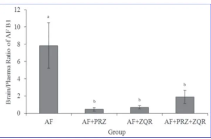
of AFB1 in the mice are shown in Fig 1. The brain/plasma
ratio in the AF group was higher than those of the groups (AF+PRZ, AF+ZQR and AF+PRZ+ZQR) that received prazosin and zosuquidar (Fig. 1, P<0.05).
No mortality was observed in the AFB1-treated groups
during the study; however, signs of toxicity, including oedema of the lower extremities, lethargy, muscular weakness, and convulsions, were observed in the AF group.
DISCUSSION
Although AFB1 was found at levels of 1-5 ppb in human
brain biopsy samples, as reported by Hooper et al.[22] no
experimental study investigated the ability of AFB1 to cross
the BBB. However, AFB1 was reported to be unable to pass
into the rat brain [23]. In this study, AFB
1 crossed the BBB after
the administration of 20 mg/kg AFB1, and the brain/plasma
ratio of AFB1 was 7.85 six hours after administration. Our
results suggest that AFB1 passed more intense to the brain
and cannot be removed immediately. We concluded that AFB1 accumulates in tissues, such as the brain, and cannot
be removed immediately due to the greater concentration of AFB1 in the brain than that in the plasma at the 6-hour
blood collection time.
Contrary to expectation in this study, prazosin administration caused a statistically significant decline in the brain concentration of AFB1 (P<0.05); however,
the decline in the plasma concentration of AFB1 was not statistically significant in the AF group (P>0.05). The brain/ plasma ratio of AFB1 in prazosin-treated mice was 0.45
(Fig. 1). Our results indicate that prazosin is an inducer of BCRP, although some authors reported that prazosin is a BCRP inhibitor and substrate [24-27]. The reason for this
contradiction may be differences in the materials used in the studies and in the employed doses of BCRP inhibitors and substrates. The drug affinity and the expression rate of BCRP differ among species, breeds, and organs [28,29].
Studies of inhibitors and inducers are typically conducted
in vitro. The majority of in vivo studies concerning the
passage of BCRP and P-gp substrates into the brain were conducted using transgenic experimental animals (bcrp/ p-gp -/-), such as mice and rats. Our study was conducted
in vivo using wild-type mice. The type of modulation (i.e.,
induction and/or inhibition) and the effectiveness of a substance on transporter proteins are reported to depend on the dose, the type of tissue, the substrate examined, and the time of administration [30,31]. Additionally, the
Fig 1. Ratios of brain concentrations to plasma concentrations of
aflatoxin B1. AF; aflatoxin B1, PRZ; prazosin, ZQR; zosuquidar. a,b Different
letters are statistically significant (P<0.05)
Şekil 1. AFB1’in beyin/plazma konsantrasyon oranları. AF; aflatoksin B1, PRZ; prazosin, ZQR; zosuquidar. a,b Farklı harfler istatistiksel açıdan
önemlidir (P<0.05)
Table 1. The plasma and brain concentrations of aflatoxin B1 at six hours
following the intraperitoneal administration of aflatoxin B1 (20 mg/kg) in
mice (n=8)
Tablo 1. Farelerde aflatoksin B1’in (20 mg/kg) intraperitoneal yolla
uygulanmasını takiben 6. saatte alınan plazma ve beyin örneklerindeki AFB1 konsantrasyonları (n=8)
Group Plasma (ng/mL) Brain (ng/g)
AF 23.45±13.84ab 181.23±89.40a
AF+PRZ 15.53±10.10b 6.69±2.81b
AF+ZQR 31.96±14.28a 24.07±19.64b
AF+PRZ+ZQR 13.37±9.50b 24.85±11.66b
a,b Different letters in the same column indicate statistically significant
difficulties of predicting in vivo relationships based on
in vitro transport assays were discussed by Enokizono
et al.[32]. Alternatively, the causes of the decreased AFB
1
concentration in the brain that is caused by prazosin may be associated with the roles of AFB1 as a P-gp substrate
and prazosin as a P-gp inducer. The inhibition of BCRP by prazosin may cause the activation of P-gp in the brain capillaries of mice. A compensatory relationship has been reported to exist between P-gp and BCRP, and P-gp has also been reported to be more prominent and inducible than BCRP at the BBB [33,34] Prazosin has been indicated to be
only an inducer of P-gp [23-27]. In addition, many BCRP and
P-gp substrates and modulators overlap [13,27,35].
In the present study similar to the results of prazosin, zosuquidar, which is a potent and selective P-gp inhibitor in
vitro, caused a non-statistically significant increase in the
plasma concentration of AFB1 but a statistically significant
decrease in the brain concentration of AFB1 (P<0.05).
Cripe et al.[34] reported that the in vivo inhibition of P-gp
by zosuquidar might lead to the activation of non-P-gp transmembrane proteins, such as BCRP. In addition, BCRP and P-gp presumably work in synergy or in a compensatory manner to restrict the passage of their concerted substrates into the CNS in mice, and P-gp can compensate for the deletion of BCRP, as suggested by Zhou et al.[33]. In addition, zosuquidar can be considered
to be an inducer of P-gp or a dual P-gp/BCRP inducer in brain capillaries. Zosuquidar is described as a specific P-gp inhibitor [13,19,35], and a number of BCRP and P-gp substrates
and modulators overlap [3,27,35].
Although the co-administration of prazosin and zosuquidar reduced the level of AFB1 in the brain, this
reduction was not statistically significant in comparison to the other two treatment groups. Thus, prazosin and zosuquidar do not show the combined effect to prevent the passage of AFB1 into the brain. This may be associated
with that prazosin or zosuquidar in combined use altere the response to the modulation of other transmembrane protein. Also, it has been reported that BCRP and P-gp work in synergy or in a compensatory manner for the efflux of their substrates and the modulation of a transmembrame protein by the inducer/inhibitor alteres the efflux effect of other transmembrane protein [33,34].
The administration of prazosin, zosuquidar and prazosin+ zosuquidar did not cause a statistically significant reduction of the plasma concentrations of AFB1 in comparison to
a single administration of AFB1 (AF group). Similar to
our findings, some previous studies reported that trans-membrane protein modulators caused no changes in the plasma concentrations or pharmacokinetics of the substrates of transmembrane proteins but did cause important changes in tissue concentrations [34,36,37]. Based
on the results of studies in this field, we believe that this difference may occur because the transmembrane proteins
in each tissue respond differently to inducers and because drugs/substances are transported by more than one transmembrane protein. Demeule et al.[38] found that
dexamethasone, which is a P-gp inducer, increased P-gp expression in the liver and the lung but reduced the expression of this molecule in the kidney. Transmembrane proteins in each tissue respond differently to inducers, as stated by Drescher et al.[39].
AFB1 is activated via the conversion into AFB1 8-9
epoxide by cytochrome 450 enzymes, especially CYP1A2 and CYP3A4, and glutathione S-transferases are the most important enzymes for detoxifying AFB1 in all species, including mice [40-42]. The plasma concentration of AFB
1 in the
ZQR-AF group was found to be significantly higher than those of the PRZ-AF and PRZ+ZQR-AF groups. The causes of this difference may be the inhibition of AFB1 metabolism
by zosuquidar and/or the induction of BCRP in excretion organs by prazosin. Van Herwaarden et al.[10] posited that
BCRP plays an important role in the renal excretion of AFB1.
P-gp inhibitors have also been reported to inhibit CYP3A activity, which plays an important role in AFB1 metabolism.
In summary, both prazosin and zosuquidar significantly reduced the brain concentration of AFB1 but not the
plasma concentration of this molecule. Thus, prazosin is a better inducer than zosuquidar for both BCRP and P-gp in brain capillaries. In addition, AFB1 may be a substrate
of both BCRP and P-gp. Inducers of transmembrane proteins, such as prazosin, can be life-saving during acute poisoning with AFB1, based on the overall health status
and brain concentrations of AFB1 in mice. The results of in
vitro studies in this issue should be confirmed with in vivo
studies of wild-type animals.
REFERENCES
1. Dalvi RR: An overview of aflatoxicosis of poultry its characteristics, prevention and reduction. Vet Res Commun, 10, 429-443, 1986. DOI: 10.1007/BF02214006
2. Oguz H: A review from experimental trials on detoxification of aflatoxin in poultry feed. Eurasian J Vet Sci, 27, 1-12, 2011.
3. Zain ME: Impact of mycotoxins on humans and animals. J Saudi Chem Soc, 15, 129-144, 2011. DOI: 10.1016/j.jscs.2010.06.006
4. Bortell R, Asquith RL, Edds GT, Simpson GF: Acute experimentally induced aflatoxicosis in the weanling pony. Am J Vet Res, 44, 2110-2114, 1983. 5. Chao TC, Maxwell SM, Wong SY: An outbreak of aflatoxicosis and boric acid poisoning in Malaysia: A clinicopathological study. J Pathol, 164, 225-233, 1991. DOI: 10.1002/path.1711640307
6. Bastaki SA, Osman N, Kochiyil J, Shafiullah M, Padmanabhan R, Abdulrazzaq YM: Toxicokinetics of aflatoxin in pregnant mice. Int J Toxicol, 29, 425-431, 2010. DOI: 10.1177/1091581810369565
7. Bailey DG: Fruit juice inhibition of uptake transport: A new type of food-drug interaction. Brit J Clin Pharmacol, 70, 645-655, 2010. DOI: 10.1111/j.1365-2125.2010.03722.x
8. Abdallah HM, Al-Abd AM, El-Dine RS, El-Halawany AM: P-glyco-protein inhibitors of natural origin as potential tumor chemosensitizers: A review. J Adv Res, 6, 45-62, 2015. DOI: 10.1016/j.jare.2014.11.008
9. An G, Mukker JK, Derendorf H, Frye RF: Enzyme- and transporter-mediated beverage-drug interactions: An update on fruit juices and
green tea. J Clin Pharmacol, 55, 1313-1331, 2015. DOI: 10.1002/jcph.563 10. Van Herwaarden AE, Wagenaar E, Karnekamp B, Merino G, Jonker JW, Schinkel AH: Breast cancer resistance protein (Bcrp1/Abcg2) reduces systemic exposure of the dietary carcinogens aflatoxin B1, IQ and Trp-P-1 but also mediates their secretion into breast milk. Carcinogenesis, 27, 123-130, 2006. DOI: 10.1093/carcin/bgi176
11. Robey RW, To KKK, Polgar O, Dohse M, Fetsch P, Dean M, Bates SE: ABCG: A perspective. Adv Drug Deliver Rev, 61, 3-13, 2009. DOI: 10.1016/j. addr.2008.11.003
12. Loe DW, Stewart RK, Massey TE, Deeley RG, Cole SP: ATP-dependent transport of aflatoxin B1 and its glutathione conjugates by the product of the multidrug resistance protein (MRP) gene. Mol Pharmacol, 51, 1034-1041, 1997.
13. Löscher W, Potscka H: Role of drug efflux transporters in the brain for drug disposition and treatment of brain diseases. Prog Neurobiol, 76, 22-76, 2005. DOI: 10.1016/j.pneurobio.2005.04.006
14. Sharom FJ: 2008: ABC multidrug transporters: Structure, function and role in chemoresistance. Pharmacogenomics, 9, 105-127, 2008. DOI: 10.2217/14622416.9.1.105
15. Giacomini KM, Huang SM, Tweedie DJ, Benet LZ, Brouwer KL, Chu X, Dahlin A, Evers R, Fischer V, Hillgren KM: International transporter consortium. Membrane transporters in drug development. Nat Rev Drug Discov, 9, 215-236, 2010. DOI: 10.1038/nrd3028
16. Eisenblatter T, Galla HJ: A new multidrug resistance protein at the blood-brain barrier. Biochem Biophys Res Commun, 293, 1273-1278, 2002. DOI: 10.1016/S0006-291X(02)00376-5
17. Aszalos A: Role of ATP-binding cassette (ABC) transporters in interactions between natural products and drugs. Curr Drug Metab, 9, 1010-1018, 2008. DOI: 10.2174/138920008786927776
18. Zhou SF: Structure, function and regulation of P-glycoprotein and its clinical relevance in drug disposition. Xenobiotica, 38, 802-832, 2008. DOI: 10.1080/00498250701867889
19. Durmus S, Xu N, Sparidans IRW, Wagenaar E, Beijnenb JH, Schinkel AH: P-glycoprotein (MDR1/ABCB1) and breast cancer resistance protein (BCRP/ABCG2) restrict brain accumulation of the JAK1/2 inhibitor, CYT387. Pharmacol Res, 76, 9-16, 2013. DOI: 10.1016/j.phrs.2013.06.009
20. AOAC (Association of Official Analytical Chemist): Official method
of analysis. Natural Toxins. Vol. 2, 17th edn., 20-22, Association of Official
Analytical Chemist, Gaithersburg, MD, USA, 2000.
21. Chiavaro E, Cacchioli C, Berni E, Spotti E: Immunoaffinity clean-up and direct fluorescence measurement of aflatoxins B1 and M1 in pig liver: Comparison with high-performance liquid chromatography determination. Food Addit Contam, 22, 1154-1161, 2005. DOI: 10.1080/ 02652030500307115
22. Hooper DG, Bolton VE, Guilford FT, Straus DC: Mycotoxin detection in human samples from patients exposed to environmental molds. Int J Mol Sci, 10, 1465-1475, 2009. DOI: 10.3390/ijms10041465
23. Gangolli S: The dictionary of substances and their effects. In, Gangolli
S (Ed): The Royal Society of Chemistry. 2nd edn., Cambridge, England, 1999.
24. Takano M, Yumoto R, Murakami T: Expression and function of efflux drug transporters in the intestine. Pharmacol Ther, 109, 137-161, 2006. DOI: 10.1016/j.pharmthera.2005.06.005
25. Liu Y, Liu H, Yang H, Wen T, Shang Y, Liu X, Xie L, Wang G: Impaired expression and function of breast cancer resistance protein (Bcrp) in brain cortex of streptozocin-induced diabetic rats. Biochem Pharmacol, 74, 1766-1772, 2007. DOI: 10.1016/j.bcp.2007.08.021
26. Juvale K. Wiese M: 4-Substituted-2-phenylquinazolines as inhibitors of BCRP. Bioorg Med Chem Lett, 22, 6766-6769, 2012. DOI: 10.1016/j. bmcl.2012.08.024
27. Takara K, Yamamoto K, Matsubara M, Minegaki T, Takahashi M, Yokoyama T, Okumura K: Effects of α-adrenoceptor antagonists on ABCG2/BCRP-mediated resistance and transport. PLoS One 7 (2): e30697. 2012. DOI: 10.1371/journal.pone.0030697
28. Tanaka Y, Slitt AL, Leazer TM, Maher JM, Klaassen CD: Tissue distribution and hormonal regulation of the breast cancer resistance protein (Bcrp/Abcg2) in rats and mice. Biochem Biophys Res Commun, 326, 181-187, 2004. DOI: 10.1016/j.bbrc.2004.11.012
29. González-Lobato L, Real R, Prieto JG, Alvarez AI, Merino G: Differential inhibition of murine Bcrp1/Abcg2 and human BCRP/ABCG2 by the mycotoxin fumitremorgin C. Eur J Pharmacol, 644, 41-48, 2010. DOI: 10.1016/j.ejphar.2010.07.016
30. Zong J, Pollack GM: Modulation of P-glycoprotein transport activity in the mouse blood-brain barrier by rifampin. J Pharmacol Exp Ther, 306, 556-562, 2003. DOI: 10.1124/jpet.103.049452
31. Cetin G, Tras B, Uney K: Effects of dexamethasone and fexofenadine
on the penetration into brain and testes tissues of levofloxacin, a P-gp substrate. 12th International Congress of the European Association for Veterinary Pharmacology and Toxicology (EAVPT 2012), July 8-12,
Noordwijkerhout-Netherlands, 2012.
32. Enokizono J, Kusuhara H, Ose A, Schinkel AH, Sugiyama Y: Quantitative investigation of the role of breast cancer resistance protein (Bcrp/Abcg2) in limiting brain and testis penetration of xenobiotic compounds. Drug Metab Dispos, 36, 995-1002, 2008. DOI: 10.1124/ dmd.107.019257
33. Zhou L, Schmidt K, Nelson FR, Zelesky V, Troutman MD: The effect of breast cancer resistance protein and P-glycoprotein on the brain penetration of flavopiridol, imatinib mesylate (Gleevec), prazosin, and 2-methoxy-3-(4-(2-(5-methyl-2-phenyloxazol-4-yl)ethoxy)phenyl) propanoic acid (PF-407288) in mice. Drug Metab Dispos, 37, 946-955, 2009. DOI: 10.1124/dmd.108.024489
34. Cripe LD, Uno H, Paietta EM, Litzow MR, Ketterling RP, Bennett JM, Rowe JM, Lazarus HM, Luger S, Tallman MS: Zosuquidar, a novel modulator of P-glycoprotein, does not improve the outcome of older patients with newly diagnosed acute myeloid leukemia: A randomized, placebo-controlled trial of the Eastern Cooperative Oncology Group 3999. Blood, 116, 4077-4085, 2010. DOI: 10.1182/blood-2010-04-277269 35. Mittapalli RK, Vaidhyanathan S, Sane R, Elmquıst WF: Impact of P-glycoprotein (ABCB1) and breast cancer resistance protein (ABCG2) on the brain distribution of a novel BRAF inhibitor: Vemurafenib (PLX4032). J Pharmacol Exp Ther, 342, 33-40, 2012. DOI: 10.1124/jpet.112.192195 36. De Lange EC, Marchand S, van den Berg D, van der Sandt IC, de Boer AG, Delon A, Bouquet S, Couet W: In vitro and in vivo investigations of fluoroquinolones: Effects of the P-glycoprotein efflux transporter on brain distribution of sparfloxacin. Eur J Pharm Sci, 12, 85-93, 2000. DOI: 10.1016/S0928-0987(00)00149-4
37. Tamai, I, Yamashita J, Kido Y, Ohnari A, Sai Y, Shıma Y, Naruhashi K, Koizumi S, Tsuji A: Limited distribition of new quinolone antibacterial agents into brain caused by multiple efflux transporters at the blood-brain barrier. J Pharmacol Exp Ther, 295, 146-152, 2000.
38. Demeule M, Jodoin J, Beaulieu E, Brossard M, Beliveau R: Dexamethasone modulation of multidrug transporters in normal tissues. Febs Letters, 442, 208-214, 1999. DOI: 10.1016/S0014-5793(98)01663-9 39. Drescher S, Glaeser H, Mürdter T, Hitzl M, Eichelbaum M, Fromm MF: P-glycoprotein-mediated intestinal and biliary digoxin transport in humans. Clin Pharmacol Ther, 73, 223-231, 2003. DOI: 10.1067/mcp. 2003.27
40. Kirby GM, Chemin I, Montesano R, Chisari FV, Lang MA, Wild VP: Induction of specific cytochrome P450s involved in aflatoxin B1 metabolism in hepatitis B virus transgenic mice. Mol Carcinogen, 11, 74- 80, 1994. DOI: 10.1002/mc.2940110204
41. Van Vleet TR, Klein PJ, Coulombe RA: Metabolism of aflatoxin B1 by normal human bronchial epithelial cells. J Toxicol Environ Health Part A, 63, 525-540, 2001. DOI: 10.1080/15287390152410156
42. Yang X, Lu H, Li Z, Bian Q, Qiu L, Li Z, Liu Q, Li J, Wang X, Wang S: Cytochrome P450 2A13 mediates aflatoxin B1-induced cytotoxicity and apoptosis in human bronchial epithelial cells. Toxicology, 300, 138-148, 2012. DOI: 10.1016/j.tox.2012.06.010