Related Surgery Original Article / Özgün Makale doi: 10.5606/ehc.2019.70076
Comparison of two-dimensional shear wave elastography and
point shear wave elastography techniques with magnetic resonance
findings in detection of patellar tendinopathy
Patellar tendinopatinin saptanmasında 2D-share wave elastografi ve point shear wave elastografi
tekniklerinin manyetik rezonans bulguları ile karşılaştırılması
Işıl Yurdaışık, MD
Department of Radiology, Istinye University Medical Park Gaziosmanpaşa Hospital, Istanbul, Turkey
Received: June 25, 2019 Accepted: August 09, 2019 Published online: October 24, 2019
Correspondence: Işıl Yurdaışık, MD. İstinye Üniversitesi Gaziosmanpaşa Medical Park Hastanesi Radyoloji Kliniği, 34250 Gaziosmanpaşa, İstanbul, Turkey. Tel: +90 212 - 979 30 00 e-mail: [email protected]
ÖZ
Amaç: Bu çalışmada point shear wave elastografi (pSWE)
ve iki boyutlu shear wave elastografi (SWE) tekniklerinin patellar tendinopati tanısında doğruluğu manyetik rezonans görüntüleme (MRG) ile karşılaştırılarak değerlendirildi.
Hastalar ve yöntemler: Çalışmaya MRG için kliniğimize
sevk edilen ve patellar tendinopati tanısı konulan 77 hasta (32 erkek, 45 kadın; ort. yaş 47.1±9.47 yıl; dağılım, 33-65 yıl) dahil edildi. Hastaların yaş ve cinsiyet gibi demografik verileri ve MRG, SWE, pSWE’de bulunan patellar tendon evreleri kaydedildi.
Bulgular: Patellar tendinopatiler 20 hastada evre I, 37 hastada
evre II ve 20 hastada evre III olarak bulundu. Ortalama SWE değeri 83.83±34.27 kPa iken ortalama pSWE değeri 79.25±33.75 kPa idi. SWE ve pSWE değerleri arasında güçlü bir ilişki vardı.
Sonuç: Makaslama dalgası elastografisi ve pSWE teknikleri
patellar tendinopatinin saptanmasında invazif olmayan ve kantitatif görüntüleme yöntemleri olarak güvenle kullanılabilir. Bulgularımız SWE ve pSWE’nin patellar tendinopati bulunan hastalarda patellar tendonun sertliğini doğrulamada yardımcı olduğunu gösterdi.
Anahtar sözcükler: Elastografi, manyetik rezonans görüntüleme, patellar tendinopati, point share wave elastografi ve share wave elastografi.
ABSTRACT
Objectives: This study aims to evaluate the accuracy of
point shear wave elastography (pSWE) and two-dimensional shear wave elastography (SWE) techniques in the diagnosis of patellar tendinopathy in comparison with magnetic resonance imaging (MRI).
Patients and methods: The study included 77 patients
(32 males, 45 females; mean age 47.1±9.47 years; range, 33 to 65 years) referred to our clinic for MRI and diagnosed with patellar tendinopathy. Patients’ demographic data such as age and gender, and patellar tendon grades found on MRI, SWE and pSWE values were recorded.
Results: Patellar tendinopathies were found as grade I in 20,
grade II in 37, and grade III in 20 patients. The mean SWE value was 83.83±34.27 kPa, while the mean pSWE value was 79.25±33.75 kPa. There was a strong correlation between SWE and pSWE values.
Conclusion: Shear wave elastography and pSWE techniques
can be safely used as non-invasive and quantitative imaging modalities in detection of patellar tendinopathy. Our results indicated that SWE and pSWE were helpful to confirm stiffness of patellar tendon in patients with patellar tendinopathy.
Keywords: Elastography, magnetic resonance imaging, patellar tendinopathy, point shear wave elastography, shear wave elastography.
Citation:
Yurdaışık I. Comparison of two-dimensional shear wave elastography and point shear wave elastography techniques with magnetic resonance findings in detection of patellar tendinopathy. Eklem Hastalik Cerrahisi 2019;30(3):275-281.
Also known as ‘Jumper’s knee’, patellar tendinopathy is a condition usually seen in sports involving movements such as repetitive jumping, landing and direction changing, and is characterized by pain localized in inferior pole of the patella and pain related to increasing demand in the knee
extensors.[1]
Various imaging modalities are used to confirm or rule out potential alternative diagnoses of anterior knee pain including patellar tendinopathy. X-rays and magnetic resonance imaging (MRI) are the classical techniques used for confirmation of the diagnosis of patellar tendinopathy. However, X-ray is not beneficial in diagnosis and evaluation particularly in early
stage of the disease.[2] Magnetic resonance imaging
provides detailed information about the other bony and ligamentous structures around the knee with
an excellent soft tissue contrast enhancement.[3]
However, although MRI is a widely used method in evaluation of musculoskeletal system disorders, it has disadvantages such as being time consuming.
Ultrasound elastography (USE) is a novel imaging method, sensitive to tissue stiffness. Recently, various clinical applications of USE have been studied, and this method has been introduced in clinical practice. This method is used in various medical fields including evaluation of breast, prostatic and hepatic lesions,
gynecological and musculoskeletal pathologies.[4]
Various techniques have been developed based on USE. Among these methods, shear wave imaging is the newest one involving application of dynamic stress to the tissue with a mechanical vibration device. Quantitative and qualitative estimations are obtained by measuring the velocity of shear wave.
Studies in the literature about the use of USE in the diagnosis of patellar tendinopathy are limited. Therefore, in this study, we aimed to evaluate the accuracy of point shear wave elastography (pSWE) and two-dimensional shear wave elastography (SWE) techniques in the diagnosis of patellar tendinopathy in comparison with MRI.
PATIENTS AND METHODS
The study included 77 patients (32 males, 45 females; mean age 47.1±9.47 years; range, 33 to 65 years) admitted to Istinye University Medical Park Gaziosmanpaşa Hospital with the complaint of anterior knee pain, referred to our clinic for MRI, and diagnosed with patellar tendinopathy on MRI between July 2018 and December 2018. Patients with a history of surgery, paraplegics, those with skeletal system abnormalities such as scoliosis or congenital
hip dislocation, patients with corticosteroid injection, those with other specific pathologies (e.g. fracture, diabetes, inflammatory, systemic or neurologic diseases, malignancy and radiculopathy) and patients who did not accept to participate were excluded. Patients’ demographic data such as age and gender, and patellar tendon grades found on MRI, SWE and pSWE values were recorded. The study protocol was approved by the Istinye University Ethics Committee. A written informed consent was obtained from each patient. The study was conducted in accordance with the principles of the Declaration of Helsinki.
All patients included in the study underwent MRI examination, and SWE and pSWE were studied in these patients simultaneously. Tendinopathies were graded on MRI findings as grade 0: normal, grade I: increased signal intensity in less than 25% of the axial cross-sectional tendon width, grade II: increased high-signal intensity in 25 to 50% of the axial cross-sectional tendon width and grade III: increased high-signal intensity occupying more than 50% of the
axial cross-sectional tendon width.[5] Elastography
examination was performed with Mindray Resona device (Shenzhen Mindray Bio-Medical Electronics Co., Ltd., Shenzhen-China) using L9 3U moderate frequency superficial probe with muscular preset. Patients were given supine position and allowed to rest for five minutes before shear imaging in order to discharge the tension on the patellar tendon. Room temperature was kept at 25°C.
All tendons were examined with patients at supine position, and knees at 20°-30° flexion in order to deactivate extensor mechanism and avoid anisotropy due to concave state of the quadriceps tendon. Length of the patellar tendon was measured and shear wave measurement was started from
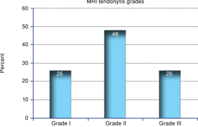
Grade I
26 26
48 MRI tendonytis grades
P erc en t 60 50 40 30 20 10 0
Grade II Grade III
Figure 1. Grades of patellar tendinopathies according to magnetic resonance imaging.
midpoint of the tendon. Elastography was applied at longitudinal plane inserting the transducer perpendicularly to the tendon. Localization of the patellar adhesion was moved to cranial of the patellar tendon, at least five-six measurements were performed in each patient, while point shear wave was measured by moving back from the same point to the cranium. Here, the device automatically measured 7 pulses from the midpoint with 3¥3 mm region of interest (ROI), and the mean value was obtained. Tendon softening on elastography was graded on colored elastogram. Color codes show stiffness of the tissues within ROI. When five stars on the left upper corner of the screen turned green, if all mapping was green, shear wave measurements were considered as 100% safe. Pascal units of the sampling area were seen in real time on the right upper corner of the screen. Elastography examinations were performed by a second radiologist who was blind to MRI outcomes and had 30-year experience. Shear wave elastography and pSWE evaluations were performed by the same radiologist to eliminate inter-observer variability.
Statistical analysis
Statistical analysis of the data was performed using the IBM SPSS version 22.0 software (IBM Corp. Armonk, NY, USA). Among the descriptive statistics, continuous variables were expressed as mean±standard deviation. Pearson’s correlation analysis was used to determine whether there was a correlation between SWE and pSWE values. P values <0.05 were considered statistically significant.
RESULTS
Of all patients, 45 (58.4%) were female and 32 (41.6%) were male. The mean age was 45.1±9.54 years in female and 49.8±9.37 years in male patients. An examination of grades of patellar tendinopathies according to MRI revealed grade I in 20 (26%), grade II in 37 (48%), and grade III in 20 (26%) patients (Figure 1).
The mean age was 40.2±8.91 years in grade I, 45.4±9.54 years in grade II, and 57.1±9.36 years in grade III patients. Grade was proportional to age, with grade of tendinopathy increased by age.
TABLE I
Magnetic resonance imaging grades, shear wave elastography and point shear wave elastography results according gen-der
Magnetic resonance imaging grades
Mean age (year) I II III SWE pSWE
Mean±SD n % n % n % Mean±SD Mean±SD
Gender
Female 45.1±9.5 9 20 23 51 13 29 88.2±32.9 82.3±33.8
Male 49.8±9.4 11 34 14 44 7 22 80.3±33.3 79.9±34.2
Total 47.1±9.5 20 26 37 48 20 26 83.8±34.3 29.3±33.8
SWE: Shear wave elastography; pSWE: Point shear wave elastography; SD: Standard deviation.
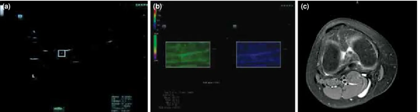
Figure 2. Point shear wave elastography (a), shear wave elastography (b) and magnetic resonance imaging (c) images of a patient with grade I patellar tendinopathy.
The mean SWE value was 83.83±34.27 kPa, while the mean pSWE value was 79.25±33.75 kPa. There was a strong correlation between SWE and pSWE values (p<0.001).
The mean SWE value was 88.17±32.88 kPa in female and 80.25±33.31 kPa in male patients, and
there was no statistically difference between genders in terms of SWE values (p>0.05). The mean pSWE value was 82.31±33.75 kPa in female and 79.94±34.16 kPa in male patients, and no statistically significant difference was found between genders in terms of pSWE values (p>0.05). Magnetic resonance imaging grades, SWE and pSWE results according gender were shown in Table I.
As grade of patellar tendinopathy found with MRI increased, pascal values found with elastography also increased. The highest SWE and pSWE values were obtained in patients with grade III patellar tendinopathy (Figure 2). Our shear wave and point shear wave values were consistent with MRI findings of patellar tendinopathy (Figure 3). Shear wave elastography and pSWE images of three patients with grade I, II and III tendinopathy were demonstrated in Figures 2, 4 and 5, respectively.
DISCUSSION
Although there are studies in the literature comparing elastography and many other methods in detection of tendinopathies, to our knowledge, there is no study comparing elastography with MRI. In our study, the results of the evaluation of patellar tendinopathy with SWE and pSWE were compatible
with clinical scoring using MRI.[6]
Figure 4. Point shear wave elastography (a), shear wave elastography (b) and magnetic resonance imaging (c) images of a patient with grade II patellar tendinopathy.
(a) (b) (c)
Figure 5. Point shear wave elastography (a), shear wave elastography (b) and magnetic resonance imaging (c) images of a patient with grade III patellar tendinopathy.
(a) (b) (c) P erc en t 140 100 120 80 60 40 20 0 Grade I SWE pSWE
Grade II Grade III
Figure 3. Shear wave elastography and point shear wave elastography values according to grades of patellar tendinopathy found with magnetic resonance imaging.
pSWE: Point shear wave elastography; SWE: Shear wave elastography. Elastography findings 44. 22 44 .6 2 80 .74 69 .1 3 13 3.1 8 13 2. 58
Patellar tendinopathy is characterized by mucoid degeneration of the patellar tendon, and includes abnormal structure of tenocytes, collagen and
vasculature.[7] Overuse of the tendon is the main factor
of the onset of patellar tendinopathy.[8] Histologic
changes are related to the absence of tendon repair (failed healing response). These changes include proliferation of tenocytes, disruption of collagen fibers, fluid accumulation between fibers, capillary proliferation and calcification. All these tissue changes induce the modification of viscoelastic features of the tendons. Elastography evaluates biomechanical and structural features of tissues by measuring their stiffness. All these changes in tissue flexibility cause alterations in tissue stiffness that are detectable with elastography. Even these changes can be detected with USE before they become visible
on conventional ultrasonography (US).[9-11] In our
study, we compared findings of SWE and pSWE techniques of USE as imaging methods in patients with patellar tendinopathy with tendon grading performed in MRI examination. Early diagnosis in tendinopathies is crucial for taking necessary medical conservative measures in order to prevent severe tendinous damage. Conventional US shows hypoechogenic regions in patellar tendinopathy. However, asymptomatic tendons may also include hypogenic areas in conventional US that are not beneficial in prediction of the development of later
symptoms.[12] So far, USE has been used for the
evaluation of Achilles tendon, quadriceps tendon, patellar tendon, epicondylar tendons and rotator cuff tendons. Achilles tendinopathy is the most studied tendinopathy including rupture and postoperative period, while the use of USE in patellar tendinopathy
is relatively new.[13]
There are studies in the literature comparing elastography techniques and other imaging modalities in the evaluation of musculoskeletal
system. In a study of Klauser et al.[14] with cadavers
comparing conventional US and elastography, it was reported that USE better depicted histological
degeneration compared to US. Porta et al.[15] compared
compression elastography (CE) and SWE techniques in healthy patellar tendons, and found good intra- and inter-observer reliability for CE. Moreover, Peltz
et al.[16] reported excellent intra-observer reliability
and moderate-excellent inter-observer reliability for SWE.
Healthy and asymptomatic patellar tendon tissue has been shown to be softer and compatible with more elastic tissue compared to tendinopathic
patellar tendons.[17] On the other hand, a study by
Ooi et al.[18] with volleyball athletes, symptomatic
tendons were found to be softer than asymptomatic ones. A higher correlation was found between softening in symptomatic patellar tendons and functional scores with USE compared to conventional US. The authors concluded that USE may increase sensitivity and accuracy of conventional US in the
diagnosis of patellar tendinopathy.[18] In a study by
Dirrichs et al.[19] comparing SWE, conventional US
and power Doppler US (PD-US) in monitoring of the healing of tendinopathies, sensitivities of US, PD-US ad SWE in detection of symptomatic tendons were found as 66%, 72% and 87.5%, respectively. A strong correlation was found between clinical scores and SWE findings. The authors reported that unlike US and PD-US, SWE could detect the process relating to healing of tendons, and it may be a helpful tool in monitoring the effects of the
treatment. Furthermore, Zhang et al.[20] evaluated
athletes with patellar tendinopathy using SWE, and found a correlation between stiffness of painful tendon and pain severity and degree of dysfunction. This indicates the utility of SWE in the diagnosis of patellar tendinopathy because of the changes in morphologic and elastic characteristics of the patellar tendon. Looking at the above-mentioned studies, it is seen that USE method using SWE and pSWE techniques is more efficient in detection of tendinopathies compared to conventional US, PD-US and compression elastography.
Another method used in the evaluation of tendinopathies is MRI. A healthy tendon is seen with low signal intensity in all sequences in MRI. The first sign of tendon abnormality in MRI is increased signal intensity first on echo images, and then on T1 weighted spin echo images. A high signal is seen around the tendon in fluid sensitive or contrast enhanced MRI. Magnetic resonance imaging changes in tendinopathy are best visualized
on axial or axial oblique images.[21] In our study,
we also used axial MRI images for grading of patellar tendinopathies. Studies comparing MRI and US techniques in detection of gluteal tendon ruptures, sensitivity of US (79-100%) was found
to be higher than MRI (33-100%).[22,23] It has been
reported that although MRI is appropriate in the differential diagnosis, US can better depict internal architecture of the tendon owing to its superior
spatial resolution.[24] Ability of US to provide a
dynamic imaging during active and passive motions can give more information about tendinopathy. USE may be valuable in improving diagnostic capability of conventional US.
Ultrasonographic elastography is increasingly finding more area of application in diagnosis and management of many diseases. However, this method has several limitations such as lack of standardization, being a subjective method, inter-observer variability of the results and ROI diameter.
The most important limitation of this study is its single-center design and relatively small number of patients. As another limitation, we were unable to compare our findings since there is no study in the literature investigating this subject.
In conclusion, we demonstrated that SWE and pSWE techniques can be safely used as non-invasive and quantitative imaging modalities in detection of patellar tendinopathy. These methods are safe and less time consuming compared to MRI, and they eliminate hesitation of some patients from MRI because of claustrophobia. Our results indicated that SWE and pSWE were helpful to confirm stiffness of patellar tendon in patients with patellar tendinopathy. In addition, our SWE and pSWE results were consistent with the grading performed with MRI. To our best knowledge, although there are studies in the literature about elastography applications in patellar tendinopathy, there is no study comparing this technique with MRI. Despite promising results, elastography has still not taken its place in musculoskeletal practice. Further studies with larger series are needed to investigate the potential role and applications of SWE and pSWE elastography techniques in the management of patellar tendinopathy.
Declaration of conflicting interests
The author declared no conflicts of interest with respect to the authorship and/or publication of this article.
Funding
The author received no financial support for the research and/or authorship of this article.
REFERENCES
1. Ferretti A, Ippolito E, Mariani P, Puddu G. Jumper's knee. Am J Sports Med 1983;11:58-62.
2. Roels J, Martens M, Mulier JC, Burssens A. Patellar tendinitis (jumper's knee). Am J Sports Med 1978;6:362-8. 3. El-Khoury GY, Wira RL, Berbaum KS, Pope TL Jr,
Monu JU. MR imaging of patellar tendinitis. Radiology 1992;184:849-54.
4. Paluch Ł, Nawrocka-Laskus E, Wieczorek J, Mruk B, Frel M, Walecki J1. Use of Ultrasound Elastography in the Assessment of the Musculoskeletal System. Pol J Radiol 2016;81:240-6.
5. Stoller DW, Tirman PF, Bredella MA, editors. Diagnostic imaging: Orthopaedics. 10th ed. North Miami: AMIRSYS; 2004.
6. Atik OŞ. Which articles do we prefer to publish? Eklem Hastalik Cerrahisi 2018;29:1.
7. Khan KM, Maffulli N, Coleman BD, Cook JL, Taunton JE. Patellar tendinopathy: some aspects of basic science and clinical management. Br J Sports Med 1998;32:346-55.
8. Glazebrook MA, Wright JR Jr, Langman M, Stanish WD, Lee JM. Histological analysis of achilles tendons in an overuse rat model. J Orthop Res 2008;26:840-6.
9. Aubry S, Nueffer JP, Tanter M, Becce F, Vidal C, Michel F. Viscoelasticity in Achilles tendonopathy: quantitative assessment by using real-time shear-wave elastography. Radiology 2015;274:821-9.
10. Khoury V, Cardinal E. “Tenomalacia”: a new sonographic sign of tendinopathy? Eur Radiol 2009;19:144-6.
11. Hsiao MY, Chen YC, Lin CY, Chen WS, Wang TG. Reduced Patellar Tendon Elasticity with Aging: In Vivo Assessment by Shear Wave Elastography. Ultrasound Med Biol 2015;41:2899-905.
12. Cook JL, Khan KM, Harcourt PR, Kiss ZS, Fehrmann MW, Griffiths L, et al. Patellar tendon ultrasonography in asymptomatic active athletes reveals hypoechoic regions: a study of 320 tendons. Victorian Institute of Sport Tendon Study Group. Clin J Sport Med 1998;8:73-7.
13. Prado-Costa R, Rebelo J, Monteiro-Barroso J, Preto AS. Ultrasound elastography: compression elastography and shear-wave elastography in the assessment of tendon injury. Insights Imaging 2018;9:791-814.
14. Klauser AS, Miyamoto H, Tamegger M, Faschingbauer R, Moriggl B, Klima G, et al. Achilles tendon assessed with sonoelastography: histologic agreement. Radiology 2013;267:837-42.
15. Porta F, Damjanov N, Galluccio F, Iagnocco A, Matucci-Cerinic M. Ultrasound elastography is a reproducible and feasible tool for the evaluation of the patellar tendon in healthy subjects. Int J Rheum Dis 2014;17:762-6.
16. Peltz CD, Haladik JA, Divine G, Siegal D, van Holsbeeck M, Bey MJ. ShearWave elastography: repeatability for measurement of tendon stiffness. Skeletal Radiol 2013;42:1151-6.
17. Coombes BK, Tucker K, Vicenzino B, Vuvan V, Mellor R, Heales L, Achilles and patellar tendinopathy display opposite changes in elastic properties: A shear wave elastography study. Scand J Med Sci Sports 2018;28:1201-8. 18. Ooi CC, Richards PJ, Maffulli N, Ede D, Schneider M,
Connell D, et al. A soft patellar tendon on ultrasound elastography is associated with pain and functional deficit in volleyball players. J Sci Med Sport 2016;19:373-8.
19. Dirrichs T, Quack V, Gatz M, Tingart M, Rath B, Betsch M, et al. Shear Wave Elastography (SWE) for Monitoring of Treatment of Tendinopathies: A Double-blinded, Longitudinal Clinical Study. Acad Radiol 2018;25:265-72.
20. Zhang ZJ, Ng GY, Lee WC, Fu SN. Changes in morphological and elastic properties of patellar tendon in athletes with unilateral patellar tendinopathy and their relationships with pain and functional disability. PLoS One 2014;9:e108337.
21. Rosenberg ZS, Beltran J, Bencardino JT. From the RSNA Refresher Courses. Radiological Society of North America. MR imaging of the ankle and foot. Radiographics 2000;20:S153-79.
22. Westacott DJ, Minns JI, Foguet P. The diagnostic accuracy of magnetic resonance imaging and ultrasonography in gluteal tendon tears--a systematic review. Hip Int 2011;21:637-45.
23. Hartgerink P, Fessell DP, Jacobson JA, van Holsbeeck
MT. Full- versus partial-thickness Achilles tendon tears: sonographic accuracy and characterization in 26 cases with surgical correlation. Radiology 2001;220:406-12.
24. Rasmussen OS. Sonography of tendons. Scand J Med Sci Sports 2000;10:360-4.