88
Acta Ortop Bras. 2020;28(2):88-91Original article
DOI: http://dx.doi.org/10.1590/1413-785220202802226166
Correspondence: Bekir Eray Kılınc. Icerenkoy District Zubeyde Hanım Street Nº: 1 B-43 PC:43752 Atasehir/Istanbul Tel nº: +905306061884. [email protected] The study was carried out at Istinye University.
All authors declare no potential conflict of interest related to this article.
Article received on7/16/2019, approved on 10/10/2019.
Citation: Kilinc BE, Oc Y, Erture RE. Treatment of midshaft clavicle fracture with superior plate placement. Acta Ortop Bras. [online]. 2020;28(2):88-91. Available from URL: http://www.scielo.br/aob.
TREATMENT OF MIDSHAFT CLAVICLE FRACTURE
WITH SUPERIOR PLATE PLACEMENT
TRATAMENTO PARA FRATURA DA DIÁFISE DA CLAVÍCULA
COM INSERÇÃO DE PLACA ÓSSEA SUPERIOR
Bekir Eray Kilinc
1, Yunus Oc
2, Ramazan Erden Erturer
31. Health Science University, Fatih Sultan Mehmet Training and Research Hospital, Orthopaedics Department, Istanbul, Turkey. 2. Health Science University, Sisli Hamidiye Training and Research Hospital, Orthopaedics Department, Istanbul, Turkey. 3. Istinye University, Faculty of Medicine, Orthopaedics Department, Istanbul, Turkey.
ABSTRACT
Objective: To evaluate the late clinical and radiological results of patients had locking plate anatomically compatible from superior surface and muscle cover on plate due to clavicle mid-region. Materials and Methods: Forty patients were included retrospec-tively. Patients had a routine right shoulder anterior posterior graph after examination. The results were assessed by returning to the patient's daily activities, Constant score, the Disability of the Arm, and Shoulder and Hand scoring, followed by radiolog-ical and clinradiolog-ical examination. Results: Fourteen (35%) patients were female and 26 (65%) were male. The mean age was 36.2 years. Twenty-six patients had right clavicle fracture and 14 patients had left. Twenty-three fractures were type 2B1 and 17 fractures were type 2B2. Mean follow-up time was 36.4 months. Radiologic union was at a mean of 9.1 ± 1.3 weeks. All patients had excellent results. The mean Constant score was 97.2 ± 1.8, the mean Disability of the Arm, and Shoulder and Hand score was 3.8 ± 2.4. Conclusion: It is possible to obtain complete union with high patient satisfaction by avoiding the complications and difficulties of the conservative treatment with the use of the anatomically compatible locking plates in superior fixation and our surgical dissection. Level of Evidence III, Retrospective Case controlled study.
Keywords: Clavicle. Midshaft Clavicle Fracture. Osteosynthesis. Plate Fixation. Superior Placement.
RESUMO
Objetivo: Avaliar os resultados clínicos e radiológicos tardios dos pacientes com placa óssea de trava anatomicamente compatível com a superfície superior e a cobertura muscular na placa devido à região média da clavícula. Materiais e Métodos: Quarenta pacientes foram incluídos retrospectivamente. Os pacientes apresentaram um gráfico ântero-posterior de rotina do ombro direito após o exame. Os resultados foram avaliados retornando às atividades diárias do paciente, escore de Constant, incapacidade do braço e escores de ombro e mão, seguidos de exame clínico e radiológico. Resultados: Quatorze (35%) pacientes eram do sexo feminino e 26 (65%) do sexo masculino. A idade média foi de 36,2 anos. Vinte e seis pacientes tiveram fratura da clavícula direita e 14 pacientes saíram. Vinte e três fraturas foram do tipo 2B1 e 17 fraturas do tipo 2B2. O tempo médio de acompanhamento foi de 36,4 meses. A união radiológica foi em média de 9,1 ± 1,3 semanas. Todos os pacientes tiveram excelentes resultados. A pontuação média constante foi de 97,2 ± 1,8, a média de incapacidade do braço e a pontuação do ombro e da mão foi de 3,8 ± 2,4. Conclusão: É possível obter união completa com alta satisfação do paciente, evitando as complicações e dificuldades do tratamento con-servador com o uso das placas ósseas de trava anatomicamente com-patíveis na fixação superior e na nossa dissecção cirúrgica. Nível de evidência III, Estudo retrospectivo controlado por caso. Descritores: Clavícula. Fratura de Clavícula de Eixo Intermediário. Fixação de Placas Ósseas. Colocação Superior.
INTRODUCTION
Clavicle fracture is a common fracture and constitutes approxi-mately 4% of all fractures in adults.1 The location of the clavicle fracture is approximately 75% clavicular and 1/3 middle part due to its thin form and direct contact with the skin. Clavicular injuries can lead to abnormal biomechanical stresses and long-term disability along the pectoral girdle.2,3
Conservative treatment and open reduction and plate fixation are used in the treatment of the currently displaced midshaft clavicle fractures. Conservative treatment was reported to have a higher union rate than open reduction and plate fixation.4 However, patients treated with open reduction and plate fixation have a better outcome than conservative treatments according to functional scores.5 Despite the reduced pain and improved
89
Acta Ortop Bras. 2020;28(2):88-91
functional recovery in patients with displaced midclavicular fractures treated with open reduction and plate fixation,6,7 infection due to the graft of a large soft tissue can lead to complications such as numbness on the skin, nonunion, delayed union, and enlarged scar tissue.8-10
Treatment of these fractures with open reduction and internal fixation preserves from nonunion, symptomatic malunion, short-ening and deformity. Studies have shown that the superior plate is biomechanically better than the anterior plate and that the locked screws are better than the unlocked ones.11-13 But superior resident plates are usually palpable under the skin and can cause skin irritation.14-19
In this study, we sought to evaluate the late clinical and radiological results of patients who underwent locking plate fixation anatomically compatible in the superior surface and muscle cover on plate due to the mid-region of the clavicle.
METHODS
Forty patients who underwent surgery between May 2009 and September 2016 with mid-diaphyseal clavicle fracture were in-cluded retrospectively in our study. All patients participating in the study signed an informed consent form. The study was conducted in agreement with the Declaration of Helsinki. Approval for our study was obtained from the institutional review board. Our study is in line with the STROCSS criteria. Displacement or shortness of more than 20 mm in patients with segmental fractures with multiple fractures or disintegration, as well as conservative treatment intolerance were indicated for surgical treatment. The study included only patients with isolated mid-diaphyseal clavicle fractures.20 Patients with pathologic fractures, open fractures, those with neurovascular injuries and/ or 2-week fractures were excluded. All patients had chest radiography to exclude possible cote and scapular injury. After diagnosis, patients were prepared for surgery by applying a shoulder-arm sling.
All patients were assessed with a routine right-shoulder anteri-or-posterior graph after a detailed physical examination. During the follow-up period, the results were clinically assessed by the patient’s return to daily activities, Constant score, the Disability of the Arm, and Shoulder and Hand scoring, followed by radiological and clinical examination of the fracture union.
Surgical Technique
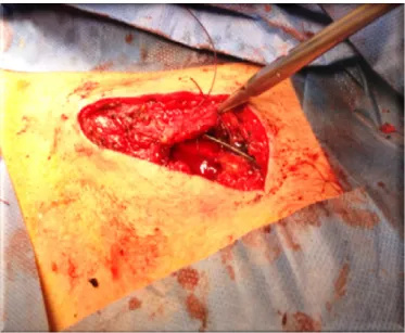
Patients were prepared for operation in beach-chair position under general or regional anesthesia. Skin incision was made approxi-mately 1 cm below the clavicle lower level (infraclavicular incision). The subcutaneous tissue was prepared without dissecting, and the preparation of skin-subcutaneous and platysma muscle together in a flap style, clavicle anterior and superior sides were elevated by approaching to proximal. Thus, the clavipectoral fascia was scrapped over the clavicle to the extent that was required and the fraction was reduced by avoiding an aggressive dissection. Temporary detection with K-wires was performed when neces-sary. In the case of the butterfly fragment, these fragments were temporarily attached to the main part with absorbable sutures (Vicryl no: 0). Rigid fixation by applying a 3.5 mm locking screw (LCP Superior Anterior Clavicle Plate) and a preformed clavicle plate with a low contact surface were performed for the all patients. At the end of the operation, clavipectoral fascia was repaired to cover the plate. Flap prepared initially from skin-subcutaneous and muscle, was completely closed on the plate in such a way that it was completely muscular (Figure 1).
Figure 1. Muscular flap. Post-operative protocol
Shoulder pendular exercises were started for patients on the first day after the surgery. Antibiotic prophylaxis continued for 2 days after the operation. Surgical wound was checked on the 3rd day, and the patients were discharged with arm sling. Patients were called for control at 4, 8 and 12 weeks postoperatively. In the fourth week, exercises of the shoulder joint movement were started when the use of the arm sling were interrupted. The radiologic examinations required to evaluate the postoperative fracture were examined by an orthopedic surgeon and a radiologist blinded for the study. Radiographically, more than 50% of the fracture lines were classified as complete union. Union was assessed by bone bridge formation between fracture fragments, sensitivity on the fracture line and clinical examination of the shoulder joint movements. The delayed union was determined by the initial radiologic callus formation that was seen after 24 weeks, and the nonunion was determined by the absence of callus and pathological movement after 24 weeks.21 Shoulder strengthening exercises have been initiated for patients that had union symptoms. Contact sports were allowed three months after the surgery.
NCSS 2007 version software (Number Cruncher Statistical System – Kaysville, Utah, USA) was used for the statistical analysis. While evaluating the data from the study, apart from using descriptive statistical methods (Mean, Standard Deviation, Median, Frequency, Rate, Minimum, Maximum), Mann Whitney U test was used for the two group comparison for the parameters with abnormal distribu-tion. Fisher-Freeman-Halton test, Fisher’s Exact test and Yates’ Continuity Correction test (Yates’ correction chi square) were used to compare qualitative data. Wilcoxon Signed Ranks test was used for intra-group comparisons of abnormal parameters. Significance was evaluated considering p < 0.05.
RESULTS
In total, 14 patients (35%) were women and 26 were men (65%). The mean age of the patients participating in the study was 36.2 (range: 22-59). Twenty-six patients had clavicle fracture on the right side and 14 patients, on the left side. According to Robinson’s classification, 23 fractures were type 2B1 and 17 fractures were type 2B2. Mean
90
Acta Ortop Bras. 2020;28(2):88-91 follow-up time was 36.4 months (range: 24-95). Fractures occurredin 20 patients due to traffic accidents, in 11 patients due to falls and in 9 patients due to sports injuries, as shown in Table 1.
Table 1. Information on patients and fractures.
Characteristic Total % Sex Women 14 35 Men 26 65 Age 36.2 Side Right 26 65 Left 14 35 Fracture type Type 2B1 23 57.5 Type 2B2 17 42.5 Injury mechanism Traffic Accident 20 50 Fall 11 27.5 Sports Injury 9 22.5
Radiologic complete union was achieved in all patients at a mean of 9.1 ± 1.3 weeks, (range: 8-13 weeks). No callus formation was observed in the fracture area of any patient. No complications such as infection or detection failure were found in the early or late period. In total, two patients described the feeling of irritation due to the plate and 7 patients were cosmetically disturbed by the incision scar. None of the patients had major complications such as infection, plate rupture or neurovascular injury. All patients reported excellent results in terms of shoulder functions. At the end of the follow-up period, the mean Constant score was found to be 97.2 ± 1.8 (range: 95-100), mean Disability of the Arm, Shoulder and Hand score was 3.8 ± 2.4 (range: 0-10), as shown in Table 2. The mean duration of the surgery was 52.2 minutes.
Table 2. Functional score results.
Outcome score Post-operative
Constant score 97.2 Disability of the Arm, Shoulder and Hand scoring 3.8 DISCUSSION
Numerous studies seeking an optimal treatment in the treatment of mid-diaphyseal clavicle fractures have been increasingly conducted. Mid-diaphyseal clavicle fractures were traditionally treated conser-vatively; however, recent studies show that nonunion, malunion, and poor shoulder functions are seen together with conservative treatment of displaced mid-diaphyseal fractures. In a comparative study between plate-treated and conservatively treated displaced mid-diaphyseal fractures, high functional outcomes, low nonunion and malunion results were found in patients treated surgically.22 Patients treated with plate fixation recovered faster and returned to their previous activity levels, and a risk of developing a symp-tomatic malunion was reported in conservatively-treated patients. Many other studies also suggest open reduction and fixation in the treatment of displaced mid-diaphyseal fractures, particularly those with 20 mm shortening, 100% displacement and bone defect.23-25 Despite the good stability, compression and mechanical fixation with plate fixation, complications such as infection and formation of scar tissue were found. Although the clavicle fixation as an
intramedullary is cosmetically acceptable, complications rates of up to 75% were reported, namely lack of rotational control, the need for a second surgical procedure to remove the implant, skin problems, and implant migration.6,26-28
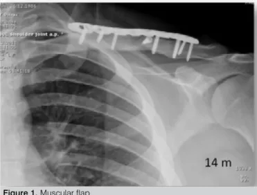
Optimal plate fixation for the treatment of mid-diaphyseal clavicle fracture is still controversial. Some studies suggest that anteroinferior plate fixation techniques are better, suggesting that plate prominence is felt less often. However, more soft tissue dissection is required for this plate fixation. In the same study, the authors suggested that the lateral bearing of the plate fixation point and the lateral screws could cause a pull-out in the placement of the superiorly positioned plate in fragmented fractures.29 This is the reason why the fixation due to an unsuccessful reduction was reported as a posterior slide of the support point and a significant force to the lateral load, causing the pull-out of the screws. The sternoclavicular joint created a tension band effect and the support point remained at the fracture fixation point in simple transverse fractures.30 In our study, 17 patients with Robinson type B2 (partial fracture) had no complications due to plate placement, which can be considered a result of the suitability of the reduction, plate fixation and fixation method for the stabilization rules, as shown in Figure 2.
In an anatomical study, the subclavian artery in the medial half of the clavicle was the closest to the posterior cortex.31,32 This is the reason why anteroinferior plate fixation can pose a great risk for neurovascular structures in the medial clavicular area. This may be considered a safe fixation method due to the reduction in the risk of a neurovascular injury caused by superior plate detection. Neurovascular injury or other major complications were not detected in any of our patients in our study when super anatomical plate detection was used in surgical treatment.
Figure 1. Muscular flap.
A previous study showed that the use of locked plate-screw in fragmented clavicle fractures increased the angular stability and decreased the effect on the bio-alloying of small fractures.21,33 Clin-ically, plate prominence inferiority due to low profile of anatomically compatible plate in mid-diaphyseal clavicle fractures is low.34 We also think that the use of preformed anatomically compatible plates in our study reduces the duration of surgery and plate tiredness risk. At the same time, without applying subcutaneous dissection after surgical incision, the approach that we provide as flap with the plate scar can provide a good cover after plate fixation and reduce plate prominence risk.
In a study conducted biomechanically, the anterior, antero-superior, and superior plating types were found to be the most important
91
Acta Ortop Bras. 2020;28(2):88-91
method for detecting axial fracture of superior plate in the detection of midshaft clavicle fractures. In the same study, no difference was found between torsional forces and resistance among all three types of plate fixation.35
Our study has some limitations. First, the study retrospective design was the main limitation, and we also included patients with wide range of age distribution. However, we included similar type of fracture and treated our patients with the same method. Our study may guide further studies on the evaluation of the superior plating treatment due to the clinical outcomes found.
We believe we have achieved excellent results with our study on the fixation of the fracture with our superior plate fixation technique
and with an early rehabilitation program applied to all patients. Furthermore, we think that we can minimize the plate prominence risk by providing the muscle flap and plate covering that we used during the surgical approach and adapt the patients to the rehabilitation period, minimizing the complaints of skin irritation in later periods.
CONCLUSION
Complications such as shortening and excessive callus formation can be observed as a result of disintegrated or multi-part midshaft clavicle fractures. It is possible to obtain complete union with high patient satisfaction by avoiding the complications with anatomically compatible locking plates in superior fixation and our surgical dissection.
AUTHORS’ CONTRIBUTIONS: Each author made significant individual contributions to this manuscript. BEK: substantial contributions to the conception
and design of the study; YO: data acquisition, analysis, and interpretation; REE: final approval of the version to be published and accountability for all aspects of the study, ensuring that questions related to the accuracy or integrity of any part of the article are appropriately investigated and resolved.
REFERENCES
1. Nowak J, Mallmin H, Larsson S. The aetiology and epidemiology of clavicular fractures: a prospective study during a two-year period in Uppsala, Sweden. Injury. 2000;31(5):353-8.
2. Hillen RJ, Burger BJ, Pöll RG, van Dijk CN, Veeger DH. The effect of expe-rimental shortening of the clavicle on shoulder kinematics. Clin Biomech. 2012;27(8):777-81.
3. Matsumura N, Ikegami H, Nakamichi N, Nakamura T, Nagura T, Imanishi N, et al. Effect of shortening deformity of the clavicle on scapular kinematics: a cadaveric study. Am J Sports Med. 2010;38(5):1000-6.
4. Robinson CM, Goudie EB, Murray IR, Jenkins PJ, Ahktar MA, Read EO, et al. Open reduction and plate fixation versus nonoperative treatment for displaced midshaft clavicular fractures: a multicenter, randomized, controlled trial. J Bone Joint Surg Am. 2013;95(17):1576-84.
5. McKee RC, Whelan DB, Schemitsch EH, McKee MD. Operative versus nono-perative care of displaced midshaft clavicular fractures: a meta-analysis of randomized clinical trials. J Bone Joint Surg Am. 2012;94(8):675-84. 6. Zlowodzki M, Zelle BA, Cole PA, Jeray K, McKee. Treatment of acute
mi-dshaft clavicle fractures: systematic review of 2144 fractures: on behalf of the Evidence-Based Orthopaedic Trauma Working Group. J Orthop Trauma. 2005;19(7):504-7.
7. Rudzki JR, Matava MJ, Paletta GA Jr. Complications of treatment of acromiocla-vicular and sternoclaacromiocla-vicular joint injuries. Clin Sports Med. 2003;22(2):387-406. 8. D’Heurle A, Le T, Grawe B, Casstevens EC, Edgington J, Archdeacon MT,
et al. Perioperative risks associated with the operative treatment of clavicle fractures. Injury. 2013;44(11):1579-81.
9. Jeong HS, Park KJ, Kil KM, Chong S, Eun HJ, Lee TS, et al. Minimally invasive plate osteosynthesis using 3D printing for shaft fractures of clavicles: technical note. Arch Orthop Trauma Surg. 2014;134(11):1551-5.
10. Sohn HS, Kim BY, Shin SJ. A surgical technique for minimally invasive plate os-teosynthesis of clavicular midshaft fractures. J Orthop Trauma. 2013;27(4):92-6. 11. Celestre P, Roberston C, Mahar A, Oka R, Meunier M, Schwartz A. Biomechanical
evaluation of clavicle fracture plating techniques: does a locking plate provide improved stability? J Orthop Trauma. 2008;22(4):241-7.
12. Partal G, Meyers KN, Sama N, Pagenkopf E, Lewis PB, Goldman A, et al. Superior versus anteroinferior plating of the clavicle revisited: a mechanical study. J Orthop Trauma. 2010;24(7):420-5.
13. Robertson C, Celestre P, Mahar A, Schwartz A. Reconstruction plates for stabilization of mid-shaft clavicle fractures: differences between nonlocked and locked plates in two different positions. J Shoulder Elbow Surg. 2009;18(2):204-9. 14. Chen CH, Chen JC, Wang C, Tien YC, Chang JK, Hung SH. Semitubular
plates for acutely displaced midclavicular fractures: a retrospective study of 111 patients followed for 2.5 to 6 years. J Orthop Trauma. 2008;22(7):463-6. 15. Court-Brown CM, Caesar B. Epidemiology of adult fractures: a review. Injury.
2006;37(8):691-7.
16. Demirhan M, Bilsel K, Atalar AC, Bozdag E, Sunbuloglu E, Kale A. Biome-chanical comparison of fixation techniques in midshaft clavicular fractures. J Orthop Trauma. 2011;25(5):272-8.
17. Eden L, Doht S, Frey SP, Ziegler D, Stoyhe J, Fehske K, et al. Biomechanical comparison of the locking compression superior anterior clavicle plate with
seven and ten hole reconstruction plates in midshaft clavicle fracture stabili-sation. Int Orthop. 2012;36(12):2537-43.
18. Ferran NA, Hodgson P, Vannet N, Williams R, Evans RO. Locked intramedullary fixation vs plating for displaced and shortened mid-shaft clavicle fractures: a randomized clinical trial. J Shoulder Elbow Surg. 2010;19(6):783-9. 19. Formaini N, Taylor BC, Backes J, Bramwell TJ. Superior versus anteroinferior
plating of clavicle fractures. Orthopedics. 2013;36(7):e898-904.
20. Agha RA, Borrelli MR, Vella-Baldacchino M, Thavayogan R, Orgill DP; STROCSS Group. The STROCSS statement: strengthening the reporting of cohort studies in surgery. Int J Surg. 2017;46:198-202.
21. Ozler T, Guven M, Kocadal AO, Ulucay C, Beyzadeoglu T, Altıntas¸ F. Locked anatomic plate fixation in displaced clavicular fractures. Acta Orthop Traumatol Turc. 2012;46(4):237-42.
22. Altamimi SA, McKee MD. Nonoperative treatment compared with plate fixation of displaced midshaft clavicular fractures. Surgical technique. J Bone Joint Surg Am. 2008;90(Suppl 2 Pt 1):1-8.
23. Jorgensen A, Troelsen A, Ban I. Predictors associated with nonunion and symptomatic malunion following non-operative treatment of displaced mi-dshaft clavicle fractures: a systematic review of the literature. Int Orthop. 2014;38(12):2543-9.
24. Lai YC, Tarng YW, Hsu CJ, Chang WN, Yang SW, Renn JH. Comparison of dynamic and locked compression plates for treating midshaft clavicle fractures. Orthopedics. 2012;35(5):e697-702.
25. Rehn CH, Kirkegaard M, Viberg B, Larsen MS. Operative versus nonoperative treatment of displaced midshaft clavicle fractures in adults: a systematic review. Eur J Orthop Surg Traumatol. 2014;24(7):1047-53.
26. Renfree T, Conrad B, Wright T. Biomechanical comparison of contemporary clavicle fixation devices. J Hand Surg Am. 2010;35(4):639-44.
27. Judd DB, Pallis MP, Smith E, Bottoni CR. Acute operative stabilization versus nonoperative management of clavicle fractures. Am J Orthop. 2009;38(7):341-5. 28. Smekal V, Irenberger A, Struve P, Wambacher M, Krappinger D, Kralinger FS.
Elastic stable intramedullary nailing versus nonoperative treatment of displaced midshaft clavicular fractures-a randomized, controlled, clinical trial. J Orthop Trauma. 2009;23(2):106-12
29. Favre P, Kloen P, Helfet DL,Werner CM. Superior versus anteroinferior plating of the clavicle: a finite element study. J Orthop Trauma. 2011;25(11):661-5. 30. Basamania CJ, Rockwood CA. Fractures of the clavicle. In: Rockwood CA,
editor. The shoulder. 4th ed. Philadelphia: WB Saunders; 2008. p. 423-8. 31. Lo EY, Eastman J, Tseng S, Lee MA, Yoo BJ. Neurovascular risks of anteroinferior
clavicular plating. Orthopedics. 2010;33(1):21.
32. Robinson L, Persico F, Lorenz E, Seligson D. Clavicular caution: an anatomic study of neurovascular structures. Injury. 2014;45(12):1867-9.
33. 33. Gautier E, Sommer C. Guidelines for the clinical application of the LCP. Injury. 2003;34(Suppl 2):B63-76.
34. 34. VanBeek C, Boselli KJ, Cadet ER, Ahmad CS, Levine WN. Precontoured plating of clavicle fractures: decreased hardware-related complications? Clin Orthop Relat Res. 2011;469(12):3337-43.
35. Huang TL, Chen WC, Lin KJ, Tsai CL, Lin KP, Wei HW. Conceptual finite element study for comparison among superior, anterior, and spiral clavicle plate fixations for midshaft clavicle fracture. Med Eng Phys. 2016;38(10):1070-5.