257
Int. J. Sec. Metabolite, Vol. 4: 3 (2017) pp. 257-263
Special Issue 1: Research Article
ISSN: 2148-6905 online Journal homepage: http://www.ijate.net/index.php/ijsm
Antibacterial activities of Calendula officinalis callus extract
Burcu ÇETİN*1, Fatih KALYONCU2, Betül KURTULUŞ1,
1Dumlupınar University, Faculty of Science &Letters, Dept. of Biology, Kütahya, Turkey 2Manisa Celal Bayar University, Faculty of Science & Letters, Dept. of Biology, Manisa, Turkey
Received: 01 May 2017 – Revised: 17 August 2017 - Accepted: 16 September 2017
Abstract: The purpose of this study was to determine the antibacterial activity of C. officinalis callus derived from
cotyledon explants. Cotyledons excised from in vitro germinated seedlings were used as explants. Explants were transferred on MS medium supplemented with benzil amino purine (BAP; 2 mg l-1), α-naphthalene-acetic acid
(NAA; 2 mg l-1) for callus studies. The cultures were maintained on the same media compositions and were
sub-cultured at an interval of 4 weeks. Callus cultures were harvested at the end of the 16th week. Calli were dried at 40̊ C in the dark for antimicrobial studies. Calendula officinalis callus extracts were tested for their antibacterial activities by using agar well diffusion method. Ethanol and chloroform extracts from these plants were assayed against nine bacteria species (Staphylococcus aureus ATCC 6538, Escherichia coli ATCC 25922, Bacillus cereus ATCC 7064, Bacillus subtilis ATCC 6633, Salmonella typhimurium CCM 5445, Proteus vulgaris ATCC 6896,
Enterococcus faecalis ATCC 29212, Enterobacter cloacae ATCC 13047, and Kocuria rhizophila ATCC 9341).
The test antibiotics penicillin G, novobiocin, amphicillin, chloramphenicol and erythromycin were used for comparison. Callus formation was observed at the end of the 5th week on cotyledon explants. C. officinalis callus extracts showed 38 mm inhibition zone against S. aureus, and chloroform extracts showed 32 mm inhibition zone against B. cereus. These results are very close to the test antibiotics used and C. officinalis is found more effective on gram positive bacteria.
Keywords: Calendula officinalis, callus, antibacterial activity, BAP, NAA. 1. INTRODUCTION
Plants are able to synthesize substances called secondary metabolites in addition to the production of carbonhydrates, proteins and fats, the primary building blocks of life, that is, primary metabolites. The discovery of the bioactive properties of secondary metabolites and their widespread use in many areas of industry such as medicine, cosmetics, paint making, and fragrance progressively increase the significance of medical and aromatic plants in the world markets [1, 2]. Biotechnological methods, especially callus culture of plant cell tissue cultures, appear as alternative methods in the production of secondary metabolites in order to solve encountered problems by wild environments [3]. In many studies callus cultures represent an alternative source for producing natural antimicrobial compounds and plant tissue cultures produce a variety of secondary metabolites, sometimes in higher percentage than the original
*Corresponding Author E-mail: [email protected]
258
plant, particularly in the polyphenolic class in which high yields have sometimes been obtained [4].
Calendula officinalis L., known for its ornamental plant characteristics, is a medicinal
plant belonging to Asteracea family. It is annual or perennial taproot plant with 20-40 cm height and has 20 varieties [5]. The chemical compounds identified in methanol extracts of C.
officinalis include polar compounds phenolic acids and flavonoid glycosides [6]. As a result of
the many studies performed with C. officinalis, the cancer [7], microbial [8], leishmanial [9], HIV [10], antioxidant [11], cytotoxic, tumor [12], viral [13], anti-inflammatory [14], edema amplifier [15], hypoglycemic [16], uterotonic [17], effects and its utilization in the treatment of venous ulcers [18], as well as the biligenic [19] pharmacologic effects of this plant have been reported.
In this study, the effect of BAP and NAA plant growth regulators applied on C. officinalis cotyledon explants and the antibacterial effect of the obtained calluses have been investigated. 2. MATERIAL and METHODS
2.1. Plant material
Calendula officinalis seeds used in the study were obtained from Hekim Sinan Medicinal
Plant Research Center of in the municipality of Kutahya, Turkey. 2.2. Medium & culture conditions
Murashige and Skoog medium was used as nutrient medium. For germination studies without any plant growth regulators and for callus studies MS medium supplement with 2 mg l-1 BAP and 2 mg l-1 NAA were respectively used [20]. They were supplemented with 100 mg l-1 myo-inositol and 30 mg l-1 sucrose. The pH of the medium was adjusted between 5.7-5.8
using 0.1 N NaOH or 0.1 N HCl before the addition of 0.7 % (w/v) agar and then the medium was autoclaved at 121°C for 15 min at 105 kPa. Cultures were maintained at 24 ± 2°C, under 16:8 photoperiod at a 4000 lux white fluorescent light, in a growth room.
2.3. Establishment of the aseptic culture
Initially, C. officinalis seed pods were unshelled, separated, and washed using distilled water. Later surface sterilization was made in 70% ethyl alcohol for 3 min and 0.5% NaOCl for 5 min, the seeds were rinsed with sterilized distilled water to remove the traces of sterilant. They were transferred on MS medium for germination.
2.4. Callus culture & induction
Calendula officinalis L. cotyledon explants were used as explants for the induction of
callus. Cotyledons were excised from aseptic seedling and were and cultured on MS medium suplemented with; 2 mg l-1 BAP and 2 mg l-1 NAA for callus studies. The cultures were maintained on same medium compositions and sub-cultured in the same medium every four weeks. The calli were harvested at the end of 16th week and were dried at 40̊ C under the dark for antimicrobial studies.
2.5. Test microorganisms and growth conditions
Test microorganisms included following bacteria: Staphylococcus aureus ATCC 6538,
Escherichia coli ATCC 25922, Bacillus cereus ATCC 7064, Bacillus subtilis ATCC 6633, Salmonella typhimurium CCM 5445, Proteus vulgaris ATCC 6896, Enterococcus faecalis
ATCC 29212, Enterobacter cloacae ATCC 13047, and Kocuria rhizophila ATCC 9341. Cultures of these bacteria were grown in Mueller Hinton broth (Oxoid) at 37oC for 24h [21].
259
Test microorganisms were obtained from the culture collection of Ege University, Faculty of Science, Basic and Industrial Microbiology Department.
2.6. Antibacterial activity assay
The dried and powdered plants were reduced to coarse powder. Two g of powder was extracted with 20 ml of ethanol and chloroform at room temperature with stirring for 3 days (125 cycles/minute). The solvents were evaporated to dryness after extraction progress. Sample solutions were prepared by dissolving the extracts in same solvents (1 ml). In vitro antibacterial studies were carried out by the agar well diffusion method against test microorganisms. Bacterial strains grown on nutrient agar at 37oC for 24 h were suspended in a saline solution
(0.85% NaCl) and adjusted to a turbidity of 0.5 MacFarland standards [106 Colony Forming Units (CFU)/ml]. Briefly, 50 microlitres (µl) inoculum (containing approximately 105 bacteria per milliliter) was added to 25 mL melted Mueller Hinton Agar (MHA) medium cooled at 45oC.
This was then poured into 90 mm diameter Petri dishes and maintained for 1h at room temperature. Small wells (6 mm diameter) were cut in the agar plate using a cork borer; 60 µl of extract concentration with a negative control (ethanol and chloroform, 60 µl) were loaded in the wells. The dishes were preincubated at 4oC for 2 h to allow uniform diffusion into the agar. After preincubation, the plates were incubated at 37oC for 24h. The antibacterial activity was evaluated by measuring the inhibition zone diameter observed. In addition, commercial antibiotics (penicillin G, novobiocin, amphicillin, chloramphenicol and erythromycin) were used as positive control to determine the sensitivity of the strains [22]. All experiments were performed in triplicate.
3. RESULTS and DISCUSSIONS
Plant tissue culture techniques enable the production of plant tissue or cells in sterile environments under controlled conditions, allowing the growth and development of the cells or tissues to be manipulated for a variety of applications. One of these methods callus cultures, could be produced pharmacologically active molecules at the desired amount and constant quality at any time in laboratory conditions [23, 24]. In this study, the antimicrobial effect of the calli obtained from the cotyledon explants of the C. officinalis plant was examined.
It is more feasible to take explants that will be used in tissue culture studies from plants grown in in vitro conditions. The risk of contamination for plants which grow under these conditions is almost nonexistent, and they regenerate much easier and can be used directly as explants [25]. Thus, the explants to be used for this study were obtained from the plantlets produced from seeds that were germinated on MS media. At the end of the surface sterilization procedure, which was performed by incubating the seeds in 70% ethyl alcohol for 3 min, in 0.5% bleach for 5 min, and rinsing 3 times with incubation for 3 min in distilled water, sterile plantlets were successfully obtained. The cotyledones to be used as explants were isolated from these plants.
Result of some studies revealed that auxins played an important role in the callus induction. Furthermore, they showed that cytokinins facilitated the effect of auxins in callus induction [26]. It has been reported that callus formation is high in many tissue culture studies in which plant growth regulators, namely BAP from among cytokines and NAA from among auxins are applied [27-29]. In the present study, 2 mg l-1 BAP ve 2 mg l-1 IBA cytokinins that
were applied caused a swelling of the explants after 5–7 d of culture, and callus induction was observed on the cotyledon explants within 5th wk of culture (Figure 1).
260
Figure 1. Calli obtained from C. officinalis cotyledon explants
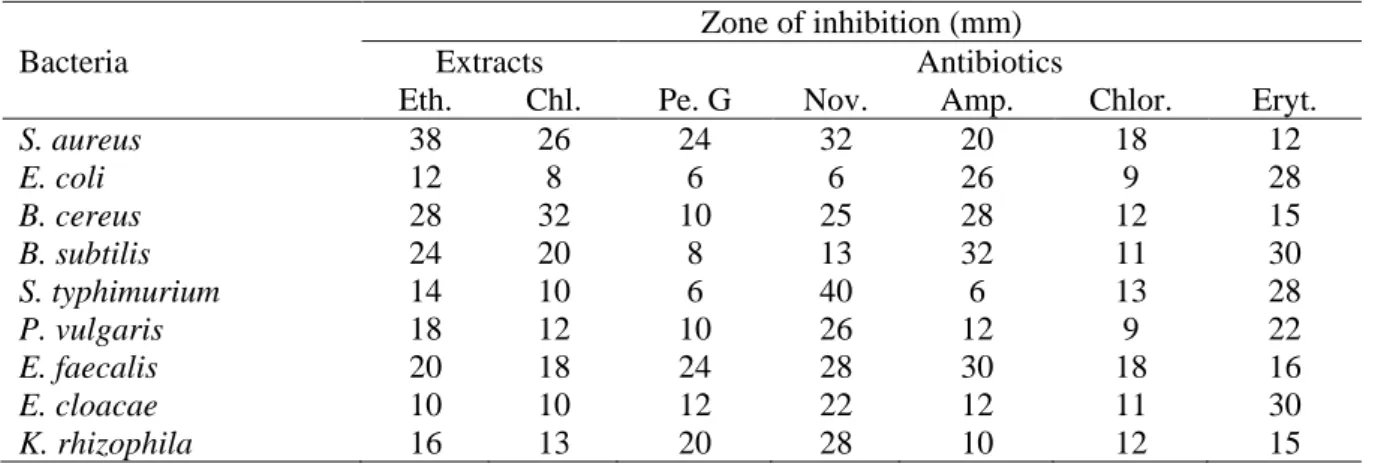
Table 1. Screening for antibacterial activity of callus from cotyledons of C. officinalis extracts against
standart microorganisms.
Zone of inhibition (mm)
Bacteria Extracts Antibiotics
Eth. Chl. Pe. G Nov. Amp. Chlor. Eryt.
S. aureus 38 26 24 32 20 18 12 E. coli 12 8 6 6 26 9 28 B. cereus 28 32 10 25 28 12 15 B. subtilis 24 20 8 13 32 11 30 S. typhimurium 14 10 6 40 6 13 28 P. vulgaris 18 12 10 26 12 9 22 E. faecalis 20 18 24 28 30 18 16 E. cloacae 10 10 12 22 12 11 30 K. rhizophila 16 13 20 28 10 12 15
Eth: Ethanol; Chl: Chloroform; Pe: Penicillin; Nov: Novobiocin; Amp: Amphicillin; Chlor: Chloramphenicol; Eryt: Erythromycin
When literature is reviewed, it is evident that many studies have been carried out in recent years on the secondary metabolite production [30-32] and on the antimicrobial effects of these metabolites in addition to studies such as cell culture, microbial growth, organogenesis, embryogenesis by callus culture method [33-37]. However, no literature has been found on the antimicrobial effect of C. officinalis calli.
In the present study, we observed in vitro antibacterial activities of ethanol and chloroform extracts of the anti-bacterial activity of cotyledon derived calli of Calendula
officinalis and standard antibiotics (Table 1.).
The extracts of C. officinalis showed various antibacterial activities against the test bacteria. All extracts studied in this work showed antibacterial activity against at least one of the test microorganisms with inhibition zones ranging from 8 to 38 mm (Table 1). Ethanol extract of C. officinalis callus showed 38 mm inhibition zone against Staphylococcus aureus and chloroform extract showed 32 mm zone against Bacillus cereus. These results are very close to the test antibiotics used and C. officinalis found more effective on gram positive bacteria.
The results of the current investigation clearly indicate that the antibacterial activity vary with the C. officinalis. Further, the active phytocompounds of this plant against some bacteria should be characterized and their toxicity should be evaluated in vivo.
Conflict of Interests
261 4. REFERENCES
[1] Rao, S. R., & Ravishankar, G. A. (2002). Plant cell cultures: chemical factories of secondary metabolites. Biotechnology advances, 20(2), 101-153.
[2] Noor, A., Bansal, V. S., & Vijayalakshmi, M. A. (2013). Current update on anti-diabetic biomolecules from key traditional Indian medicinal plants. Current science, 721-727. [3] Erkoyuncu, M. T., & Yorgancılar, M. (2016). Bitki Doku Kültürü Yöntemleri İle Sekonder
Metabolitlerin Üretimi. Selçuk Tarım Bilimleri Dergisi, 2(1), 66-76.
[4] Bahri-Sahloul, R., Ben Fredj, R., Boughalleb, N., Shriaa, J., Saguem, S., Hilbert, J. L., & Harzallah-Skhiri, F. (2014). Phenolic composition and antioxidant and antimicrobial activities of extracts obtained from Crataegus azarolus L. var. aronia (Willd.) Batt. ovaries calli. Journal of Botany, 2014.
[5] Davis, P. H. (1982). Flora of Turkey and the Aegean Islands. Vol. 8. Univ. Pres. Edinburgh. [6] Arora, D., Rani, A., & Sharma, A. (2013). A review on phytochemistry and ethnopharmacological aspects of genus Calendula. Pharmacognosy reviews, 7(14), 179. [7] Teiten, M. H., Gaascht, F., Dicato, M., & Diederich, M. (2013). Anticancer bioactivity of
compounds from medicinal plants used in European medieval traditions. Biochemical
pharmacology, 86(9), 1239-1247.
[8] Iauk, L., Lo Bue, A. M., Milazzo, I., Rapisarda, A., & Blandino, G. (2003). Antibacterial activity of medicinal plant extracts against periodontopathic bacteria. Phytotherapy
Research, 17(6), 599-604.
[9] Nabi, S., Ahmed, N., Khan, M. J., Bazai, Z., Yasinzai, M., & Al-Kahraman, Y. M. S. A. (2012). In vitro antileishmanial, antitumor activities and phytochemical studies of methanolic extract and its fractions of Juniperus Excelsa Berries. World Applied Sciences
Journal, 19(10), 1495-1500.
[10] Kalvatchev, Z., Walder, R., & Garzaro, D. (1997). Anti-HIV activity of extracts from Calendula officinalis flowers. Biomedicine & pharmacotherapy, 51(4), 176-180.
[11] Ćetković, G. S., Djilas, S. M., Čanadanović-Brunet, J. M., & Tumbas, V. T. (2004). Antioxidant properties of marigold extracts. Food Research International, 37(7), 643-650. [12] Boucaud-Maitre, Y., Algernon, O., & Raynaud, J. (1988). Cytotoxic and antitumoral
activity of Calendula officinalis extracts. Pharmazie, 43(3), 220-221.
[13] De Tommasi, N., Conti, C., Stein, M. L., & Pizza, C. (1991). Structure and in vitro antiviral activity of triterpenoid saponins from Calendula arvensis. Planta medica, 57(03), 250-253. [14] Hamburger, M., Adler, S., Baumann, D., Förg, A., & Weinreich, B. (2003). Preparative purification of the major anti-inflammatory triterpenoid esters from Marigold (Calendula officinalis). Fitoterapia, 74(4), 328-338.
[15] Zitterl-Eglseer, K., Sosa, S., Jurenitsch, J., Schubert-Zsilavecz, M., Della Loggia, R., Tubaro, A., … & Franz, C. (1997). Anti-oedematous activities of the main triterpendiol esters of marigold (Calendula officinalis L.). Journal of ethnopharmacology, 57(2), 139-144.
[16] Marukami, T., Kishi, A., & Yoshikawa, M. (2001). Medicinal flowers. IV. Marigold.(2): Structures of new ionone and sesquiterpene glycosides from Egyptian Calendula officinalis. Chemical and pharmaceutical bulletin, 49(8), 974-978.
[17] Shipochliev, T. (1981). Uterotonic action of extracts from a group of medicinal plants.
Veterinarno-meditsinski nauki, 18(4), 94-98.
[18] Duran, V., Matic, M., Jovanovć, M., Mimica, N., Gajinov, Z., Poljacki, M., & Boza, P. (2005). Results of the clinical examination of an ointment with marigold (Calendula
262
officinalis) extract in the treatment of venous leg ulcers. International journal of tissue
reactions, 27(3), 101-106.
[19] Ugulu, I., Baslar, S., Yorek, N., & Dogan, Y. (2009). The investigation and quantitative ethnobotanical evaluation of medicinal plants used around Izmir province, Turkey. Journal
of Medicinal plants research, 3(5), 345-367.
[20] Murashige, T., & Skoog, F. (1962). A revised medium for rapid growth and bio assays with tobacco tissue cultures. Physiologia plantarum, 15(3), 473-497.
[21] Oskay, M., & Sarı, D. (2007). Antimicrobial screening of some Turkish medicinal plants.
Pharmaceutical Biology, 45(3), 176-181.
[22] Kalyoncu, F., & Oskay, M. (2008). Antimicrobial activities of four wild mushroom species collected from Turkey. In Mushroom biology and mushroom products. Proceedings of the
Sixth International Conference on Mushroom Biology and Mushroom Products, Bonn, Germany, 29 September-3 October, 2008 (pp. 31-35). GAMU GmbH, Institut für
Pilzforschung.
[23] Dias, M. I., Sousa, M. J., Alves, R. C., & Ferreira, I. C. (2016). Exploring plant tissue culture to improve the production of phenolic compounds: A review. Industrial Crops and
Products, 82, 9-22.
[24] Shukla, M. R., Singh, A. S., Piunno, K., Saxena, P. K., & Jones, A. M. P. (2017). Application of 3D printing to prototype and develop novel plant tissue culture systems.
Plant Methods, 13(1), 6.
[25] Kocaçalışkan, İ. (2008). Bitki Doku Kültürleri (Organ, Doku ve Hücre)(ISBN: 978-975-8201-47-6). DPÜ, Fen Edebiyat Fakültesi, Biyoloji Bölümü, Kütahya.
[26] Ghasempour, H., Soheilikhah, Z., Zebarjadi, A. R., Ghasempour, S., & Karimi, N. (2014). In vitro micro propagation, callus induction and shoot regeneration in safflower L. cv. Lesaf.
[27] Ilahi, I. H. S. A. N., Jabeen, M., & Sadaf, S. N. (2007). Rapid clonal propagation of Chrysanthemum through embryogenic callus formation. Pak. J. Bot, 39(6), 1945-1952. [28] Patel, R. M., & Shah, R. R. (2009). Regeneration of Stevia plant through callus culture.
Indian journal of pharmaceutical sciences, 71(1), 46.
[29] Wani, M., Pande, S., & More, N. (2010). Callus induction studies in Tridax procumbens L. International Journal of Biotechnology Applications, 2(1), 11-14.
[30] Jain, S. C., Pancholi, B., & Jain, R. (2012). In-vitro Callus Propagation and Secondary Metabolite Quantification in Sericostoma pauciflorum. Iranian journal of pharmaceutical
research: IJPR, 11(4), 1103.
[31] Szopa, A., & Ekiert, H. (2014). Production of biologically active phenolic acids in Aronia melanocarpa (Michx.) Elliott in vitro cultures cultivated on different variants of the Murashige and Skoog medium. Plant growth regulation, 72(1), 51-58.
[32] Yildirim, A. B., & Turker, A. U. (2014). Effects of regeneration enhancers on micropropagation of Fragaria vesca L. and phenolic content comparison of field-grown and in vitro-grown plant materials by liquid chromatography-electrospray tandem mass spectrometry (LC–ESI-MS/MS). Scientia Horticulturae, 169, 169-178.
[33] Chiruvella, K. K., Mohammed, A., Dampuri, G., Ghanta, R. G., & Raghavan, S. C. (2007). Phytochemical and antimicrobial studies of methyl angolensate and luteolin-7-O-glucoside isolated from callus cultures of Soymida febrifuga. International journal of biomedical
science: IJBS, 3(4), 269.
[34] Cetin, B., & Kalyoncu, F. (2011). Antimicrobial Activity of Extracts from the Callus Culture of Rubia tinctorum L. Journal of Pure And Applied Microbiology, 5(2), 903-907.
263
[35] Ahire, M. L., Ghane, S. G., Lokhande, V. H., Suprasanna, P., & Nikam, T. D. (2011). Micropropagation of Uraria picta through adventitious bud regeneration and antimicrobial activity of callus. In Vitro Cellular & Developmental Biology-Plant, 47(4), 488.
[36] Magda, M. A., El Nour, M. E., Hassan, A. A. E. L., & Ezzdeen, L. T. (2015). Antibacterial activities of seeds, leaves and callus (hypocotyls and cotyledons) extracts of Jatropha curcas L. Int. J. Biosci, 6(11), 58-63.
[37] Rohela, G. K., Bylla, P., Korra, R., & Reuben, C. (2016). Phytochemical Screening and Antimicrobial Activity of Leaf, Stem, Root and their Callus Extracts in Rauwolfia tetraphylla. International Journal of Agriculture & Biology, 18(3).