https://doi.org/10.1177/1470320320928872 Journal of the Renin-Angiotensin-Aldosterone System
April-June 2020: 1 –9 © The Author(s) 2020 Article reuse guidelines: sagepub.com/journals-permissions DOI: 10.1177/1470320320928872 journals.sagepub.com/home/jra
Creative Commons Non Commercial CC BY-NC: This article is distributed under the terms of the Creative Commons Attribution-NonCommercial 4.0 License (https://creativecommons.org/licenses/by-nc/4.0/) which permits non-commercial use, reproduction and distribution of the work without further permission provided the original work is attributed as specified on the SAGE and Open Access pages (https://us.sagepub.com/en-us/nam/open-access-at-sage).
Introduction
Coronaviruses (CoV) are the enveloped RNA viruses that can cause enzootic fatal infections primarily in birds and mammals.1–4 Moreover, CoV has the ability to infect humankind.5–7 The presenting symptoms and signs of the infection could be chills, night sweats, persistent cough, diarrhea, and fever. However, the viral infection could cause a wide variety of clinical presentations, ranging from upper respiratory infections (URI) to lower respiratory infections (LRI), including bronchitis, pneumonia, and severe acute respiratory syndrome (SARS). CoV affects the respiratory system and specifically the lungs.5,8,9 Furthermore, the viral infection can also affect the gastro-intestinal tract, as well as the neurological and hepatic systems.10 The severe acute respiratory syndrome (SARS) epidemic in 2003 and the Middle East respiratory
syndrome (MERS) in 2012 revealed that CoV can be fatal when they cross the species barrier and infect humans.6 A newly identified CoV called SARS-CoV-2 (formerly
In vitro analysis of the renin–angiotensin
system and inflammatory gene transcripts
in human bronchial epithelial cells after
infection with severe acute respiratory
syndrome coronavirus
Can Turk
1, Seyhan Turk
2, Elif Sena Temirci
3,
Umit Yavuz Malkan
4and İbrahim C. Haznedaroglu
5Abstract
Introduction: Severe acute respiratory syndrome coronavirus 2 (SARS-CoV-2) is a recently identified coronavirus family member that triggers a respiratory disease similar to severe acute respiratory syndrome coronavirus (SARS-CoV). SARS-CoV and SARS-CoV-2 are very similar to each other in many respects, such as structure, genetics, and pathobiology. We hypothesized that coronaviruses could affect pulmonary tissues via integration with the critical immune genes after their interaction with renin–angiotensin system (RAS) elements. The aim of the present bioinformatics study was to assess expression changes of the RAS and non-RAS genes, particularly immune response genes, in the lung epithelial cells after infection with SARS-CoV.
Methods: Linear regression, hierarchical clustering, pathway analysis, and network analysis were performed using the E-GEOD-17400 data set.
Results: The whole-genome expression data of the lung epithelial cells infected with SARS-CoV for 12, 24, and 48 hours were analyzed, and a total of 15 RAS family and 29 immune genes were found to be highly correlated with the exposure time to the virus in the studied groups.
Conclusion: RAS genes are important at the initiation of the infections caused by coronavirus family members and may have a strong relationship with the exchange of immune genes in due course following the infection.
Keywords
RAS, ANPEP, ACE2, EGFR, IGF2R, SARS-CoV, SARS-CoV-2, immune system genes Date received: 22 February 2020; accepted: 25 April 2020
1 Department of Medical Microbiology, Lokman Hekim University,
Faculty of Medicine, Turkey
2 Department of Biochemistry, Hacettepe University, Faculty of
Pharmacy, Turkey
3 Department of Molecular Biology and Genetics, Bilkent University,
Faculty of Science, Turkey
4 Department of Haematology, Dışkapı Yıldırım Beyazıt Training and
Research Hospital, University of Health Sciences, Turkey
5 Department of Haematology, Hacettepe University, Faculty of
Medicine, Turkey
Corresponding author:
Seyhan Turk, Department of Biochemistry, Hacettepe University, Faculty of Pharmacy, Ankara, 06532, Turkey.
Email: [email protected]
entitled nCoV) appeared within the last month of 2019 in the city of Wuhan, China.11 Very early findings suggested that the virus passed from a specific animal species to humans. However, some studies then revealed that SARS-CoV-2 could also be passed from person to person, lead-ing to serious respiratory diseases such as acute respiratory distress syndrome similar to that seen in the SARS-CoV infection.12,13 SARS-CoV-2 is an enveloped, single-stranded, positive-sense RNA (beta-) CoV. The same as SARS-CoV and MERS-CoV, the SARS-CoV-2 genome can encode structural proteins such as the spike glycopro-tein, non-structural proteins, and accessory proteins. The spike glycoprotein, in which this virus has encoding func-tions, plays an important role in the interactions of the virus and the cell receptor during viral entry.6 More spe-cifically, a spike protein that is associated with the enve-lope of SARS-CoV-2 binds first to the host receptor and then mediates the entry of the CoV into the host cells by fusing viral and host membranes.14–16 Once the fusion has taken place, the RNA virus begins to replicate its genome in the cell and makes new virions that will be secreted to infect other cells.17–19
There is a clear pathobiological association between CoV and the renin–angiotensin system (RAS). SARS-CoV (a member of the CoV family that emerged in 2002), which is very similar to SARS-CoV-2, and angiotensin convert-ing enzyme 2 (ACE2) interactions have been extensively studied.13,16 There is a fundamental atomic interaction between the spike protein receptor binding domain of the virus and the host receptor of ACE2. SARS-CoV infec-tions lead to ACE2 down-regulation by the binding of the SARS-CoV spike protein to ACE2.20 Thus, the SARS-CoV family, including SARS-SARS-CoV-2, utilizes ACE2 as a critical receptor to enter target host cells.13,16
The main task of ACE2 is to hydrolyze angiotensin II (Ang II) to form angiotensin (1–7) (Ang-(1–7)) that binds to the G-coupled protein receptor MAS to antagonize Ang II–mediated cellular effects. This function is very impor-tant since the increased Ang II levels are estimated to cause increased ACE2 activity. ACE2 is one of the central RAS enzymes that regulate blood pressure, fluid/electrolyte balance, and systemic vascular resistance in the tissue RAS. ACE2 is also critical for the RAS, which functions for the regulation of cell growth/proliferation, inflamma-tion/cytokine production, blood pressure, and as a homeo-static regulator of vascular function. The RAS is highly active within the lungs where SARS-CoV-2 is estimated to be the primary target organ. Ang II can cause pulmonary vasoconstriction in response to hypoxia, which is impor-tant in patients with pneumonia or lung injury. Locally increased Ang II production increases vascular permea-bility, which facilitates pulmonary edema. ACE2, which plays a role in regulating the Ang II as well as Ang-(1–7) levels, may be important for those pathobiological lung events.13,21,22 ACE2 is an important negative directing
factor for the severity of lung injury, and SARS-CoV spike protein–associated ACE2 inhibition has been shown to contribute to the severity of lung pathologies.20 In addition, numerous genes are involved within the interactions of ACE2, the RAS, and the pulmonary microenvironment.
The activation of the local pulmonary RAS can affect the genesis of lung damage through multiple mechanisms, such as increments in vascular permeability and alterations in alveolar epithelial cells. The pathogenesis of the CoV is a rather complicated process, which may also include local tissue RAS.13,21
The main purpose of this present in silico genomic study was to assess how the expressions of the RAS gene family changes after cellular infection with SARS-CoV in the lung epithelial cell culture. We hypothesized that CoV could affect pulmonary tissues via integration with the critical immune genes of the affected cellular micro-environment after their interaction with RAS elements. We also aimed to figure out whether these genomic alter-ations are related to the exposure time of the virus. In order to understand the pathobiology of SARS-CoV and SARS-CoV-2 infection better, since they share similari-ties within many aspects we examined the gene expres-sion changes of other gene families as well. Elucidation of the interactions between SARS-CoV-2 and RAS genes and the description of the related genomic mechanisms are very important in the fight against this fatal viral global infection.
Methods
Obtaining and normalizing whole-genome
expression data of human bronchial epithelial
cells infected with SARS-CoV
Gene expression data of human bronchial epithelial cells treated with SARS-CoV for 12, 24, and 48 hours were obtained from Array Express (GSE17400).23 These data were generated by Yoshikawa et al. to characterize the dynamic, spatial, and temporal changes of the gene expres-sion induced by SARS-CoV using microarray technology. In order to use the obtained data in other targeted anal-yses, the raw data were normalized by robust multi-sequence analysis in accordance with the procedure in the Affy package in R. These data consist of 23,344 genes (54,675 probe sets). In addition, each gene has three repeated expression data values for 12, 24, and 48 hours, respectively.
Comparison of RAS gene transcripts in 12-,
24- and 48-hour SARS-CoV-infected groups
The whole normalized gene expression data of lung epi-thelial cells infected with SARS-CoV for 12, 24, and 48 hours were compared between different groups in order to
determine significantly and differentially expressed RAS family genes. The mean value of RAS gene transcripts was determined for each group infected with SARS-CoV for 12, 24, and 48 hours. The comparison was made between the 12- and 24-hour groups as well as between the 12- and 48-hour groups, based on the RAS gene transcripts.
p-Values were calculated for each gene using the t-test.
The genes belonging to the RAS family with a statistically significant difference among the groups were determined (p⩽0.05).
Linear regression analysis of the genes (other
than the RAS genes) with the highest
variance among the groups
In order to determine the genes whose expression is highly correlated with the exposure time to the virus, we per-formed linear regression analysis. Genes with a standard deviation >0.9 were identified between the groups using Microsoft Excel (2016). Pearson product–moment correla-tion coefficient values (r) were calculated for these genes. Genes with a Pearson’s r >0.95 were selected for use in the subsequent analyses.
Hierarchical clustering, pathway analysis, and
network analysis
In order to determine whether the identified genes could distinguish between the virus and infected groups, to iden-tify the network connections between these genes and to demonstrate the pathways in which these genes were involved, we performed hierarchical clustering, pathway analysis, and network analysis.
Genes with the highest variation and Pearson’s r-value between groups were hierarchically clustered using the
similarity metric parameter and the Euclidean distance Gene Cluster v3.0 program as a full link.24 Using the DAVID online tool, the pathways related to the genes were determined to elaborate on the pathway data resulting from the analysis further.25,26
In addition, the GeneMANIA application in Cytoscape was used to indicate the network and pathway relationship between the selected genes and other genes.27,28
Results
Among the three groups of human bronchial epithelial cells with different exposure times to SARS-CoV (12-, 24- and 48-hour groups), we identified the changes in expression of the RAS gene transcripts as well as the trend in expression of the most differentially expressed genes among groups other than the RAS family.
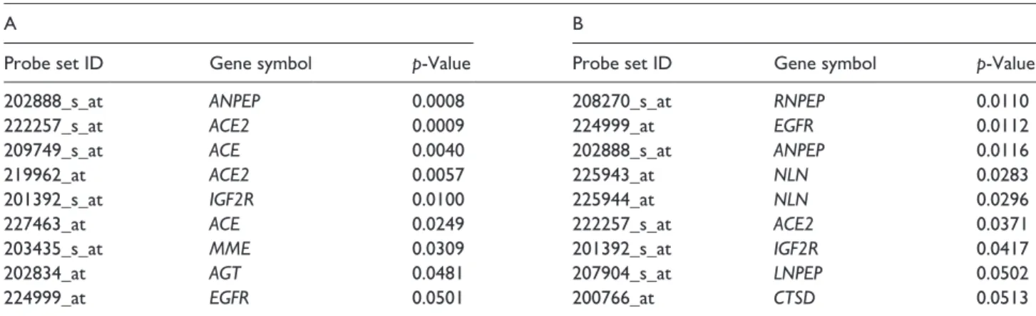
A total of seven RAS signaling pathway genes (nine probe sets; alanyl aminopeptidase (ANPEP), ACE2, angiotensin converting enzyme (ACE), insulin-like growth factor 2 receptor (IGF2R), angiotensinogen (AGT), epidermal growth factor receptor (EGFR) and membrane metalloendopeptidase (MME)) showed significantly different expression values between the 12-hour group and the 24-hour group. Comparing the 12-hour and 48-hour groups, eight genes (nine probe sets; arginyl aminopeptidase (RNPEP), epidermal growth factor receptor (EGFR), ANPEP, neurolysin (NLN), ACE2, IGF2R, leucyl and cysteinyl aminopepti-dase (LNPEP) and cathepsin D (CTSD)) were statisti-cally significantly expressed (Table 1). Four common genes (IGF2R, ANPEP, ACE2, and EGFR) in these two separate groups were identified (Figure 1).
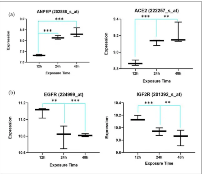
Figure 2 shows the correlation of the expression and exposure time to the virus of the statistically significant
Table 1. The list of the RAS family genes whose expression shows a significant difference between 12-hour vs 24-hour (A) and
12-hour vs 48-hour (B) is depicted. Seven of the RAS signaling pathway genes showed different expression values in 12-hour vs 24-hour group, while 8 of the genes found significantly expressed in the 12-hour vs 48-hour group.
A B
Probe set ID Gene symbol p-Value Probe set ID Gene symbol p-Value
202888_s_at ANPEP 0.0008 208270_s_at RNPEP 0.0110
222257_s_at ACE2 0.0009 224999_at EGFR 0.0112
209749_s_at ACE 0.0040 202888_s_at ANPEP 0.0116
219962_at ACE2 0.0057 225943_at NLN 0.0283
201392_s_at IGF2R 0.0100 225944_at NLN 0.0296
227463_at ACE 0.0249 222257_s_at ACE2 0.0371
203435_s_at MME 0.0309 201392_s_at IGF2R 0.0417
202834_at AGT 0.0481 207904_s_at LNPEP 0.0502
224999_at EGFR 0.0501 200766_at CTSD 0.0513
ANPEP: alanyl aminopeptidase; ACE2: angiotensin converting enzyme 2; ACE: angiotensin converting enzyme; IGF2R: insulin-like growth factor 2 receptor; MME: membrane metalloendopeptidase; AGT: angiotensinogen; EGFR: epidermal growth factor receptor; RNPEP: arginyl aminopeptidase; NLN: neurolysin; LNPEP: leucyl and cysteinyl aminopeptidase; CTSD: cathepsin D.
Figure 1. Venn diagram showing common genes between the 12-hour and 24-hour groups and the 12-hour and 48-hour groups.
Four genes were found to be common to both groups. As mentioned, these genes consistently showed statistically meaningful expression differences according to exposure time to the virus.
Figure 2. Two out of four common differentially expressed renin–angiotensin system (RAS) genes show a positive correlation
with exposure time to the virus (a), while the other common genes, epidermal growth factor receptor (EGFR) and insulin-like growth factor 2 receptor (IGF2R), show a negative correlation (b).
four common members of RAS genes found in the two groups (Figure 2).
In order to figure out the role of the non-RAS genes, we determined the most variant genes among these three groups. The standard deviation values of 29 genes (36 probe sets) were found to be >0.9. Linear regression anal-ysis revealed that all these genes had a Pearson’s r >0.95 and were highly correlated with the exposure time to the virus (Table 2).
Likewise, all of these genes showed a positive correla-tion with exposure time to the virus. The correlacorrela-tion figure for each gene that was found to be highly correlated to
virus infection exposure time is presented in the Supplemental Material.
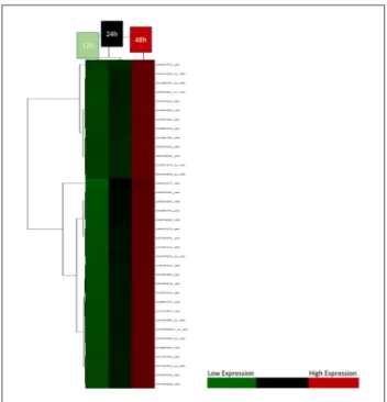
In addition, as shown in Figure 3, these genes were able to be clustered for the 12-, 24- and 48-hour groups separately.
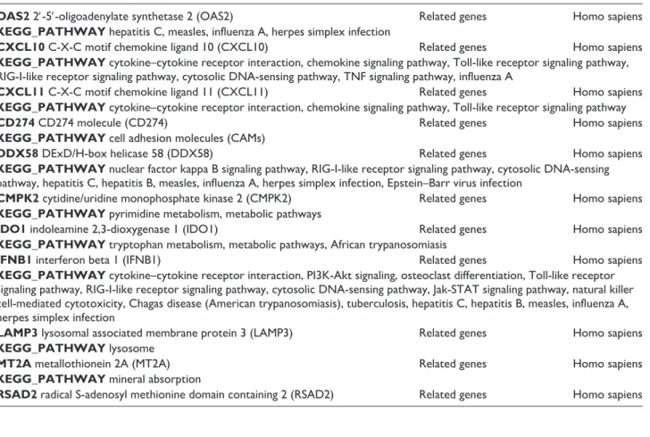
Table 3 shows the pathway analysis of all the signifi-cant genes. With the aim of discovering the relationship of these genes with each other and with other genes, a net-work analysis was performed (Figure 4).
Discussion
CoV-2 was found to be mostly similar to the SARS-CoV at the amino-acid level, despite some differences. Based on the phylogenetic analysis on all genomes of vari-ous viruses, SARS-CoV-2 is in the same beta-CoV clone as SARS-CoV, SARS-like bat CoV, and MERS-CoV. SARS-CoV-2 has the highest similarity with SARS-like bat CoV and is less associated with MERS-CoV.29
The mechanism of SARS-CoV and its association with the renin–angiotensin pathway had been defined in previ-ous studies. The first genetic proof of the ACE2 and SARS-CoV receptor relationship was reported by Kuba et al.20 Our results showed that in lung epithelial cells, ACE2 gene expression increased between 12 and 24 hours and remained at the same level between 24 and 48 hours after infection with SARS-CoV. Complementary to our findings, Kuba et al. demonstrated a decrement in the
Table 2. The most variant genes between all groups. 29 genes
(36 Probe Sets) found to have >0.9 standard deviation value. All of these genes are highly correlated with exposure time to viruses an most of them are immune genes.
Probe set ID Gene symbol SD Pearson’s r
211122_s_at CXCL11 1.68295417 0.99402069 210163_at CXCL11 1.67995636 0.99308306 213293_s_at TRIM22 1.64689117 0.99812764 214079_at DHRS2 1.38291569 0.98065023 242625_at RSAD2 1.3619956 0.99998811 208173_at IFNB1 1.32200238 0.96674605 213797_at RSAD2 1.29016111 0.99987245 210029_at IDO1 1.28419322 0.98922768 226702_at CMPK2 1.23620496 0.99368587 204972_at OAS2 1.20086416 0.99494266 205660_at OASL 1.18935939 0.99589149 214022_s_at IFITM1 1.17991713 0.99401632 201601_x_at IFITM1 1.17122332 0.99420957 206553_at OAS2 1.14649914 0.99866159 204533_at CXCL10 1.11355998 0.99132404 204994_at MX2 1.10795097 0.99999559 228152_s_at DDX60L 1.08126568 0.98604577 206157_at PTX3 1.06159153 0.97359778 228617_at XAF1 1.04957699 0.99912443 219863_at HERC5 1.0436716 0.99976953 204747_at IFIT3 1.04191315 0.99999404 217502_at IFIT2 1.01930351 0.99727623 236285_at KLHDC7B 1.01815991 0.99341293 205569_at LAMP3 1.01338642 0.98242768 226757_at IFIT2 1.0132658 0.99994894 229450_at IFIT3 1.00446864 0.99482924 219211_at USP18 0.99619048 0.9993661 218943_s_at DDX58 0.94794454 0.99979879 218986_s_at DDX60 0.94081343 0.99961176 210797_s_at OASL 0.94028093 0.99930186 1559883_s_at SAMHD1 0.93726862 0.9997964 219684_at RTP4 0.93351655 0.99821178 226603_at SAMD9L 0.93102808 0.99815238 212185_x_at MT2A 0.92870941 0.98679396 227458_at CD274 0.92087529 0.98185747 230036_at SAMD9L 0.90113608 0.99960499 SD: standard deviation.
Figure 3. Hierarchical cluster of most variant genes between
three groups. As shown, determined genes were able to classify the groups clearly. In the 12-hour group, these genes show low expression, while in the 48-hour group, they all show high expression.
Table 3. Pathways related to non-RAS differentially expressed genes. Most of non-RAS differentially expressed genes were found
to be related immune the system and pathways which could be involved during the course of the viral infectious disease.
OAS2 2′-5′-oligoadenylate synthetase 2 (OAS2) Related genes Homo sapiens
KEGG_PATHWAY hepatitis C, measles, influenza A, herpes simplex infection
CXCL10 C-X-C motif chemokine ligand 10 (CXCL10) Related genes Homo sapiens
KEGG_PATHWAY cytokine–cytokine receptor interaction, chemokine signaling pathway, Toll-like receptor signaling pathway,
RIG-I-like receptor signaling pathway, cytosolic DNA-sensing pathway, TNF signaling pathway, influenza A
CXCL11 C-X-C motif chemokine ligand 11 (CXCL11) Related genes Homo sapiens
KEGG_PATHWAY cytokine–cytokine receptor interaction, chemokine signaling pathway, Toll-like receptor signaling pathway
CD274 CD274 molecule (CD274) Related genes Homo sapiens
KEGG_PATHWAY cell adhesion molecules (CAMs)
DDX58 DExD/H-box helicase 58 (DDX58) Related genes Homo sapiens
KEGG_PATHWAY nuclear factor kappa B signaling pathway, RIG-I-like receptor signaling pathway, cytosolic DNA-sensing
pathway, hepatitis C, hepatitis B, measles, influenza A, herpes simplex infection, Epstein–Barr virus infection
CMPK2 cytidine/uridine monophosphate kinase 2 (CMPK2) Related genes Homo sapiens
KEGG_PATHWAY pyrimidine metabolism, metabolic pathways
IDO1 indoleamine 2,3-dioxygenase 1 (IDO1) Related genes Homo sapiens
KEGG_PATHWAY tryptophan metabolism, metabolic pathways, African trypanosomiasis
IFNB1 interferon beta 1 (IFNB1) Related genes Homo sapiens
KEGG_PATHWAY cytokine–cytokine receptor interaction, PI3K-Akt signaling, osteoclast differentiation, Toll-like receptor
signaling pathway, RIG-I-like receptor signaling pathway, cytosolic DNA-sensing pathway, Jak-STAT signaling pathway, natural killer cell-mediated cytotoxicity, Chagas disease (American trypanosomiasis), tuberculosis, hepatitis C, hepatitis B, measles, influenza A, herpes simplex infection
LAMP3 lysosomal associated membrane protein 3 (LAMP3) Related genes Homo sapiens
KEGG_PATHWAY lysosome
MT2A metallothionein 2A (MT2A) Related genes Homo sapiens
KEGG_PATHWAY mineral absorption
RSAD2 radical S-adenosyl methionine domain containing 2 (RSAD2) Related genes Homo sapiens
Figure 4. Co-expression and pathway analysis of non-RAS differentially expressed genes. The figure shows that 28/29 genes create
a strong co-expression network. On the other hand, the program was able to connect some of the genes and predict some related genes in pathway network analysis.
ACE2 protein level in the lungs of SARS-CoV-infected mice on day 2. They also reported that the addition of SARS-CoV spike into mice exacerbates acute lung failure in vivo that can be reduced by inhibiting the renin–angio-tensin pathway.20
Moreover, SARS-CoV-2 uses a similar mechanism as SARS-CoV. The structural analyses showed that like SARS-CoV, SARS-CoV-2 also utilizes ACE2 as the host receptor.16
Based on the evidence with regard to the similarity between SARS-CoV and SARS-CoV-2, we used data from Yoshikawa et al. In their study, all the genome expression data of the confluent 2B4 cells which were infected with SARS-CoV and grown in T-75 flasks for 12, 24 and 48 hours were used to characterize the dynamic, spatial and temporal changes of the gene expressions caused by SARS-CoV. The study was performed three times at each time point in order to meet the minimum number required for the application of statistical algorithms, and a total of nine arrays were given for SARS-CoV.23
Our current study was carried out to understand the bio-logical mechanism of SARS-CoV-2 infection better by focusing on the similarity of CoV-2 and SARS-CoV in terms of structure, biological function, and the pathology of the infection. We proposed a hypothesis based on the genomic results obtained in the present study under three key headings (Figure 5).
Initial phase
According to the results of the analysis, there was a signifi-cant up-regulation of ACE2 and ANPEP genes in human bronchial epithelial cells within 12 and 24 hours of infec-tion. Many previous studies indicated that SARS-CoV also uses ACE2 as a receptor to enter host cells, as in SARS-CoV-2.30 The results of our present study showed the addi-tional possible function of ANPEP (Figure 5).
The ANPEP gene acts as a receptor, especially for HCoV-229E, another member of the CoV family. This virus is predicted to induce the infection by triggering conformational changes in the spike glycoprotein, which interacts with the host ANPEP receptor and activates membrane fusion.31 Moreover, ANPEP (CD13) can act as a receptor in SARS-CoV and induce growth inhibition and apoptosis by infecting hematopoietic stem/progenitor cells.32 These findings are in concordance with our results demonstrating the up-regulation in the ACE2 and ANPEP as exposure time to the virus increased.
As suggested in many previous studies, ACE2 forms Ang-(1–7) from Ang II and binds to the MAS receptor, the specific receptor of Ang-(1–7), and then inhibits inflam-matory, vascular and cellular growth mechanisms.33 In this case, it may be useful to use the agonistic peptides of Ang-(1–7) to change the functioning of ACE2 in favor of the infected host in the initial phase of infection.
Propagating phase
Based on our results, in this phase, as the exposure time to SARS-CoV increases, EGFR and IGF2R, two receptors with key roles in the RAS signaling pathway, were signifi-cantly down-regulated in the infected human bronchial epithelial cells.
Habib et al. proposed that apoptosis occurs in various cell types that require an active tyrosine kinase but do not require EGFR autophosphorylation sites by experimen-tally increasing the level of EGFR expression. The expres-sion of a predominant negative RAS mutant in cells that over-express the EGFR leads to significantly enhanced EGFR-induced apoptosis.34 On the other hand, IGF2R, acting as a G-coupled protein receptor, could lead to the activation of the mitochondria-mediated apoptosis path-way, when insulin-like growth factor-II (IGF2) binds to this receptor.35 Thus, those two receptors, which are asso-ciated with cellular apoptosis, are expected to be down-regulated as the infection time of virus-infected cells progresses. This event could give the chance for the virus to have sufficient time to continue replication and increase its copy number. This phase of the viral infection might be important, particularly for hypertensive subjects.
Complicating phase
In this in silico genomic study, 29 genes (36 probe sets) were detected which showed noteworthy up-regulation in 24- versus 48-hour groups. Almost all of these are genes associated with the immune system, innate immune response, and adaptive immune response.
The relevant biological analyzers of these genes whose expressions are concordant with cell exposure time to the virus were explored via the pathway analyzers of the 29 genes. The genes involved in specific pathways related to
Figure 5. Three important phases associated with SARS-CoV
infection. At the initial and propagating phases of infection, some RAS family genes were up-regulated while some others were down-regulated. At the complicating phase, there is an increase in the expression of some key immune system genes.
the critical pathological events of CoV infection, including SARS-CoV and SARS-CoV-2, such as the Toll-like recep-tor (TLR) signaling pathway.
TLRs are essential sensor molecules of the host innate immune system. Various TLRs are involved in early inter-actions of invasive viruses and host cells, which affect viral pathogenesis and regulate viral replication as well as host responses. Initiation of antiviral immune responses with TLR agonists has been shown to provide protection from many different viruses, including hepatitis B virus, influenza virus, some HIV strains, and CoV. Furthermore, non-structural protein 3 of the SARS-CoV interacts with IRF3 through the papain-like protease domain. After that, the associations of the TLR and RLR pathways occur, and this binding prevents the nuclear translocation as well as the phosphorylation of IRF3.36
It has previously been observed that he TLR signal through the TRIF adapter protein protects mice from lethal SARS-CoV. Thus, a balanced immune response that works both in TRIF and MyD88-guided pathways pro-vides the most effective host-cell intrinsic antiviral defense responses against severe SARS-CoV, and the absence of any branch of TLR signaling leads to SARS-CoV being fatal.37 Another important pathway is associated with the interferon beta 1 (IFNB1) gene identified as a result of pathway analysis is the natural killer (NK) cell-mediated cytotoxicity pathway. Interferons are considered as the screams of the affected cells during the active virus attack. IFNB1 deficiency results in partial suppression of the sterol pathway in macrophages during viral infections, thereby associating the regulation of the lipid metabolism pathway with interferon antiviral defense responses.38 Furthermore, NK cells are important in immune defense against virus infections. Enlarged virus replication and more serious diseases during encephalomyocarditis virus, Coxsackie virus, and Theiler’s murine encephalitis virus infections are linked with the reduction of NK cells or low levels of NK cell cytolytic function. NK cells are corre-spondingly associated with direct inhibition of virus repli-cation and stimulation of liver damage during mouse hepatitis virus (MHV) infection. It is not known whether NK cells play a direct antiviral role in human infections with picornaviruses or CoV such as SARS.39
Lysosome, which has an important path in CoV infec-tion, is another pathway related to the pathway analysis performed. Enveloped viruses must fuse with a host-cell membrane so as to transport their genomes to the host cell. Although some viruses fuse with the plasma membrane, many viral infections are associated with endocytosis before fusion. In the endosomal microenvironment, a par-ticular marker induces conformational changes in viral fusion proteins, leading to viral and host membrane fusion.40 According to the De Haan et al. study, the proteins known to be important for late endosomal maturation and endosome–lysosome fusion deeply promoted infection of cells with mouse hepatitis CoV (MHV).41 In another study of SARS-CoV, SARS-CoV accessory protein open reading
frames (SARS 3a) were oligomerized by dynamically inserting them into late endosomal, lysosomal and trans-Golgi network membranes.42 Furthermore, the chemotactic cytokines are engaged in a great deal of biological pro-cesses. Besides microbial infection, the infected cells have strong chemokine signals. Those signals could play signifi-cant roles in both innate and adaptive immune responses that control the growth of the invading pathogen.43 Therefore, the presence of chemokine signaling pathways parallels the other results obtained. The main limitation of this study is the lack of uninfected control data, which is subject to further experimental studies.
Conclusion
In conclusion, if the results obtained in the current study are validated by in vitro experiments and clinical samples, it can be suggested that the disruption of RAS genes may be important for the initial management of the CoV infec-tions, particularly SARS-CoV-2. The results of the current study help us to understand better the pathobiology of SARS-CoV and other similar CoV family members such as SARS-CoV-2. Based on our results, the interactions between the SARS-CoV-2 and RAS genes, resulting in immune-related genomic disruption, lead to acquired immune deficiency states. It is hoped that critical local RAS-affecting drugs such as MAS agonists, soluble ACE2 and Ang-(1–7) will be used for the modulation of SARS-CoV-2 and RAS pathological interactions for the improve-ment of immune genomic states in patients infected with SARS-CoV-2 in future trials.
Declaration of conflicting interests
The author(s) declared no potential conflicts of interest with respect to the research, authorship, and/or publication of this article.
Funding
The author(s) received no financial support for the research, authorship, and/or publication of this article.
Supplemental material
Supplemental material for this article is available online.
ORCID iDs
Seyhan Turk https://orcid.org/0000-0003-3843-4173
Elif Sena Temirci https://orcid.org/0000-0001-5944-6718
Umit Yavuz Malkan https://orcid.org/0000-0001-5444-4895
References
1. Dhama K, Pawaiya RVS, Chakrabort S, et al. Coronavirus infection in equines: a review. Asian J Anim Vet Adv 2014; 9: 164–176.
2. Fan Y, Zhao K, Shi ZL, et al. Bat coronaviruses in China.
Viruses 2019; 11: 210.
3. Fehr AR and Perlman S. Coronaviruses: an overview of their replication and pathogenesis. Methods Mol Biol 2015; 1282: 1–23.
4. Huang C, Wang Y, Li X, et al. Clinical features of patients infected with 2019 novel coronavirus in Wuhan, China.
Lancet 2020; 395: 497–506.
5. Habibzadeh P and Stoneman EK. The novel coronavirus: a bird’s eye view. Int J Occup Environ Med 2020; 11: 65–71. 6. Jonsdottir HR and Dijkman R. Coronaviruses and the
human airway: a universal system for virus–host interaction studies. Virol J 2016; 13: 24.
7. Zhou P, Yang XL, Wang XG, et al. A pneumonia outbreak associated with a new coronavirus of probable bat origin.
Nature 2020; 579: 270–273.
8. Schoeman D and Fielding BC. Coronavirus envelope pro-tein: current knowledge. Virol J 2019; 16: 69.
9. Wang M, Zhou Y, Zong Z, et al. A precision medicine approach to managing Wuhan coronavirus pneumonia.
Precis Clin Med 2020; 3: 14–21.
10. Zhu N, Zhang D, Wang W, et al. A novel coronavirus from patients with pneumonia in China, 2019. N Engl J Med 2020; 382: 727–733.
11. World Health Organization. Novel coronavirus (2019-nCoV)
situation report – 22. Geneva: World Health Organization,
2020.
12. Li Q, Guan X, Wu P, et al. Early transmission dynamics in Wuhan, China, of novel coronavirus-infected pneumonia. N
Engl J Med 2020; 382: 1199–1207.
13. Wu Y. Compensation of ACE2 function for possible clini-cal management of 2019-nCoV-induced acute lung injury.
Virol Sin. Epub ahead of print 7 February 2020. DOI:
10.1007/s12250-020-00205-6.
14. Letko M and Munster V. Functional assessment of cell entry and receptor usage for lineage B β-coronaviruses, including 2019-nCoV. bioRxiv. Epub ahead of print 22 January 2020. DOI: 10.1101/2020.01.22.915660.
15. Li F. Structure, function, and evolution of coronavirus spike proteins. Annu Rev Virol 2016; 3: 237–261.
16. Wan Y, Shang J, Graham R, et al. Receptor recognition by novel coronavirus from Wuhan: an analysis based on dec-ade-long structural studies of SARS. J Virol 2020; 94. 17. Kruse RL. Therapeutic strategies in an outbreak scenario
to treat the novel coronavirus originating in Wuhan, China.
F1000Res 2020; 9.
18. Kuhn JH, Li W, Choe H, et al. Angiotensin-converting enzyme 2: a functional receptor for SARS coronavirus. Cell
Mol Life Sci 2004; 61: 2738–2743.
19. Zhou P, Yang X-L, Wang X-G, et al. A pneumonia outbreak associated with a new coronavirus of probable bat origin.
Nature 2020; 579: 270–273.
20. Kuba K, Imai Y, Rao S, et al. A crucial role of angiotensin converting enzyme 2 (ACE2) in SARS coronavirus-induced lung injury. Nat Med 2005; 11: 875–879.
21. Tikellis C and Thomas MC. Angiotensin-converting enzyme 2 (ACE2) is a key modulator of the renin angiotensin system in health and disease. Int J Pept 2012; 2012: 256294. 22. Varagic J, Ahmad S, Nagata S, et al. ACE2: angiotensin II/
angiotensin-(1–7) balance in cardiac and renal injury. Curr
Hypertens Rep 2014; 16: 420.
23. Yoshikawa T, Hill TE, Yoshikawa N, et al. Dynamic innate immune responses of human bronchial epithelial cells to severe acute respiratory syndrome-associated coronavirus infection. PLoS One 2010; 5: e8729.
24. De Hoon MJ, Imoto S, Nolan J, et al. Open source clustering software. Bioinformatics 2004; 20: 1453–1454.
25. Huang Da W, Sherman BT and Lempicki RA. Systematic and integrative analysis of large gene lists using DAVID bioinformatics resources. Nat Protoc 2009; 4: 44–57. 26. Huang Da W, Sherman BT and Lempicki RA. Bioinformatics
enrichment tools: paths toward the comprehensive functional analysis of large gene lists. Nucl Acids Res 2009; 37: 1–13. 27. Shannon P, Markiel A, Ozier O, et al. Cytoscape: a software
environment for integrated models of biomolecular interac-tion networks. Genome Res 2003; 13: 2498–2504.
28. Warde-Farley D, Donaldson SL, Comes O, et al. The GeneMANIA prediction server: biological network integra-tion for gene prioritizaintegra-tion and predicting gene funcintegra-tion.
Nucl Acids Res 2010; 38: W214–220.
29. Wu A, Peng Y, Huang B, et al. Genome composition and divergence of the novel coronavirus (2019-nCoV) originat-ing in China. Cell Host Microbe 2020; 27: 325–328. 30. Hoffmann M, Kleine-Weber H, Krüger N, et al. The novel
coronavirus 2019 (2019-nCoV) uses the SARS-coronavirus receptor ACE2 and the cellular protease TMPRSS2 for entry into target cells. bioRxiv. Epub ahead of print 31 January 2020. DOI: 10.1101/2020.01.31.929042.
31. Liang W, Feng Z, Rao S, et al. Diarrhea may be underes-timated: a missing link in 2019 novel coronavirus. Gut. Epub ahead of print 26 February 2020. DOI: 10.1136/ gutjnl-2020-320832.
32. Yang M, Li C, Li K, et al. Hematological findings in SARS patients and possible mechanisms. Int J Mol Med 2004; 14: 311–315.
33. Simoes e Silva AC, Silveira KD, Ferreira AJ, et al. ACE2, angiotensin-(1–7) and Mas receptor axis in inflammation and fibrosis. Br J Pharmacol 2013; 169: 477–492.
34. Högnason T, Chatterjee S, Vartanian T, et al. Epidermal growth factor receptor induced apoptosis: potentiation by inhibition of Ras signaling. FEBS Lett 2001; 491: 9–15. 35. Chen WK, Kuo WW, Hsieh DJ, et al. CREB negatively
reg-ulates IGF2R gene expression and downstream pathways to inhibit hypoxia-induced H9c2 cardiomyoblast cell death. Int
J Mol Sci 2015; 16: 27921–27930.
36. Lester SN and Li K. Toll-like receptors in antiviral innate immunity. J Mol Biol 2014; 426: 1246–1264.
37. Totura AL, Whitmore A, Agnihothram S, et al. Toll-like receptor 3 signaling via TRIF contributes to a protective innate immune response to severe acute respiratory syn-drome coronavirus infection. mBio 2015; 6: e00638-15. 38. Dantoft W, Robertson KA, Watkins WJ, et al. Metabolic
regu-lators Nampt and Sirt6 serially participate in the macrophage interferon antiviral cascade. Front Microbiol 2019; 10: 355. 39. Waggoner SN, Reighard SD, Gyurova IE, et al. Roles of
natural killer cells in antiviral immunity. Curr Opin Virol 2016; 16: 15–23.
40. Millet JK and Whittaker GR. Physiological and molecular triggers for SARS-CoV membrane fusion and entry into host cells. Virology 2018; 517: 3–8.
41. Burkard C, Verheije MH, Wicht O, et al. Coronavirus cell entry occurs through the endo-/lysosomal pathway in a prote-olysis-dependent manner. PLoS Pathog 2014; 10: e1004502. 42. Yue Y, Nabar NR, Shi CS, et al. SARS-coronavirus open
reading frame-3a drives multimodal necrotic cell death. Cell
Death Dis 2018; 9: 904.
43. Skinner D, Marro BS and Lane TE. Chemokine CXCL10 and coronavirus-induced neurologic disease. Viral Immunol 2019; 32: 25–37.