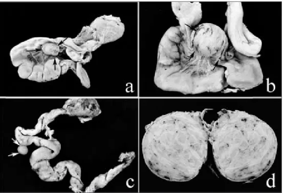
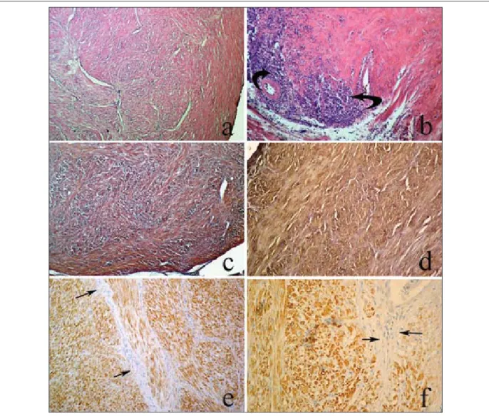
Leiomyomas of oviduct and ıts ventral ligament of hens: ımmunohistochemical evaluation and the plasma concentration levels of 17 β-oestradiol and progesterone
Tam metin
Şekil
Benzer Belgeler
Bu çalıĢmada, OECD ülkeleri için 2007-2018 yılları arasında sanayi katma değerinin yüksek teknoloji ürün ihracatı üzerine etkisi panel veri analizi
Slogan olarak kullanımının dışında, ihtiyaç terimi, her zaman, az ya da çok ölçülen ihtiyacın karşısındaki bazı sosyal normları ve standardları ima eder..
İkinci önemli husus, Cumhuriyet’in ilk yılla- rında bir öte duygusuna, sonsuzluğa, yani kısaca metafiziğe susamış bir okuyucu ve aydın beklentisini karşılamış
Kýrýk bulgusu olan hastalarýn yaþlarýnýn anlamlý olarak daha yüksek olduðu ve femur boyun T deðerlerinin daha düþük olduðu fakat farkýn istatistiksel olarak
In contrast, the proof of the conjecture for the case (g,d) = (1,4) found in [ 1] makes essential use of the real structure, since an elliptic curve with eight ordinary cusps in P 1 ×
Ancak, bunu tam olarak ortaya koymak mümkün değildir, çünkü Origo yazarı pagan imparatorlara karşı hiç de sempatik bir dil kullanmamakta, bilakis Galerius, Severus,
Bu çalışmada, endokrin bozucuözelliğe sahip olan antibiyotik bileşiklerinden ülkemizde ve Kuzey Avrupa ülkeleri genelinde yaygın bir kullanıma sahip olan β-laktam
Ahmet Bey’in geçmişinde yaşadığı olaylar sonrasında kendini fiziksel, duygusal ve düşünsel anlamda soyutlaması ile birlikte odak figürün kendini ayrı bir
