1 Differential Expressions of Cancer-Associated Genes and Their Regulatory miRNAs in
Colorectal Carcinoma
Murat Kara1, Onder Yumrutas2, Onder Ozcan3, Ozgur Ilhan Celik4, Esra Bozgeyik5, Ibrahim
Bozgeyik2*, Sener Tasdemir6
1Department of Medical Genetics, Faculty of Medicine, Mugla Sıtkı Kocman University,
Mugla, Turkey
2Department of Medical Biology, Faculty of Medicine, Adiyaman University, Adiyaman,
Turkey
3Department of General Surgery, Faculty of Medicine, Mugla Sıtkı Kocman University,
Mugla, Turkey
4Department of Medical Pathology, Faculty of Medicine, Mugla Sıtkı Kocman University,
Mugla, Turkey
5Department of Medical Biology, Faculty of Medicine, University of Gaziantep, Gaziantep,
Turkey
6Department of Medical Genetics, Faculty of Medicine, Ataturk University, Erzurum, Turkey
*Corresponding Author
Ibrahim BOZGEYIK
Phone: +904162231690
Adiyaman University, Faculty of Medicine, Department of Medical Biology
Adiyaman, Turkey
Version of Record: https://www.sciencedirect.com/science/article/pii/S0378111915005077 Manuscript_edb4a2bf75bf5e09289b9c20e6b94770
ABSTRACT
Colorectal cancer is one of the frequently seen malignancy in the world. To date, several oncogenes and tumor suppressor gene have been identified and linked to colorectal cancer pathogenesis. Although recent advances in the diagnosis and therapy of colorectal cancer are promising, identifying novel genetic contributors is still high priority. In the present study, expression profile of some cancer-related genes and their regulatory miRNA molecules were evaluated by using a high-throughput real-time PCR method. For the study, a total of 54 patients diagnosed with CRC and normal colon tissue samples of 42 healthy controls were included. For the expression analysis, total RNA was extracted from FFPE tissue samples and converted to cDNA. All expression analysis were assessed by using Fludigm Microfluidic Dynamic Array chips for 96 samples and the reactions were held in Fludigm BioMark™ HD System Real-Time PCR. As a result of the study, expression of the ADAMTS1, FHIT, RUNX1, RUNX3 and WWOX genes were shown to be significantly altered in CRC tissues in contrast to normal tissue samples. Moreover, 378a-3p, 155-5p, 193b-3p, miR-96-5p, miR-17-5p, miR-27a-3p, miR-133b, miR-203a, miR-205-5p, miR-34c-5p, 130a-3p, 301a-130a-3p, 132-130a-3p, 222-130a-3p, 34a-5p, 21-5p, 29a-3p and miR-29b-3p were found to be significantly deregulated in CRC. Consequently, results of the current study strongly suggest the involvement of novel cancer-related genes and their regulatory miRNA in CRC physiopathology.
Introduction
Colorectal cancer (CRC) is a complex heterogeneous disease and responsible for the 10 % of the cancer-related deaths [1]. Molecular pathogenesis of CRC involves gradual accumulation of mutations in the DNA repair genes, proto-oncogenes, and tumor suppressor genes [2]. Despite recent promising diagnostic and therapeutic methods, CRC-related mortality is very high all over the world. Hence, identifying novel biomarkers with diagnostic and therapeutic potentials for the diagnosis of CRC and developing alternative treatment methods is very important.
Moreover, it is believed that cancer arose from the accumulating mutations of oncogenes and tumor suppressor genes. These facts about the cancer pathogenesis have been changed by the discovery of novel regulatory molecules, called miRNAs [3-5]. miRNAs are a family of non-coding regulatory RNAs and regulate gene expression by targeting mRNAs [5]. These tiny RNA molecules play key roles in a variety of biological processes including cell proliferation, differentiation, angiogenesis, and apoptosis and they also participate in the invasion and metastasis of cancer cells [3-8].
Also, a growing number of evidence suggests that deregulated miRNA expressions are one of the important hallmarks of cancers including the CRC [4, 6, 7, 9]. miRNAs with oncogenic potentials have been well characterized in CRC [10]. For instance, miR-21-5p, miR-29-3p, and miR-148-3p were reported to be associate with CRC [11]. Additionally, expression of miR-29a-3p was shown to be elevated in CRC [12]. In addition, it is reported that expression levels of miR-148a-3p were significantly decreased in advanced stages of CRC [13].
Accordingly, it is well known that increased or decreased expression of a miRNA leads to deregulation of its target genes and results in complete deregulation of the regarding pathway. Thus, determining the interrelation between cancer-associated genes and miRNAs is very important and will provide much information for the CRC pathology. Thus, in the present study, we aimed to determine the expression levels of some cancer related genes and their regulatory miRNAs in FFPE tissue samples of CRC patients and normal FFPE tissues.
Material and Methods
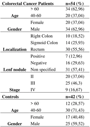
In this study, cancerous tissue samples of 54 patients who were diagnosed with CRC and normal colon tissue samples of 42 healthy controls were included. The demographic characteristics and clinical findings of patients and controls were presented in Table 1. The study was ethically approved by the local ethics committee of Mugla Sitki Kocman University (accession number: 17.02.2015/02) in accordance with the ethical standards of Helsinki Declaration. All study participants gave a written informed consent prior to inclusion in the study. 5–20 μm thick, formalin-fixed, paraffin-embedded tissue sections of patients and controls were obtained from the pathology unit and stored at -20 until RNA isolations.
Determination of validated miRNA regulators
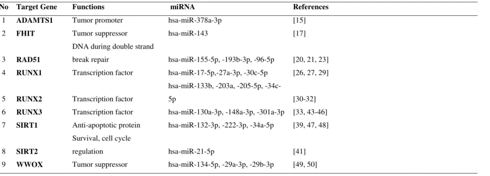
Determination of validated miRNAs that target A disintegrin and metalloproteinase with thrombospondin motifs 1 (ADAMTS1), fragile histidine triad protein (FHIT), repair of DNA double strand breaks RAD51, Runt-related transcription factor 1-3 (RUNX1-3) Sirtuin 1-2 (SIRT1-2), WW domain-containing oxidoreductase (WWOX) genes were carried out by literature scanning of earlier studies. Validated miRNAs of selected genes were presented in Table 2.
Isolation and Quantification of total RNA from FFPE Tissues
Formalin-fixed, paraffin-embedded (FFPE) tissue samples were obtained from the archive of pathology unit. Total RNA isolation from FFPE samples were carried out by using miRNeasy FFPE Kit (QIAGEN Sample & Assay Technologies, Germany) according to instructions of the manufacturer. The concentration and purity of RNA samples were determined by using a spectrophotometer (NanoDrop ND-100, Thermo Fisher Scientific Inc. Wilmington, USA). Concentrations of RNA samples were adjusted according to spectrophotometric measurements and equal aliquots of samples were stored at -80 oC.
cDNA Synthesis of RNA samples
The reverse transcriptions reactions were achieved by using Qiagen miScript II RT Kit (QIAGEN Sample & Assay Technologies, Germany). Single-stranded cDNA synthesis were carried out in accordance with the manufacturer's recommended protocols. Equal aliquots of cDNA samples were stored at -80 oC until gene expression analysis.
The expression levels of selected genes and miRNAs were held in high-throughput Fludigm BioMark™ HD System Real-Time PCR. Nano-technology based Fludigm Microfluidic Dynamic Array chips (Fluidigm, South San Francisco, Calif., USA) for 96 samples were used for the expression analysis. Briefly, diluted cDNA samples and reaction mixtures were mixed and loaded on chips. Also, primers pairs for the amplification of selected mRNAs and miRNAs were loaded on chips. The cDNA samples and primers were allowed to mix on Dynamic Array chips by the help of IFC (integrated fluidic circuits) machine (Fluidigm, South San Francisco, Calif., USA). Finally, chips were placed into Fludigm BioMark™ HD System Real-Time PCR for the analysis.
Collection of Expression data and Statistical analysis
Pre-analysis and collection of expression results were attained by using Fluidigm Real-Time PCR Analysis software (Fluidigm, South San Francisco, Calif., USA). Expression levels of mRNAs were normalized with Beta-2-microglobulin (B2M) gene. For the normalization of miRNA expressions SNORD61, SNORD68, SNORD72, and SNORD95 small RNAs were used. In the analysis of qRT-PCR results, 2-ΔCt (ΔCt = Target gene – Reference gene) formula was used. Resulting data were statistically tested by using GraphPad Prism (v6.02) program and Wilcoxon signed rank test. Additionally, fold-change analysis were carried out for both mRNA and miRNA expressions. Fold-changes were analyzed by using online program of RT²
Profiler PCR Array Data Analysis Version 3.5
(http://pcrdataanalysis.sabiosciences.com/pcr/arrayanalysis.php?target=upload). P values for all statistical tests were two-tailed and p <0.05 accepted as statistically significant.
Results
Differential expression of cancer-associated genes
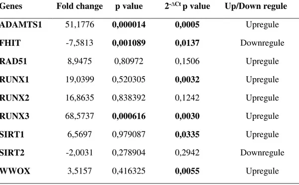
To determine the expression levels of ADAMTS1, RAD51, RUNX1, RUNX2, RUNX3, SIRT1, SIRT2, FHIT and WWOX gene a high-throughput real-time PCR method was used. As a result of the expression analysis of selected genes, ADAMTS1, RAD51, RUNX1, RUNX2, RUNX3, SIRT1, and WWOX were shown to be significantly upregulated in CRC tissues as compared to normal tissues samples (Figure 1, Table 3, Figure 2). In particular, a 51-fold increase was detected in ADAMTS1 gene by fold-change analysis (p=0,000014). In addition, fold-change elevations of RAD51, RUNX1, RUNX2, SIRT1, and WWOX were found to be 9, 19, 17, 6, and 3, respectively (Figure 1, Table 3, Figure 2). In addition to,
expression increase in RUNX3 gene was found to be 68-fold. On the other hand, FHIT and SIRT2 genes were found to be downregulated in CRC tissues as compared to normal tissues (Figure 1, Figure 2). Fold-change analysis of FHIT and SIRT2 genes revealed 7.5 and 2 increase, respectively (Table 3).
Differential expression of miRNAs that target cancer-associated genes
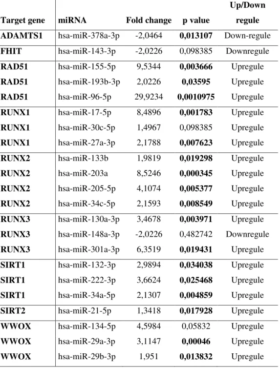
miRNAs that target selected cancer associated genes were analyzed for expression changes. Expression changes of miRNAs were presented in Figure 3 and Table 4. Expression of hsa-miR-378a-3p which targets ADAMTS1 was found to be downregulated in CRC tissues (Table 4). Additionally, expression levels of hsa-miR-143-3p which target FHIT and hsa-miR-148a-3p which target RUNX3 were found to be downregulated in CRC as compared to normal tissue samples (Table 4).
Moreover, validated miRNAs of RAD51 gene, hsa-miR-155-5p, -193b-3p, and -96-5p were found to be upregulated in CRC tissues. Also, expressions of hsamiR175p, 27a3p, and -30c-5p which targets RUNX1 were upregulated in CRC tissues. In addition, expression levels of hsa-miR-133b, -203a, -205-5p, and -34c-5p which targets RUNX2 were determined to be upregulated. In addition to, miRNA regulators of RUNX3, hsamiR130a3p and hsamiR -301a-3p were found to be upregulated in CRC tissues. Expressions of hsa-miR-132-3p, -222-3p, -34a-5p which targets SIRT1 were found to be upregulated. Also, miRNAs of miR-134-5p, -29a-3p, -29b-3p that targets WWOX were shown to be upregulated. Lastly, hsa-miR-21-5p which targets SIRT2 were shown to be upregulated in CRC tissues (Figure 3, Table 4).
Discussion
Previous studies indicated that increased ADAMTS1 activity stimulates tumor promoter events such as increased proliferative signaling, inhibition of apoptosis and increased metastatic potential [14]. They also reported tumor suppressor properties of ADAMTS1. However, in present study, expression levels of ADAMTS1 were found to be elevated in CRC tissues. Consistent with the findings of pro-tumorigenic functions of ADAMTS1, our study results strongly suggest ADAMTS1 as a tumor promoter gene in CRC. Also, in a study, miR-378-3p was found to be negative regulator of ADAMTS1 in granulosa cells [15]. Our study results revealed that miR-378-3p was downregulated in CRC tissues as compared to normal tissues. Suggesting that miR-378-3p may have a key role in the CRC pathogenesis through
regulating ADAMTS1. Downregulation of miR-378-3p results in the upregulation of one of its targets, ADAMTS1. Yet, these findings must be supported by the future investigations of miRNA mimic and inhibitor studies.
FHIT is one of the tumor suppressor gene which is commonly downregulated and/or lost in various types of malignancies [16]. Consistent with the results of the previous reports, in our study, the expression of FHIT gene was shown to be significantly downregulated in CRC tissues. Indicating that FHIT may have important roles in the pathogenesis of CRC. Also, Lin
et. al. reported that FHIT is one of the direct target of miR-143-3p [17]. Also, expression level
of miR-143-3pwas also found to be insignificantly downregulated in CRC tissues.
Deregulation of DNA repair system plays a central role in the carcinogenesis [18]. DNA double-strand break repair and homologous recombination protein Rad51 was reported to be frequently overexpressed in cancers [19]. Consistent with the previous reports, RAD51 was upregulated in CRC tissues compared to normal tissues. However, expression changes in RAD51 gene was found to be statistically insignificant. In addition, several miRNAs that target Rad51 were found to be upregulated in CRC tissues. Wang et. al. reposted miR-96-5p as a direct target of RAD51. Overexpression studies of miR-96-5p resulted significant downregulation of RAD51 gene [20]. However, in our study, expression of miR-96-5p was found to be significantly overexpressed in CRC tissues. Indicating potential role of this miRNA in the CRC pathogenesis, but not dependent to RAD51 expression. In another study, RAD51 gene was found to be one of the target of miR-155 [21]. Also, miR-155 is well known of fine-tuner of inflammatory responses and frequently deregulated in various human cancers [22]. In breast cancer, overexpression of miR-155 was correlated with downregulation RAD51 gene expression [21]. Consistent with previous studies, miR-155 is found to be significantly upregulated in CRC tissues. Indicating this miRNA plays key role in the CRC progression and may be associated with the cancer related inflammation (CRI). However, RAD51 overexpression may be independent from miR-155 overexpression. As a result, these findings have to be confirmed by the independent future studies to clearly understand RAD51 and miR-155 interaction. Furthermore, inhibition of miR-193b expression was found to be associated with the RAD51 overexpression in ovarian cancer [23]. Also, it was reported that miR-193b overexpression decreased the RAD51 expression [23]. In our study, miR-193b was found to be significantly overexpressed in CRC tissues. Suggesting possible clinical significance of this miRNA in CRC physiopathology. However, RAD51 overexpression seems not to be related with miR-193b overexpression.
Runt-related transcription factors (RUNXs) have been implicated in tumor development [24]. There are three RUNX genes in mammalian cells designed as RUNX1-3 and they have tissue specific expression patterns [25]. They have been shown to play key roles in the process of carcinogenesis [25]. Also, several studies reported the relationship between RUNX genes and cancer-associated miRNAs [26-29]. It was reported that miR-17-5p targets 3′UTR of RUNX1 gene and negatively regulates its expression. In particular, anti-miR-27 transduction experiments revealed significant upregulation of RUNX1 in granulocytes [27] . In the present study, expressions of both miR-17-5p and its target RUNX1 were found to be upregulated. However, statistical analysis of RUNX1 expression changes was insignificant. On the other hand, elevated expression of miR-17-5p was shown to be statistically significant. Suggesting an important role of miR-17-5p in the colorectal carcinogenesis, yet this expression increase is independent from RUNX1. Similarly, expression of miR-30c-5p which targets RUNX1 [26] was found to be increased in CRC tissues. However, miR-30c-5p expression change was statistically insignificant.
Interestingly, expression levels of miR-203a, miR-133b, miR-205-5p and miR-34c-5p was found to be elevated in CRC tissues. All of these miRNAs are known to target RUNX2 gene. Studies investigated that downregulation of miR-203a is correlated with increased cell proliferation, metastasis and invasion. [30]. Also, previous studies reported miR-203a as potential biomarker in metastatic prostate cancer [30]. In addition, miR-133b was reported to inhibit RUNX2 translation [31]. In addition to, Zhang et. al. reported that miR-34c-5p strongly inhibits RUNX2 and miR-205-5p moderately inhibits RUNX2 translation [32]. In our study, expression levels of RUNX2 were found to be insignificantly upregulated in CRC tissues. Suggesting that miR-203a, miR-133b, miR-205-5p and miR-34c-5p miRNAs have important roles in CRC progression. However, their mechanism of action is not related to RUNX2 gene.
Furthermore, expression of RUNX3 was found to be upregulated in our study. miR-130a, miR-148a and miR-301a-3p were reported to be target RUNX3 gene. In a study, ectopic expression of miR-148a was reported to be interfere with proliferation of gastric cancer cells [33]. As it is correlated with overexpression of RUNX3, miR-148a expression is downregulated in CRC tissues in our study. However, this expression change was statistically insignificant. On the other hand, expression of miR-130a and miR-301a-3p were shown to be significantly upregulated in our study. Indicating potential role of these miRNAs in CRC physiopathology.
Silent information regulator 1 (SIRT1) is a well known member of sirtuin family of genes [34]. SIRT1 is NAD-dependent histone deacetylase which is activated in response to oxidative and genotoxic stresses [35]. Also, it is known to participate in a variety of physiological processes including cell proliferation, inflammatory responses, cell cycle and cell migration [35]. SIRT1 have been reported to have dual functions in tumor development [35-37]. In some studies, increased SIRT1 activity was reported to be associate with tumor promoting events [37, 38]. As consistent with these findings, SIRT1 expression was shown to be elevated in our study. However, this expression change was statistically insignificant. On the other hand, SIRT1 targeting miRNAs; miR-132, miR-222 and miR-34a-5p were found to be significantly upregulated in CRC tissues. In previous studies, increased miR-222 expression was reported in prostate cancer cell lines (PC3 and LNcaP) [39]. Downregulation of miR-222 was shown to inhibit cell proliferation, migration and increase apoptotic cell death [39]. Also, miR-34a activity was reported to have tumor suppressor effects in breast cancer [40]. All in all, these miRNAs appears to be significant regulators of CRC pathology.
Another well characterized member of the sirtuin family of genes is SIRT2 and it was reported to be downregulated in glioma tissues and various cancer cell lines [41]. Also, miR-21 have been shown to target SIRT2 and regulate its expression [41]. In our study, an inverse relationship was found SIRT2 and miR-21-5p expression. In particular, expression of SIRT2 was reduced and miR-21-5p was increased in CRC tissues. Indicating deregulation of SIRT2-miR-21-5p axis may contribute to CRC physiopathology.
Lastly, WW domain-containing oxidoreductase (WWOX) is known to be tumor suppressor gene [42]. In our study, WWOX expression was found to be insignificantly upregulated. Several WWOX targeting miRNAs have been shown to be associate with cancers. In a study, miR-134 was shown to be upregulated in head and neck squamous cell carcinoma (HNSCC) and negatively regulate WWOX expression. Also, miR-29s (-29a,-29b) were shown to be upregulated in lung cancer cell lines and their expression inversely associated with WWOX expression. In our study, mir-29a-3p and miR-29b-3p were found to be significantly upregulated and miR-134-5p insignificantly upregulated in CRC tissues. Suggesting that mir-29a-3p and miR-29b-3p are the important contributors of CRC physiopathology.
In conclusion, significant interactions between miRNAs and their target genes were determined. The findings of the present study will shed light into future upcoming studies to understand more about CRC physiopathology. On the other hand, there are several limitations of our study. One of them is the lack of protein expression data and the validation of the protein expression of selected genes will be great of interest for the future investigators. Also, the results of the present study have to be supported by the expression of selected genes and miRNAs in CRC cell lines. Moreover, in the future studies the results of this study have to be confirmed in an independent cohort form different ethnic groups. In addition to, miRNA mimicry and inhibitory studies in cell lines will be more informative for to understand these miRNA-mRNA interactions.
Acknowledgements
This study was supported by a project from the Scientific Research Projects Management Unit of Mugla Sıtkı Kocman University (Grant number: 13/152).
Declaration of interest
The authors report no conflicts of interest.
References
1. Siegel, R., D. Naishadham, and A. Jemal, Cancer statistics, 2012. CA: a cancer journal for clinicians. 62(1): p. 10-29.
2. Centelles, J.J., General aspects of colorectal cancer. ISRN oncology, 2012. 2012. 3. Adams, B.D., A.L. Kasinski, and F.J. Slack, Aberrant regulation and function of
microRNAs in cancer. Curr Biol, 2014. 24(16): p. R762-76.
4. Ahmed, F.E., miRNA as markers for the diagnostic screening of colon cancer. Expert review of anticancer therapy, 2014. 14(4): p. 463-485.
5. Bartel, D.P., MicroRNAs: target recognition and regulatory functions. Cell, 2009. 136(2): p. 215-233.
6. Hong, L., et al., Angiogenesis-related microRNAs in colon cancer. Expert opinion on biological therapy, 2013. 13(1): p. 77-84.
7. Rossi, S., et al., microRNAs in colon cancer: a roadmap for discovery. FEBS letters, 2012. 586(19): p. 3000-3007.
8. Zhou, J.-J., et al., MicroRNA regulation network in colorectal cancer metastasis. World journal of biological chemistry, 2014. 5(3): p. 301.
9. Faber, C., T. Kirchner, and F. Hlubek, The impact of microRNAs on colorectal cancer. Virchows Archiv, 2009. 454(4): p. 359-367.
10. Schetter, A.J. and C.C. Harris. Alterations of microRNAs contribute to colon
carcinogenesis. in Seminars in oncology. 2011: Elsevier.
11. Dong, Y., J. Yu, and S.S.M. Ng, MicroRNA dysregulation as a prognostic biomarker
in colorectal cancer. Cancer management and research, 2014. 6: p. 405.
12. Fu, J., et al., Identifying microRNA-mRNA regulatory network in colorectal cancer by
a combination of expression profile and bioinformatics analysis. BMC systems
biology, 2012. 6(1): p. 68.
13. Takahashi, M., et al., The clinical significance of MiR-148a as a predictive biomarker
in patients with advanced colorectal cancer. PloS one, 2012. 7(10): p. e46684.
14. Arao Tan, I., C. Ricciardelli, and D.L. Russell, The metalloproteinase ADAMTS1: A
comprehensive review of its role in tumorigenic and metastatic pathways.
International Journal of Cancer, 2013. 133(10): p. 2263-2276.
15. Toms, D., et al., Progesterone receptor expression in granulosa cells is suppressed by
microRNA-378-3p. Molecular and cellular endocrinology, 2015. 399: p. 95-102.
16. Waters, C.E., et al., The FHIT gene product: tumor suppressor and genome
“caretaker†. Cellular and Molecular Life Sciences, 2014. 71(23): p. 4577-4587.
17. Lin, Y.-x., et al., microRNA-143 protects cells from DNA damage-induced killing by
downregulating FHIT expression. Cancer biotherapy & radiopharmaceuticals, 2011.
26(3): p. 365-372.
18. Pitroda, S.P., et al., DNA repair pathway gene expression score correlates with repair
proficiency and tumor sensitivity to chemotherapy. Science translational medicine,
2014. 6(229): p. 229ra42-229ra42.
19. Hine, C.M., et al., Regulation of Rad51 promoter. Cell Cycle, 2014. 13(13): p. 2038-2045.
20. Wang, Y., et al., MiR-96 downregulates REV1 and RAD51 to promote cellular
sensitivity to cisplatin and PARP inhibition. Cancer research, 2012. 72(16): p.
4037-4046.
21. Gasparini, P., et al., Protective role of miR-155 in breast cancer through RAD51
targeting impairs homologous recombination after irradiation. Proceedings of the
22. O'Connell, R.M., et al., MicroRNA-155 is induced during the macrophage
inflammatory response. Proceedings of the National Academy of Sciences, 2007.
104(5): p. 1604-1609.
23. Choi, Y.E., et al., MicroRNAs down-regulate homologous recombination in the G1
phase of cycling cells to maintain genomic stability. eLife, 2014. 3.
24. Slattery, M.L., et al., Associations between genetic variation in RUNX1, RUNX2,
RUNX3, MAPK1, and eIF4E and risk of colon and rectal cancer: additional support for a TGF-β signaling pathway. Carcinogenesis, 2010: p. bgq245.
25. Ito, Y., S.-C. Bae, and L.S.H. Chuang, The RUNX family: developmental regulators in
cancer. Nature Reviews Cancer, 2015. 15(2): p. 81-95.
26. Ben-Ami, O., et al., A regulatory interplay between miR-27a and Runx1 during
megakaryopoiesis. Proceedings of the National Academy of Sciences, 2009. 106(1): p.
238-243.
27. Feng, J., et al., MicroRNA-27 enhances differentiation of myeloblasts into
granulocytes by post-transcriptionally downregulating Runx1. British journal of
haematology, 2009. 145(3): p. 412-423.
28. Fischer, J.A., et al., miR-17 deregulates a core RUNX1-miRNA mechanism of CBF
acute myeloid leukemia. Molecular Cancer, 2015. 14(1): p. 7.
29. Rossetti, S. and N. Sacchi, RUNX1: A microRNA hub in normal and malignant
hematopoiesis. International journal of molecular sciences, 2013. 14(1): p. 1566-1588.
30. Viticchie, G., et al., MiR-203 controls proliferation, migration and invasive potential
of prostate cancer cell lines. Cell Cycle, 2011. 10(7): p. 1121-1131.
31. Li, Z., et al., A microRNA signature for a BMP2-induced osteoblast lineage
commitment program. Proceedings of the National Academy of Sciences, 2008.
105(37): p. 13906-13911.
32. Zhang, Y., et al., A program of microRNAs controls osteogenic lineage progression by
targeting transcription factor Runx2. Proceedings of the National Academy of
Sciences, 2011. 108(24): p. 9863-9868.
33. Chen, Z., et al., MicroRNA-148a: a potential therapeutic target for cancer. Gene, 2014. 533(1): p. 456-457.
34. Yuan, H., L. Su, and W.Y. Chen, The emerging and diverse roles of sirtuins in
cancer: a clinical perspective. OncoTargets and therapy, 2013. 6: p. 1399.
35. Longo, V.D. and B.K. Kennedy, Sirtuins in aging and age-related disease. Cell, 2006. 126(2): p. 257-268.
36. Jung, W., et al., SIRT1 Expression is associated with good prognosis in colorectal
cancer. Korean journal of pathology, 2013. 47(4): p. 332-339.
37. Kim, J.R., et al., Expression of SIRT1 and DBC1 is associated with poor prognosis of
soft tissue sarcomas. PloS one, 2013. 8(9): p. e74738.
38. Masui, T., et al., Expression of METH-1 and METH-2 in pancreatic cancer. Clinical cancer research, 2001. 7(11): p. 3437-3443.
39. Yang, X., et al., Down-Regulation of mir-221 and mir-222 Restrain Prostate Cancer
Cell Proliferation and Migration That Is Partly Mediated by Activation of SIRT1. PloS
one, 2014. 9(6): p. e98833.
40. Li, L., et al., MiR-34a inhibits proliferation and migration of breast cancer through
down-regulation of Bcl-2 and SIRT1. Clinical and experimental medicine, 2013. 13(2):
p. 109-117.
41. Li, Y., et al., Sirt2 suppresses glioma cell growth through targeting NF-κB–miR-21
axis. Biochemical and biophysical research communications, 2013. 441(3): p.
661-667.
42. Lin, J.-T., et al., WWOX suppresses prostate cancer cell progression through cyclin
D1-mediated cell cycle arrest in the G1 phase. Cell Cycle, 2015. 14(3): p. 408-416.
43. Lai, K.W., et al., MicroRNA-130b regulates the tumour suppressor RUNX3 in gastric
cancer. European Journal of Cancer, 2010. 46(8): p. 1456-1463.
44. Wang, M., et al., Overexpressed miR-301a promotes cell proliferation and invasion by
targeting RUNX3 in gastric cancer. Journal of gastroenterology, 2013. 48(9): p.
1023-1033.
45. Xu, Q., et al., MicroRNA-130a regulates autophagy of endothelial progenitor cells
through Runx3. Clinical and Experimental Pharmacology and Physiology, 2014.
41(5): p. 351-357.
46. Zuo, J., et al., MicroRNA-148a can regulate runt-related transcription factor 3 gene
expression via modulation of DNA methyltransferase 1 in gastric cancer. Molecules
and cells, 2013. 35(4): p. 313-319.
47. Vinall, R.L., et al., MiR-34a chemosensitizes bladder cancer cells to cisplatin
treatment regardless of p53†Rb pathway status. International Journal of Cancer,
2012. 130(11): p. 2526-2538.
48. Zhang, L., et al., MiR-132 inhibits expression of SIRT1 and induces pro-inflammatory
processes of vascular endothelial inflammation through blockade of the SREBP-1c metabolic pathway. Cardiovascular Drugs and Therapy, 2014. 28(4): p. 303-311.
49. Fabbri, M., et al., MicroRNA-29 family reverts aberrant methylation in lung cancer by
targeting DNA methyltransferases 3A and 3B. Proceedings of the National Academy
of Sciences, 2007. 104(40): p. 15805-15810.
50. Liu, C.J., et al., miR-134 induces oncogenicity and metastasis in head and neck
carcinoma through targeting WWOX gene. International Journal of Cancer, 2014.
Table 1: Demographic and clinical characteristics of patients and controls
Colorectal Cancer Patients n=54 (%) Age > 60 34 (62,96) 40-60 20 (37,04) Gender Female 20 (37,04) Male 34 (62,96) Localization Right Colon 10 (18,52) Sigmoid Colon 14 (25,93) Rectum 30 (55,56) Lenf nodule Positive 7 (12,96) Negative 16 (29,63) Non specified 31 (57,41) Stage II 20 (37,04) III 25 (46,3) IV 9 (16,67) Controls n=42 (%) Age > 60 12 (28,57) 40-60 30 (71,43) Gender Female 17 (40,48) Male 25 (59,52)
Table 2. Selected cancer-associated genes and their validated miRNAs
No Target Gene Functions miRNA References
1 ADAMTS1 Tumor promoter hsa-miR-378a-3p [15]
2 FHIT Tumor suppressor hsa-miR-143 [17]
3 RAD51
DNA during double strand
break repair hsa-miR-155-5p, -193b-3p, -96-5p [20, 21, 23]
4 RUNX1 Transcription factor hsa-miR-17-5p,-27a-3p, -30c-5p [26, 27, 29]
5 RUNX2 Transcription factor
hsa-miR-133b, -203a, -205-5p,
-34c-5p [30-32]
6 RUNX3 Transcription factor hsa-miR-130a-3p, -148a-3p, -301a-3p [33, 43-46]
7 SIRT1 Anti-apoptotic protein hsa-miR-132-3p, -222-3p, -34a-5p [39, 47, 48]
8 SIRT2
Survival, cell cycle
regulation hsa-miR-21-5p [41]
9 WWOX Tumor suppressor hsa-miR-134-5p, -29a-3p, -29b-3p [49, 50]
Table 3. Fold change analysis of selected genes
Genes Fold change p value 2-∆Ct p value Up/Down regule
ADAMTS1 51,1776 0,000014 0,0005 Upregule FHIT -7,5813 0,001089 0,0137 Downregule RAD51 8,9475 0,80972 0,1506 Upregule RUNX1 19,0399 0,520305 0,0032 Upregule RUNX2 16,8635 0,838392 0,1242 Upregule RUNX3 68,5737 0,000616 0,0030 Upregule SIRT1 6,5697 0,979087 0,0335 Upregule SIRT2 -2,0031 0,278904 0,2942 Downregule WWOX 3,5157 0,416325 0,0055 Upregule
Table 4. Fold change analysis of miRNAs and representation of their target mRNAs.
Target gene miRNA Fold change p value
Up/Down regule ADAMTS1 hsa-miR-378a-3p -2,0464 0,013107 Down-regule FHIT hsa-miR-143-3p -2,0226 0,098385 Downregule RAD51 hsa-miR-155-5p 9,5344 0,003666 Upregule RAD51 hsa-miR-193b-3p 2,0226 0,03595 Upregule RAD51 hsa-miR-96-5p 29,9234 0,0010975 Upregule RUNX1 hsa-miR-17-5p 8,4896 0,001783 Upregule RUNX1 hsa-miR-30c-5p 1,4967 0,098385 Upregule RUNX1 hsa-miR-27a-3p 2,1788 0,007623 Upregule
RUNX2 hsa-miR-133b 1,9819 0,019298 Upregule
RUNX2 hsa-miR-203a 8,5246 0,000345 Upregule
RUNX2 hsa-miR-205-5p 4,1074 0,005377 Upregule RUNX2 hsa-miR-34c-5p 2,1593 0,008549 Upregule RUNX3 hsa-miR-130a-3p 3,4678 0,003971 Upregule RUNX3 hsa-miR-148a-3p -2,0226 0,482742 Downregule RUNX3 hsa-miR-301a-3p 6,3519 0,019431 Upregule SIRT1 hsa-miR-132-3p 2,9894 0,034038 Upregule SIRT1 hsa-miR-222-3p 3,6624 0,025468 Upregule SIRT1 hsa-miR-34a-5p 2,1307 0,004859 Upregule SIRT2 hsa-miR-21-5p 1,3418 0,017928 Upregule
WWOX hsa-miR-134-5p 4,5984 0,05832 Upregule
WWOX hsa-miR-29a-3p 3,1147 0,00046 Upregule
Figure legends
Figure 1. Representation of the expression of selected genes. ADAMTS1, RAD51, RUNX1-3 SIRT1 and WWOX genes were shown to be upregulated and FHIT and SIRT2 were shown to be downregulated. P: Colorectal cancer patients, C: Normal colon tissue. *p<0.05 accepted as significant.
Figure 2. Representation of the expression levels of selected genes in CRC tissues group according to the fold-change analysis. Results were compared to control group. p<0.05 accepted as significant.
Figure 3. Representation of the differential expression of miRNAs in CRC tissues as compared to control group. These miRNAs are the validated targets of selected genes. p<0.05 accepted as significant.