Can Hematologic Parameters be an Indicator of
Metabolic Disorders Accompanying Rosacea?
Asli Akin Belli
1, Asude Kara
1, Seyran Ozbas Gok
21Mugla Sitki Kocman University Training and Research Hospital, Department of
Dermatology, Mugla, Turkey; 2Artvin Hopa State Hospital, Department of Dermatology,
Artvin, Turkey
Corresponding author: Asli Akin Belli, MD
Mugla Sitki Kocman University, Department of Dermatology 48000, Mugla
Turkey
ABSTRACT Recently, diverse hematologic parameters have been used as an in-dicator of the presence or severity of inflammatory and cardiovascular diseases. Our aim was to investigate the ratios of neutrophils to lymphocytes (NL), mono-cytes to high-density lipoprotein (HDL) cholesterol (MHC), and platelets to lym-phocytes (PL) in patients with rosacea in comparison with the control group and determine whether there was a correlation between these ratios and metabolic disorders in patients with rosacea. We conducted a case-control study on 61 pa-tients with rosacea and 60 healthy controls between January 2015 and January 2016 at the Dermatology Outpatient Clinic, Mugla, Turkey. Demographic data, biochemical parameters, hematologic parameters and ratios, the presence of metabolic syndrome (MS), and the presence of insulin resistance (IR) in the par-ticipants were recorded. Sixty one patients with rosacea (16 men, 45 women) and 60 controls (13 men, 47 women) were included in the study. The NL ratio, mean levels of low-density lipoprotein (LDL) and total cholesterol, triglyceride, C-reactive protein (CRP), systolic and diastolic blood pressures, and the presence of IR were significantly higher in patients with rosacea than in controls. In the rosacea group, the MHC ratio was significantly higher in patients with rosacea with IR and MS. Moreover, only the MHC ratio was an independent predictor of MS according to univariate logistic regression analysis. The cutoff value of MHC on admission for predicting MS in patients with rosacea was 0.013.The higher levels of NL ratio and IR in the rosacea group corroborate the previous studies demonstrating a high level of cardiovascular risk factors in patients with rosa-cea. The MHC ratio may be used as a simple and inexpensive method to predict metabolic disorders in patients with rosacea.
KEY WORDS: hematologic parameters, insulin resistance, metabolic syndrome, rosacea
INTRODUCTION
Various hematologic parameters or ratios have re-cently been used as an indicator of the presence or severity of inflammatory diseases. Insulin resistance, coronary artery disease, hypertension, and inflam-matory skin diseases such as psoriasis and Behçet’s disease have been evaluated (1-5). Rosacea is also a chronic inflammatory skin disease (6). It has been pro-posed that inflammation related to the excessive re-sponse of the innate immune system by various trig-gers plays a key role in the pathogenesis of rosacea
(7-11). In recent studies, dyslipidemia, hypertension, and cardiovascular risk factors have been reported to be high in patients with rosacea (6,12,13). Further-more, there was a very recent report on the increased frequency of insulin resistance and some parameters of metabolic syndrome in patients with rosacea (14).
To the best of our knowledge, there is no study in-vestigating the hematological ratios in patients with rosacea or the relationship between these ratios and metabolic disorders accompanying rosacea.
Received: February 22, 2016 Accepted: May 15, 2017
Our aim was to investigate the neutrophil-to-lym-phocyte (NL), monocyte-to-high-density lipoprotein (HDL) cholesterol (MHC), and platelet-to-lymphocyte (PL) ratios in patients with rosacea in comparison with the control group and determine whether there was a correlation between these ratios and metabolic syndrome and insulin resistance in patients with ro-sacea.
METHODS
We conducted a case-control study on 61 pa-tients with rosacea and 60 healthy controls (matched for age, sex, and body mass index) between January 2015 and January 2016 at the Dermatology Outpa-tient Clinics of the Mugla Sitki Kocman University Training and Research Hospital. Ethic Committee ap-proval was obtained before the study from the local hospital Ethic Committee. The diagnosis of rosacea was based on characteristic clinical findings accord-ing to the National Rosacea Society criteria. The con-trols were selected randomly from the patients who were admitted to the outpatient clinic with various
skin problems other than rosacea. Inclusion criteria for the participants were absence of known diabetes mellitus, coronary artery disease, hyperlipidemia, and chronic inflammatory disease (including inflamma-tory skin diseases).
Age, sex, body mass index (BMI), lipid profile (to-tal cholesterol, triglyceride, low-density lipoprotein, and high-density lipoprotein), fasting blood glucose (FBG), insulin, C-reactive protein (CRP), the ratios of NL, MHC, and PL (obtained from the complete blood count), systolic and diastolic blood pressure levels, and the presence of metabolic syndrome (MS) and insulin resistance (IR) were recorded.
BMI was calculated using the formula weight (kg) / height (m2). Lipid parameters, insulin, and FBG
lev-els were studied after a 12-hour fasting period. Based on the diagnostic criteria of the International Diabe-tes Federation (IDF-2005), MS was defined as waist circumference >94cm in men and >80cm in women plus at least two of the following criteria: triglyceride value >150 mg/dL or specific treatment for this lipid abnormality, high density lipoprotein; <40 mg/dL in
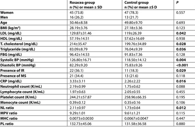
Table 1. Comparison of the demographic, biochemical, metabolic, and hematologic parameters in the
ro-sacea and control groups
Rosacea group
n (%) or mean ± SD Control groupn (%) or mean ±S D P Women Men 45 (73.8)16 (26.2) 47 (78.3) 13 (21.7) 0.557 Age 50.46±8.58 49.80±9.70 0.693 BMI (kg/m2) 28.19±3.76 27.18±3.36 0.123 LDL (mg/dL) 129.87±31.46 119±26.39 0.042 HDL (mg/dL) 57.19±14.51 57.62±16.69 0.938 T. cholesterol (mg/dL) 214±35.47 199.76±34.89 0.028 Triglyceride (mg/dL) 83.09±8.79 76.04±9.39 0.036 FBG (mg/dL) 96.42±14.53 91.83±7.36 0.128 Systolic BP (mmHg) 126.80±16.71 118.50±14.12 0.004 Diastolic BP (mmHg) 82.29±9.20 75.83±9.26 <0.001 Presence of IR 22 (36.1) 11 (18.3) 0.029 Presence of MS 21 (34.4) 13 (21.6) 0.118 CRP (mg/dL) 3.33±3.11 2.26±2.22 0.016 Neutrophil count (K/mL) 2.19±0.99 1.75±0.62 0.088 Lymphocyte count (K/mL) 1.97±0.63 2.05±0.55 0.455 Platelet count (K/mL) 244.21±57.87 258.96±66.35 0.195 Monocyte count (K/mL) 0.39±0.12 0.35±0.16 0.106 NL ratio 2.11±0.97 1.73±0.64 0.012 MPV ratio 9.29±1.01 9.61±1.21 0.115 MHC ratio 0.0073±0.0030 0.0067±0.0047 0.447 PL ratio 132.73±45.06 131.58±36.58 0.880
Chi-square test, Student’s t-test, and Mann-Whitney U test. BMI: body mass index; FBG: fasting blood glucose; BP: blood pres-sure; IR: insulin resistance; MS: metabolic syndrome; CRP: C-reactive protein; NL: neutrophil/lymphocyte; MHC: monocyte/ high-density lipoprotein (HDL) cholesterol; LDL: high-density lipoprotein; PL: platelet/lymphocyte; MPV: mean platelet vol-ume; SD: Standard Deviation
men and <50 mg/dL in women or specific treatment for this lipid abnormality, blood pressure ≥130/85 mmHg or antihypertensive treatment, and fasting blood glucose ≥100 mg/dL or diagnosed diabetes mellitus (15).
Insulin resistance was calculated using the homeo-stasis model assessment of insulin resistance (HOMA-IR) according to the following formula: [fasting insulin level (uIU/mL) × fasting glucose level (mg/dL)/405]. A value of >2.7 was considered to indicate IR.
For the data analysis, the statistical program “SPSS for windows 20.0” was used. The chi-square test was used for the analysis of qualitative data. The distribu-tion of variables was checked with the Kolmogorov-Smirnov test. The Student’s t-test and Mann-Whitney
U-test were used for variables with normal and ab-normal distribution, respectively. P<0.05 was consid-ered significant.
RESULTS
Sixty one rosacea patients (16 men, 45 women; age range 35-74 years) and 60 healthy controls (13 men, 47 women; age range 33-78 years) were included in the study. The duration of the disease varied between 3 months to 20 years (mean ± Standard Deviation:
Table 2. Comparison of hematologic parameters in patients with or without IR and MS
IR (+) n=22 Mean ± SD IR (-) n=39 Mean ± SD P MS (+) n=21 Mean ± SD MS (-) n=40 Mean ± SD P NL ratio 2.15±1.04 2.08±0.95 0.088 2.39±1.09 1.96±0.89 0.088 MHC ratio 0.0080±0.0034 0.0069±0.0027 0.008 0.0087±0.0031 0.0066±0.0027 0.007 PL ratio 140.43±54.44 128.39±38.92 0.505 138.11±59.73 129.92±35.65 0.505 Neutrophil count (K/mL) 3.74±0.90 3.88±1.59 0.448 2.39±1.09 1.96±0.89 0.088 Lymphocyte count (K/mL) 2.01±0.85 1.95±0.47 0.387 1.87±0.88 2.02±0.45 0.387 Platelet count (K/mL) 252.32±62.16 239.64±55.62 0.084 226.52±54.19 253.50±58.22 0.084 MPV (fl) 9.03±0.99 9.44±1.01 0.376 9.45±1.01 9.21±1.02 0.376 Monocyte count (K/mL) 0.39±0.12 0.39±0.12 0.840 0.40±0.11 0.39±0.13 0.840 Student’s t-test and Mann-Whitney U test. IR: insulin resistance; MS: metabolic syndrome; NL: neutrophil/lymphocyte; MHC: monocyte/ high-density lipoprotein (HDL) cholesterol; PL: platelet/lymphocyte; MPV: mean platelet volume; SD: Standard Deviation
Figure 1. Correlation between the neutrophil–to-lympho-cyte (NL) ratio and C-reactive protein (CRP) levels in pa-tients with rosacea.
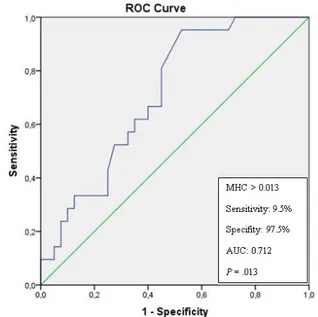
Figure 2. Receiver operating the characteristics curve of monocyte count to high-density lipoprotein (HDL) choles-terol (MHC) ratio for predicting metabolic syndrome in pa-tients with rosacea (AUC: Area under the curve)
1.75±3.53 years) in patients with rosacea. Of the pa-tients, 29 (47.5%) had the erythematotelangiectatic type of rosacea, while 31 (50.8%) had papulopustular type and one (1.6%) had the phymatous type.
Among the hematological ratios, the NL ratio of the rosacea group was significantly higher than in the control group (P=0.012). The NL ratio levels were cor-related with CRP levels (P<0.05) (Figure 1). Other than the NL ratio, the mean levels of LDL, total cholesterol, triglyceride, CRP, systolic and diastolic blood pres-sures, and presence of IR were significantly higher in patients with rosacea than those of controls (P<0.05) (Table 1).
When we evaluated only the rosacea group, the MHC ratio was significantly higher in the patients with MS and IR (P=0.007 and P=0.008, respectively) (Table 2). There was no correlation between rosacea subtype and hematological ratios (P>0.05). In addi-tion, only the MHC ratio was an independent predic-tor of MS in the rosacea group, according to univari-ate logistic regression analysis (P=0.015, OR: 1.54, CI: 1.723-1.371). The cutoff value of the MHC ratio on admission for predicting MS in patients with rosacea was 0.013 with 9.5% sensitivity and 97.5% specificity (area under the curve 0.712, P=0.013) (Figure 2).
DISCUSSION
Diverse hematologic ratios have recently become popular for predicting the course of cardiovascular diseases, various cancers, and inflammatory diseas-es. Since rosacea is an inflammatory disease with a known association with cardiovascular diseases, we conducted the current study to determine whether the hematological ratios may be used to predict met-abolic disorders accompanying rosacea. We found that the NL ratio of patients with rosacea was signifi-cantly higher than that of controls, and that the MHC ratio significantly higher in patients with rosacea with MS and IR.
Although the pathogenesis of rosacea has not been completely elucidated, increased levels of cat-helicidin peptides (LL-37), kallikrein-5 (KLK5), toll-like receptor 2 (TLR2), endoplasmic reticulum (ER) stress, oxidative stress, and pro-inflammatory cytokines (TNF-α, IL-8, and IL-1β) have been found in rosacea-effected skin (7-11). Similar pathogenetic mecha-nisms such as ER stress, oxidative stress, high LL-37 levels, and high inflammatory cytokines have been demonstrated in the development of atherosclerosis, MS, and cardiovascular diseases (16-19). Although Egeberg et al. did not find an association between rosacea and increased risk of adverse cardiovascular outcomes or death, an association between rosacea
to cardiovascular diseases, MS, and IR have been re-ported in some studies (6,12-14,20).
Similarly to rosacea, psoriasis and Behçet’s dis-ease are chronic inflammatory skin disdis-eases which are associated with metabolic syndrome and cardio-vascular diseases. Moreover, psoriasis and Behcet’s disease are also systemic diseases, which is not the case with rosacea. The hematologic ratios have been used in psoriasis and Behcet’s disease to determine underlying inflammation or cardiovascular risk fac-tors. Yurtdas et al. found that the NL ratio was sig-nificantly higher and the only predictor of subclini-cal atherosclerosis in patients with psoriasis (4). Sen
et al. have reported similar high NL ratios in patients
with psoriasis and a positive correlation between the NL ratio and CRP levels (21). Furthermore, both the NL and PL ratios have been noted as strong predic-tors of psoriatic arthritis in a study involving patients with psoriasis, psoriatic arthritis, and healthy controls (22). In another two studies, the NL ratio and carotid intima media thickness have been found to be high in patients with Behçet’s disease (23,24). These re-sults corroborate the hypothesis that inflammation and endothelial dysfunction have an important role in the pathogenesis of Behçet’s disease, and that NL ratio may be used to evaluate the disease activity. Additionally, Rifaioglu et al. noticed that the NL ratio was significantly increased in patients with active Be-hçet’s disease (5). Alan et al. have suggested that NL ratio may be a diagnostic criterion of Behçet’s disease based on the results of their study (25).
The hematologic ratios, particularly NL and MHC ratios, were first used for cardiovascular diseases, metabolic disorders, and some cancers. Several stud-ies showed that NL ratio is an inexpensive and easy indicator of atherosclerosis, hypertension, and in-flammation (2,3). Furthermore, Lou et al. noticed that the NL ratio and IR were significantly associated (1). Buyukkaya et al. also found a relationship between NL ratio and MS, as well as severity of MS. In a cross-sectional study consisting of 1070 individuals, in-creased NL ratio levels were significantly related to chronic diseases such as hypertension and diabetes mellitus (26). It has been proposed that neutrophils increase damage to the endothelium by interacting with endothelial tissue and that they play a key role in the development of inflammation, and thus athero-sclerosis (2).
MHC ratio has been found to be high in patients with slow coronary flow, as reported by Canpolat et al (27). In another study, 340 patients with chronic renal disease were followed for about 33 months, finding that cardiovascular events were more common in
patients with high MHC ratio (28). Monocytes interact with damaged endothelium and induce over-expres-sion of pro-inflammatory cytokines and adheover-expres-sion molecules. Contrarily, HDL cholesterol has anti-in-flammatory, anti-oxidant, and vasodilator effects (27). Thus, MHC ratio has been considered a good indica-tor of inflammaindica-tory conditions such as cardiovascular events. In order to provide a fair comparison, since a relationship between these hematological ratios and obesity has been previously reported, our study in-cluded BMI-matched subjects in the rosacea and control groups. When the rosacea and control groups were compared, the NL ratios of the rosacea group were significantly higher than those of control group. However, only MHC ratio was an independent predic-tor of MS in patients with rosacea.
CONCLUSION
The presence of high cardiovascular risk factors among patients with rosacea in recent studies en-couraged us to perform the current study. The high-er levels of NL ratio and IR we found in the rosacea group corroborate the previous studies, demonstrat-ing the presence of high cardiovascular risk factors in patients with rosacea. In the rosacea group, MHC ra-tio was significantly higher in patients with MS and IR. MHC ratio may be used as a simple and inexpensive method to indicate metabolic disorders accompany-ing rosacea. Further studies are needed to duplicate our results and to evaluate the usefulness of hemato-logic parameters for predicting metabolic disorders in rosacea.
References:
1. Lou M, Luo P, Tang R, Peng Y, Yu S, Huang W, et al. Relationship between neutrophil-lymphocyte ra-tio and insulin resistance in newly diagnosed type 2 diabetes mellitus patients. BMC Endocr Disord 2015;15:9.
2. Balta S, Celik T, Mikhailidis DP, Ozturk C, Demirkol S, Aparci M, et al. The Relation Between Athero-sclerosis and the Neutrophil-Lymphocyte Ratio. Clin Appl Thromb Hemost 2016;22:405-11. 3. Belen E, Sungur A, Sungur MA, Erdoğan G.
In-creased Neutrophil to Lymphocyte Ratio in Pa-tients With Resistant Hypertension. J Clin Hyper-tens (Greenwich) 2015;17:532-7.
4. Yurtdaş M, Yaylali YT, Kaya Y, Ozdemir M, Ozkan I, Aladağ N. Neutrophil-to-lymphocyte ratio may predict subclinical atherosclerosis in patients with psoriasis. Echocardiography 2014;31:1095-104. 5. Rifaioglu EN, Bülbül Şen B, Ekiz Ö, Cigdem
Dogram-aci A. Neutrophil to lymphocyte ratio in Behçet’s disease as a marker of disease activity. Acta Der-matovenerol Alp Pannonica Adriat 2014;23:65-7. 6. Hua TC, Chung PI, Chen YJ, Wu LC, Chen YD, Hwang
CY, et al. Cardiovascular comorbidities in patients with rosacea: A nationwide case-control study from Taiwan. J Am Acad Dermatol 2015;73:249-54.
7. Casas C, Paul C, Lahfa M, Livideanu B, Lejeune O, Alvarez-Georges S, et al. Quantification of Demo-dex folliculorum by PCR in rosacea and its rela-tionship to skin innate immune activation. Exp Dermatol 2012;21:906-10.
8. Melnik BC. Endoplasmic reticulum stress: key promoter of rosacea pathogenesis. Exp Dermatol 2014;23:868-73.
9. Two AM, Wu W, Gallo RL, Hata TR. Rosacea: Part I. Introduction, categorization, histology, pat-hogenesis, and risk factors. J Am Acad Dermatol 2015;72:749-58.
10. Yamasaki K, Gallo RL. Rosacea as a Disease of Ca-thelicidins and Skin Innate Immunity. J Investig Dermatol Symp Proc 2011;15:12-5.
11. Takci Z, Bilgili SG, Karadag AS, Kucukoglu ME, Se-lek S, Aslan M. Decreased serum paraoxonase and arylesterase activities in patients with rosacea. J Eur Acad Dermatol Venereol 2015;29:367-70. 12. Duman N, Ersoy Evans S, Atakan N. Rosacea and
cardiovascular risk factors: a case control study. J Eur Acad Dermatol Venereol 2014;28:1165-9. 13. Rainer BM, Fischer AH, Luz Felipe da Silva D, Kang
S, Chien AL. Rosacea is associated with chronic systemic diseases in a skin severity-dependent manner: Results of a case-control study. J Am Acad Dermatol 2015;73:604-8.
14. Akin Belli A, Ozbas Gok S, Akbaba G, Etgu F, Do-gan G. The relation of rosacea to insulin resistance and metabolic syndrome. Eur J Dermatol 2016 1;26:260-4.
15. Alberti KG, Zimmet P, Shaw J; IDF Epidemiology Task Force Consensus Group. The metabolic syndrome--a new worldwide definition. Lancet 2005;366:1059-62.
16. Edfeldt K, Agerberth B, Rottenberg ME, Gud-mundsson GH, Wang XB, Mandal K, et al. Involve-ment of the antimicrobial peptide LL-37 in human atherosclerosis. Arterioscler Thromb Vasc Biol 2006;26:1551-7.
17. Benachour H, Zaiou M, Samara A, Herbeth B, Pfis-ter M, Lambert D, et al. Association of human ca-thelicidin (hCAP-18/LL-37) gene expression with
cardiovascular disease risk factors. Nutr Metab Cardiovasc 2009;19:720-8.
18. Meigs J, Larson M, Fox C. Association of Oxidative Stress, Insulin Resistance, and Diabetes Risk Phe-notypes. The Framingham Offspring Study. Dia-betes Care 2007;30:2529-35.
19. Kim OK, Jun W, Lee J. Mechanism of ER Stress and Inflammation for Hepatic Insulin Resistance in Obesity. Ann Nutr Metab 2015;67:218-27. 20. Egeberg A, Hansen PR, Gislason GH, Thyssen JP.
Assessment of the risk of cardiovascular disease in patients with rosacea. J Am Acad Dermatol. 2016;75:336-9.
21. Sen BB, Rifaioglu EN, Ekiz O, Inan MU, Sen T, Sen N. Neutrophil to lymphocyte ratio as a measure of systemic inflammation in psoriasis. Cutan Ocul Toxicol 2014;33:223-7.
22. Kim DS, Shin D, Lee MS, Kim HJ, Kim do Y, Kim SM, et al. Assessments of neutrophil to lympho-cyte ratio and platelet to lympholympho-cyte ratio in Kor-ean patients with psoriasis vulgaris and psoriatic arthritis. J Dermatol 2016;43:305-10.
23. Yuksel M, Yildiz A, Oylumlu M, Turkcu FM, Bilik MZ, Ekinci A, et al. Novel markers of endothelial dysfunction and inflammation in Behçet’s disease patients with ocular involvement: epicardial fat thickness, carotid intima media thickness, serum
ADMA level, and neutrophil-to-lymphocyte ratio. Clin Rheumatol 2016;35:701-8.
24. Ozturk C, Balta S, Balta I, Demirkol S, Celik T, Turker T, et al. Neutrophil-lymphocyte ratio and carotid-intima media thickness in patients with Behçet di-sease without cardiovascular involvement. Angi-ology 2015;66:291-6.
25. Alan S, Tuna S, Türkoğlu EB. The relation of neu-trophil-to-lymphocyte ratio, platelet-to-lympho-cyte ratio, and mean platelet volume with the presence and severity of Behçet’s syndrome. Ka-ohsiung J Med Sci 2015;31:626-31.
26. Buyukkaya E, Karakas MF, Karakas E, Akçay AB, Tanboga IH, Kurt M, et al. Correlation of neutrophil to lymphocyte ratio with the presence and seve-rity of metabolic syndrome. Clin Appl Thromb He-most 2014;20:159-63.
27. Canpolat U, Çetin EH, Cetin S, Aydin S, Akboga MK, Yayla C, et al. Association of Monocyte-to-HDL Cholesterol Ratio with Slow Coronary Flow is Linked to Systemic Inflammation. Clin Appl Th-romb Hemost 2016;22:476-82.
28. Kanbay M, Solak Y, Unal HU, Kurt YG, Gok M, Ce-tinkaya H, et al. Monocyte count/HDL choleste-rol ratio and cardiovascular events in patients with chronic kidney disease. Int Urol Nephrol 2014;46:1619-25.