Aydin, E et al 2016 Temporal Lobe Parenchyma Herniation into the Transverse Sinus: MRI Findings in a Case. Journal of the Belgian Society of Radiology, 100(1): 7, pp. 1-3, DOI: http://dx.doi.org/10.5334/jbr-btr.1001
* Başkent University, TR
[email protected], [email protected], [email protected]
CASE REPORT
Temporal Lobe Parenchyma Herniation into the
Transverse Sinus: MRI Findings in a Case
Elçin Aydin
*, Hasan Yerli
*and Esin Gezmiş
*Brain parenchyma herniation into dural venous sinus which is a uncommon entity, can cause dural venous sinus filling and simulate sinus thrombosis and other pathologies. It is isointense to brain parenchyma on all sequences by magnetic resonance imaging, surrounded by a cerebrospinal fluid rim and is seen to be contiguous with brain tissue on images. We report a rare case with spontaneous occult herniation of tem-poral lobe tissue into the left transverse sinus that may associated with headache.
Keywords: brain; herniation; MRI; venous sinus; headache
There are several causes of dural venous sinus filling defe-cts including arachnoid granulations, sinus thrombosis, tumours, intrasinus septa (fibrotic bands) and hypopla-sia or aplahypopla-sia of dural sinuses. Brain herniation with sur-rounding cerebrospinal fluid (CSF) into the dural venous
sinuses is a uncommon entity and can simulate aforemen-tioned pathologies and variations causing dural venous sinus filling. It was recently described on magnetic reso-nance imaging (MRI) and it is also named as ‘‘encephalo-cele” and “invagination”. Although its clinical significance is controversial, it is suggested that brain herniations may cause some symptoms suchs as headache, dizziness, syn-cope and imbalance. We report a rare case with headache and spontaneous occult herniation of temporal lobe tis-sue into the left transverse sinus.
Case report
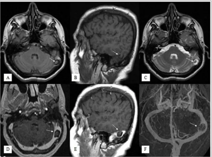
A-50-year-old female presented with history of headache for a long time. Her neurological examination was nor-mal. The laboratory results were within normal limits. Brain MRI demostrated a small herniation of a temporal lobe with surrounding CSF rim into the left transverse sinus (Figures 1A–E, arrows). The herniation material
was isointense to brain parenchyma on all sequences in the contiguous brain tissue images. T2-weighted axial image showed loss of signal void in the left transverse sinus ( Figure 1C). After contrast media administration,
no pathological parenchymal or meningeal opacification was seen, the herniation tissue was seen to bulge into left transverse sinus and it was caused the narrowing of
the sinus (Figure 1D,E). On MR venography imaging,
there was left transverse sinus stenosis but no venous thrombosis (Figure 1F).
Discussion
Brain parenchyma herniations into a dural venous sinus are rare entities which can be seen as filling defects of dural sinuses on radiological images [1]. Brain paren-chyma herniations are different from classic encephaloce-les that composed of meninges and brain located outside of the skull. Suggested main mechanisms regarding with classic encephalocele in the literature are non-union of ossification centres in bones or variations of bone thick-ness that may cause brain tissue herniation by pressure of the brain tissue or CSF [2]. Calvarial defects can be seen in radiological images in the form of classical herniation in the skull level. Conversely, there is no bone defect in the brain parenchyma herniation unlike the classical enceph-alocele. The mechanism of brain parenchyma herniations as it is in the classic encephaloceles is not clear. Progres-sive dural thinning secondary to elevated CSF pressure, inflammation, aging, and erosive arachnoid granulations are among the etiologies thought to be responsible [3].
Classical encephalocele can sometimes cause CSF leak-age, epilepsy, meningitis and ear disturbances such as hear-ing loss, otorrhea and otitis media [3]. On the other hand, brain herniation into dural sinuses may associated with some different symptoms such as headache, syncope, diz-ziness and imbalance although the relationship between herniated brain and symptoms are indefinite [3]. Our patient was complaining about headache. In literature, it is reported some cases with headache and brain hernia-tion into dural sinuses. Battal et al. described four brain herniations into transvers sinuses and they observed that two of them had histories of headaches [4]. Karatag et al.
Aydin et al: Temporal Lobe Parenchyma Herniation into the Transverse Sinus Art. 7, page 2 of 3
describe a case of temporal lob herniation into the sig-moid sinus which had a history of headache [1]. The ten-sion of duramater and vessels due to the pressure of the herniation may cause headache but there is not enough information in the literature and exact pathogenesis is unclear. Long-term follow-up studies are needed to be understood if symptoms are in relation with radiological findings or not.
The main differential diagnosis for the lesions causing dural venous sinus filling defect includes dural sinus thrombosis, arachnoid granulations and tumor . The MR signals depends on clot age in the dural sinus thrombo-sis. Acute thrombosis is seen as isointense, hypointense and hyperintense on T1, T2 and T2*-weighted images, respectively. Subacute sinus thrombosis shows hyper-intense signals on T1, T2 and T2*-weighted images. In the chronic stage, thrombosis shows isointense sig-nals on T1-weigted images and moderately hyperin-tense signals on T2-weighted images and postcontrast T1-weighted images shows thick enhancing duramater. Arachnoid granulations are always isointense to CSF on all MR images [5]. Tumours can cause mass affect and its differentiation from the herniation may not
difficult. Brain tissue herniation is also a rare cause cre-ating venous sinus filling defect but must be kept in mind. MRI is the best choice to confirm the diagnosis. It is isointense to brain parenchyma on all sequences by magnetic resonance imaging, surrounded by a cerebro-spinal fluid rim and is seen to be contiguous with brain tissue on images [6].
In conclusion, it should be considered that brain
her-niation can be one of the potential cause of filling defects within the dural venous sinuses. Herniation are probably incidental findings, however, it may be associated with headache based our case and other cases defined in litera-ture. There is need to collect more numbers of cases for determination possible relationship.
Competing Interests
The authors declare that they have no competing interests.
References
1. Karatag, O, Cosar, M, Kizildag, B, et al. Dural sinus
filling defect: intrasigmoid encephalocele. BMJ Case
Rep. 2013; 2013 pii: bcr2013201616. DOI: http://
dx.doi.org/10.1136/bcr-2013-201616
Figure 1: Fluid Attenuated Inversion Recovery axial (A), T1-weighted sagittal (B), T2-weighted axial (C),
contrast-enhanced T1-weighted axial (D) and sagittal (E) images show a small herniation of temporal lobe parenchyma with surrounding CSF into left transverse sinus, that was isointense to brain parenchyma on all sequences (arrows). No pathological enhancement is seen but the brain herniation sac is causing moderate stenosis in the left transverse sinus. On venography imaging (F), there was left transverse sinus stenosis but no venous thrombosis.
Aydin et al: Temporal Lobe Parenchyma Herniation into the Transverse Sinus Art. 7, page 3 of 3
2. Wilkins, RH, Radtke, RA and Burger, PC.
Spon-taneous temporal encephalocele: case report.
J Neurosurg. 1993; 78: 492–498. DOI: http://
dx.doi.org/10.3171/jns.1993.78.3.0492. PMid: 8433155.
3. Wind, JJ, Caputy, AJ and Roberti, F. Spontaneous
encephaloceles of the temporal lobe. Neurosurgical
Focus. 2008; 25: 11. DOI: http://dx.doi.org/10.3171/
FOC.2008.25.12.E11. PMid: 19035698.
4. Battal, B and Castillo, M. Brain herniations into the
dural venous sinuses or calvarium: MRI of a recently recognized entity. Neuroradiol J. 2014; 27: 55–62.
DOI: http://dx.doi.org/10.15274/NRJ-2014-10006. PMid: 24571834; PMCid: PMC4202845.
5. Liang, L, Korogi, Y, Suguhara, T, et al. Normal
struc-tures in the intracranial dural sinuses: delineation with 3D contrast-enhanced magnetization prepared rapid acquisition gradient-echo imaging sequence.
AJNR Am J Neuroradiol. 2002; 1739–1746. PMid:
12427634.
6. Kollar, C, Hohnston, I, Parker, G and Harper, C.
Dural arteriovenous fistula in association with het-erotopic brain nodule in the transverse sinus. AJNR
Am J Neuroradiol. 1993; 19: 1126–1128.
How to cite this article: Aydin, E, Yerli, H and Gezmiş, E 2016 Temporal Lobe Parenchyma Herniation into the Transverse Sinus: MRI Findings in a Case. Journal of the Belgian Society of Radiology, 100(1): 7, pp. 1–3, DOI: http://dx.doi.org/10.5334/jbr-btr.1001 Published: 29 January 2016
Copyright: © 2016 The Author(s). This is an open-access article distributed under the terms of the Creative Commons Attribution 4.0 International License (CC-BY 4.0), which permits unrestricted use, distribution, and reproduction in any medium, provided the original author and source are credited. See http://creativecommons.org/licenses/by/4.0/.