215 Interesting Image / İlginç Görüntü
Turkish Journal of Thoracic and Cardiovascular Surgery 2020;28(1):215-216
http://dx.doi.org/doi: 10.5606/tgkdc.dergisi.2020.18620
Successful management of acute pulmonary embolism complicated
with cardiopulmonary shock by implementing point-of-care ultrasonography
and extracorporeal membrane oxygenation
Kardiyopulmoner şok ile komplike olan akut pulmoner embolinin yatak başı ultrasonografi ve ekstrakorporeal membran oksijenasyonu ile başarılı yönetimi
Umut Kocabaş1, Hakan Altay1, Gülay Eren2, Vedat Bakuy3, Deniz Süha Küçükaksu3
Received: August 17, 2019 Accepted: October 23, 2019 Published online: January 23, 2020
1Department of Cardiology, Başkent University Istanbul Health Application and Research Center, Istanbul, Turkey
2Department of Anesthesiology and Reanimation, Başkent University Istanbul Health Application and Research Center, Istanbul, Turkey 3Department of Cardiovascular Surgery, Başkent University Istanbul Health Application and Research Center, Istanbul, Turkey
Correspondence: Umut Kocabaş, MD. Başkent Üniversitesi İstanbul Sağlık Uygulama ve Araştırma Merkezi, Kardiyoloji Kliniği, 34662 Üsküdar, İstanbul, Türkiye. Tel: +90 507 - 997 49 99 e-mail: [email protected]
©2020 All right reserved by the Turkish Society of Cardiovascular Surgery.
Kocabaş U, Altay H, Eren G, Bakuy V, Küçükaksu DS. Successful management of acute pulmonary embolism complicated with cardiopulmonary shock by imple-menting point-of-care ultrasonography and extracorporeal membrane oxygenation. Turk Gogus Kalp Dama 2020;28(1):215-216
Cite this article as:
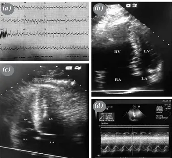
Figure 1. On admission, (a) 12-lead-electrocardiography showing
sinus tachycardia along with S wave in lead I, Q wave and inverted T wave in lead III, (b) point-of-care transthoracic cardiac
ultrasound during cardiopulmonary resuscitation showing severe right ventricular dilatation, (c) after 24 hours bedside transthoracic
echocardiography showing complete recovery of right ventricular chamber size and (d) right ventricular systolic function with a
tricuspid annular plane systolic excursion of 22 mm.
RV: Right ventricle; RA: Right atrium; LV: Left ventricle; LA: Left atrium.
(a)
(c)
(d) (b)
A 26-year-old man with a history of tibial fracture 10 days ago presented to emergency department with acute respiratory failure and cardiogenic shock. Initial systolic blood pressure was 80/40 mmHg and the heart rate was 126 bpm. Blood gas analysis showed hypoxia, hypocarbia, and lactic acidosis. Electrocardiography revealed sinus tachycardia along with S wave in lead I and Q wave and inverted T wave in lead III (Figure 1a). During diagnostic work-up, cardiopulmonary arrest occurred and the patient was intubated and cardiopulmonary resuscitation (CPR) was initiated. As the patient was unresponsive to the initial CPR with intravenous (IV) vasopressor therapy, point-of-care transthoracic cardiac ultrasonography was performed concurrently with CPR which revealed severe right ventricular dilatation with poor systolic function, moderate tricuspid regurgitation (TR), and a systolic pulmonary artery pressure (sPAP) of 50 mmHg (Figure 1b). Point-of-care lower extremity venous Duplex ultrasound also showed deep venous thrombosis in the left lower extremity. Based on these findings, acute pulmonary embolism (PE) was suspected and IV unfractionated heparin plus systemic thrombolytic therapy (TT) with infusion of tissue plasminogen activator at a rate of 50 mg/h for two hours were started immediately. At 60 min of CPR, TT was discontinued and the patient transferred to the
216
Turk Gogus Kalp Dama 2020;28(1):215-216
operating room under CPR and femoro-femoral veno-arterial extracorporeal membrane oxygenation (ECMO) (Medos Medizintechnik AG, Stolberg, Germany) was initiated with a 23-Fr venous and 17-Fr arterial cannula with restoration of systemic blood flow and oxygen delivery (4.5 L/min, 3,600 rpm). After ECMO insertion, IV unfractionated heparin infusion therapy was started with activated partial thromboplastin time monitoring. The next day, pulmonary angiography was performed under ECMO support in the catheterization laboratory which demonstrated filling defects of the left lobar arteries, compatible with acute PE. After 24 hours, bedside transthoracic echocardiography showed complete recovery of the right ventricular function with a tricuspid annular plane systolic excursion 24 mm and mild TR with a sPAP of 35 mmHg (Figure 1c, d). The hemodynamically stable patient was weaned from ECMO after 32 hours. After weaning from mechanical ventilation and deep sedation, the patient woke up with right hemiparesis. Cranial computed tomography demonstrated left middle cerebral artery territory infarction due to possible ECMO-related thromboembolism. During the hospital stay, the patient was followed with rivaroxaban 20 mg/day and stroke rehabilitation program. At the end of 20 days, the patient was discharged from hospital with mild neurological sequela. A written informed consent was obtained from the patient.
Point-of-care ultrasonography including transthoracic cardiac ultrasound and lower extremity venous Duplex ultrasound in the emergency department are helpful imaging methods to urgently diagnose acute PE. The presence of right heart dilatation, right ventricle systolic dysfunction with TR, and elevated sPAP on point-of-care transthoracic cardiac
ultrasound and deep venous thrombosis on point-of-care lower extremity venous Duplex ultrasound allow to start emergency treatment in case of suspected of PE with cardiogenic shock.[1] Management of
PE guidelines recommends TT in patients with massive PE complicated by cardiopulmonary shock.[2] On the other hand, ECMO is suggested
to provide cardiopulmonary circulatory support in patients with high-risk PE complicated with sustained hemodynamic and respiratory failure, despite initial TT and vasopressor therapy.[3]
In conclusion, combined use of point-of-care ultrasonography, thrombolytic therapy, and extracorporeal membrane oxygenation are important diagnostic and therapeutic strategies in acute massive PE complicated with cardiopulmonary shock.
Declaration of conflicting interests
The authors declared no conflicts of interest with respect to the authorship and/or publication of this article.
Funding
The authors received no financial support for the research and/or authorship of this article.
REFERENCES
1. Squizzato A, Galli L, Gerdes VE. Point-of-care ultrasound in the diagnosis of pulmonary embolism. Crit Ultrasound J 2015;7:7.
2. Konstantinides SV, Torbicki A, Agnelli G, Danchin N, Fitzmaurice D, Galiè N, et al. 2014 ESC guidelines on the diagnosis and management of acute pulmonary embolism. Eur Heart J 2014;35:3033-69.
3. Al-Bawardy R, Rosenfield K, Borges J, Young MN, Albaghdadi M, Rosovsky R, et al. Extracorporeal membrane oxygenation in acute massive pulmonary embolism: a case series and review of the literature. Perfusion 2019;34:22-8.