Effects of therapeutic ultrasound and exercise on
pain, function, and isokinetic shoulder rotator
strength of patients with rotator cuff disease
Pınar Doruk analan, MD1)*, Berrın leBleBıcı, MD1), MehMet aDaM, MD1)
1) Department of Physical Medicine and Rehabilitation, Faculty of Medicine, Baskent University Adana
Research and Education Center: Kazım Karabekir Cad. 4227. Sok. No 37 Yüreğir, Adana, Turkey
Abstract. [Purpose] The aim of this prospective, double-blind, randomized, placebo-controlled study was to evaluate the effects of therapeutic continuous ultrasound (US) on patients with rotator cuff disease. [Subjects and Methods] Twenty-two patients were treated with a standard physiotherapy program consisting of a hot-pack, trans-cutaneous electrical nerve stimulation (TENS), exercises, and US that was supervised by the same physiotherapist. The patients were randomly assigned to either a group that received true US (n=11) or one that received sham US (n=11). [Results] There were significant differences between the pre- and post-intervention UCLA Shoulder Rating Scale and Constant-Murley Shoulder Outcome scores, VAS, and external rotation (ER) peak torque 60°/second val-ues for both the true and sham US groups. However, the between-group differences were not statistically significant. [Conclusion] In patients with rotator cuff disease, physiotherapy interventions effectively treat the pain, improve the clinical status, and increase the muscle strength of the shoulder ER at a low constant angular velocity, with functional improvement being seen immediately after treatment. However, at the end of the intervention, the US had yielded no additional efficacy to the physiotherapy treatment regimen of the patients with rotator cuff disease. Key words: Rotator cuff disease, Therapeutic ultrasound, Shoulder rehabilitation
(This article was submitted Jun. 8, 2015, and was accepted Jul. 9, 2015)
INTRODUCTION
Rotator cuff disease is the most common cause of shoul-der pain, particularly among the middle-aged and elshoul-derly. It is the most commonly diagnosed cause of shoulder dysfunc-tion being the causative factor in 74% of cases of shoulder pain. With increasing age, the tendons that form the rotator cuff undergo progressive degenerative changes which can lead to partial or full-thickness tearing of the tendons, result-ing in pain, weakness and dysfunction in the rotator cuff and scapular muscles, tightness in the soft tissues, and postural abnormalities1–5).
Rotator cuff disease can possibly be treated successfully with conservative management, but this depends upon the gender of the patient and tear size as well as the degree of atrophy2). Conservative management aims to control pain,
improve patients’ range of motion (ROM), and enhance the scapulothoracic rhythm and function. Conservative manage-ment also includes patient education and the use of analge-sics, non-steroidal anti-inflammatory drugs (NSAIDs), and injections, functional rehabilitation, and manual therapy along with physiotherapy modalities such as therapeutic
ultrasound (US), which is used to promote healing and regeneration in inflamed tissue, reduce pain and muscle spasms, and improve ROM1–3, 6–8). However, some studies
have found that the use of US does not significantly improve shoulder problems3, 7–14).
The aim of this study was to evaluate the effects of therapeutic continuous US on pain, functional status, and isokinetic shoulder rotator strength in patients with shoulder pain due to rotator cuff disease.
SUBJECTS AND METHODS
This prospective, randomized, double-blinded, placebo-controlled study involved of 22 patients with rotator cuff disease (11 males, 11 females; age range, 52–81 years) who were treated at the Physical Medicine and Rehabilitation Department of Baskent University Hospital in Adana, Tur-key. A physiotherapist randomly assigned half of the patients to a true US group and the other 11 to a sham US group. Selection was done by drawing lots. These assignments were unknown to both the patients and the physiatrist working with them. This study was approved by the local ethics com-mittee of Baskent University, and written informed consent was obtained from all of the patients.
The inclusion criteria of the study were: the presence of unilateral shoulder pain and limited active movement for at least four weeks prior to the study, normal passive shoulder movement, a diagnosis of rotator cuff disease by magnetic resonance imaging (MRI), and no physiotherapy for the shoulder in the four weeks prior to the study. In addition,
*Corresponding author. Pınar Doruk Analan (E-mail: [email protected]; [email protected]) ©2015 The Society of Physical Therapy Science. Published by IPEC Inc. This is an open-access article distributed under the terms of the Cre-ative Commons Attribution Non-Commercial No DerivCre-atives (by-nc-nd) License <http://creativecommons.org/licenses/by-nc-nd/3.0/>.
potential study subjects needed to have at least two of the following positive impingement signs: positive results in the Speed, Neer, or Hawkins tests or the drop arm, lift-off, or su-praspinatus isolation tests; a resistant, painful shoulder dur-ing internal and external rotation (IR and ER); or pain durdur-ing the abduction of the shoulder with a painful arch8, 15, 16). The
consecutively referred patients who met these criteria were informed about the details of the study.
Patients were excluded if they had any of the following: a history of acute trauma, surgery or a fracture to or in the proximity of the shoulders, ROM limitation in the upper extremities, a neurological deficit in the upper extremities, an underlying inflammatory rheumatic disease, or signs of cervical pathologies that might be associated with shoulder pain.
The rotator cuff pathologies were graded in three stages according to the results of MRI, with stage 1 indicating acute inflammation with either tendonitis or bursitis, stage 2 signifying chronic inflammation either with or without degeneration, and stage 3 representing patients with a rupture of the cuff2). All the patients were evaluated before
and immediately after rehabilitation (20th day) by the same physiatrist using the visual analog scale (VAS), University of California, Los Angeles (UCLA) Shoulder Rating Scale, and the Constant-Murley Shoulder Outcome score as well as isokinetic measurements of the shoulder ER and IR muscles.
The Constant-Murley Shoulder Outcome score assesses the range of functional active movement of a normal, dis-eased, or treated shoulder. It is comprised of subjective assessments of pain (15 points) and activities of daily living (ADL) (20 points) together with objective measures of ROM (40 points) and shoulder strength (25 points) that are added and have a maximum possible score of 10015, 17).
The UCLA Shoulder Rating Scale includes an evalua-tion of pain and a patient satisfacevalua-tion rating together with functional active forward flexion, and strength of forward flexion assesment performed with manual muscle testing. The maximum possible total score is 35 points, with higher scores indicating better shoulder function17, 18).
To perform the isokinetic evaluation, we used a Biodex System 3 Pro™ dynamometer (Biodex Medical Systems, Shirley, NY, USA) to test the isokinetic strength of the shoulder ER and IR muscles at constant angular velocities of 60°/second, and 180°/second via a concentric/concentric mode, and these tests were performed by the same phys-iatrist. To standardize the measurements, we created a new test protocol for the isokinetic mode menu of the machine. Before the testing of each patient began, the dynamometer was calibrated, and its position was established according to the standard measurement technique in which the device was tilted 50° and oriented 20° and the seat was tilted 85° and oriented 15°. In addition, before the isokinetic muscle testing, 1 mL intra-articular 2% prilocaine was injected into the affected shoulder to enable painless movement. The pa-tients were then seated on a supine position in the isokinetic dynamometer chair at 80° hip flexion and 90° knee flexion with their ankles unrestricted. Their trunks and legs were sta-bilized using straps across the chest, waist, and upper thighs. Muscle strength was tested with the shoulder in a modified neutral position (45° abduction and 30° flexion), and the
elbow flexed at 90°. The axis of rotation was adjusted with the axis alignment being longitudinal through the head of the shaft of the humerus and the center of the glenohumeral joint in the horizontal plane. The elbow axis was aligned parallel to the entrance axis of the dynamometer, and the neutral hand position was adjusted as indicated by the user’s manual. Next, the movement range was set to a painless 90° shoulder rotation around the modified neutral position (45° of IR and 45° of ER). Gravity correction was also used for the testing position. All of these isokinetic protocols were performed exactly according to the manufacturer’s recom-mendations.
After explaining the procedure to the patients, they were familiarized with it by performing three submaximal and five maximal repetitions at each speed. In addition, a 30-second rest period was provided between the familiarization trials and the testing. The isokinetic evaluation at 60°/second and 180°/second consisted of five and 10 repetitions of maximal effort for each respective movement. A 10-second rest pe-riod was allowed after each measurement with a five-minute rest between the testing of the two shoulders.
We also measured the isokinetic strength at peak torque (PT) of ER and IR at each speed and used the highest torque value (Newton meters) recorded during the isokinetic efforts in the analysis.
The true US group received continuous US via a Gymna Combi 200 device for two-channel electrotherapy (Gym-naUniphy N.V. Bilzen, Belgium) operated at a frequency of 1.1 MHz and at an intensity of 1.5 W/cm2. The transducer
head had an area of 4.7 cm2 and an effective radiating area
of 4.1 cm2. While sitting on a table, the patients placed an
arm on their lap with their hand in a supinated position. Using slow circular movements, the physiotherapist applied the transducer head over the glenohumeral joint, covering an area of approximately 10 cm2 for a five-minute period.
For the sham US group, the intervention was performed with the same device set to the “off” mode, and the transducer head applied to the same area in the same way with the same transmission gel.
Afterwards, transcutaneous electrical nerve stimulation (TENS) and hot packs were applied to the patients of both groups for 20 minutes each, and then for 30 minutes, the pa-tients perfromed exercises that included active and passive shoulder ROM, Codmann exercises, capsular stretching, isometric and isotonic exercises for strengthening the rotator cuff, biceps, and deltoid muscles, and scapulohumeral stabil-ity exercises. These exercises were performed at the reha-bilitation clinic and supervised by the same physiotherapist. At the start of the therapy, or when a patient had severe pain or discomfort, passive restricted ROM exercises and gentle stretching were used. At a later phase or when discomfort or pain had lessened, exercise shifted toward active ROM exercises, and gradually isometric and dynamic resistance exercises were added, resulting in a longer duration of the exercise session. TENS (STT-500, Stratek, Korea) was ap-plied with a frequency of 100 Hz and current of 8.4 mA over the glenohumeral joint for 20 minutes. All of the patients received the same physiotherapy program from the same physiotherapist.
for Windows version 17.0 software program (SPSS Inc., Chi-cago, IL, USA). For each continuous variable, distribution normality was checked using the Kolmogorov-Smirnov and Shapiro-Wilk tests and histograms. Comparisons between groups were performed using the Mann-Whitney U test for the data that was not normally distributed. In addition, the pre- and post-intervention data were analyzed via repeated measures analysis, and p values of < 0.05 were considered to be statistically significant.
RESULTS
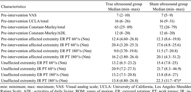
The true and sham US groups were evaluated accord-ing to age, gender, affected extremity, stage of rotator cuff pathology, and duration of symptoms (Table 1). No statistically significant differences were found. In addition, the pre-intervention values of the VAS, UCLA Shoulder Rating Scale, and Constant-Murley Shoulder Outcome score, and the isokinetic muscle testing measurements of the affected extremity were similar for both groups (Table 2). Furthermore, there was no significant correlation between the patients’ pathology grades based on the MRI and the pre-intervention values of their isokinetic muscle strength, VAS, Constant-Murley Shoulder Outcome, and the UCLA Rating Scale scores (p>0.05).
Moreover, the pre-and post-intervention evaluation of the scores from the same instruments showed statisti-cally significant improvement in both the true and sham US groups, but the differences in these scores between the two groups were not statistically significant (p>0.05). The ADL subgroup scores of the Constant-Murley Shoulder Outcome test remained the same in the sham group, whereas those in the US group showed improvementbut the difference was not statistically significant in the ADL scores between the two groups (p>0.05). Moreover, when the pre- and post-in-tervention isokinetic measurements of the affected extremity were compared, only the ER PT 60°/second score improved in the two groups, but the difference was not at a statistically significant (p>0.05) (Table 3).
DISCUSSION
Several factors, such as poor posture, weakness and dys-function of the rotator cuff and scapular muscles, decreased ROM in the shoulder complex, degeneration and inflamma-tion of tendons and bursae, acromial dysmorphology, cap-sular tightness, and glenohumeral instability, are commonly believed to contribute to rotator cuff disease3, 8, 15, 17, 19). For
this reason, the rehabilitation of this disease aims to control pain, improve ROM, and provide scapulothoracic rhythm
Table 1. Clinical characteristics of the study population
Characteristics True ultrasound group Sham ultrasound group Age (years) (mean ± SD) 52.9 ± 11.1 57.1 ± 15.0 Females/ Males (%) 5/6 (45.5/54.5) 6/5 (54.5/45.5) Stage 2/Stage 3 radiological grade (%) 5/6 (45.5/54.50) 6/5 (54.5/45.5) Right/left affected extremity (%) 5/6 (45.5/54.5) 9/2 (81.8/18.2) Duration of symptoms (months) (mean± SD) 27.5 ± 37.0 18.1 ± 33.1 SD: standard deviation
Table 2. VAS, UCLA Shoulder Rating Scale, and Constant-Murley Shoulder Outcome scores and isokinetic measure-ments of the study population
Characteristics True ultrasound group Median (min–max) Sham ultrasound group Median (min–max)
Pre-intervention VAS 7 (2–10) 7 (5–9)
Pre-intervention UCLA/total 16 (6–26) 16 (9–31) Pre-intervention Constant-Murley/total 63 (25–89) 72 (26–79) Pre-intervention Constant-Murley/ADL 12 (8–20) 12 (6–20) Pre-intervention affected extremity ER PT 60°/s (Nm) 12.4 (4.60–26.8) 12.3 (8.6–19.8) Pre-intervention affected extremity IR PT 60°/s (Nm) 20.4 (3.20–25.3) 17.6 (4.8–25.6) Pre-intervention affected extremity ER PT 180°/s (Nm) 9.0 (3.70–19.8) 11.5 (7–20.8) Pre-intervention affected extremity IR PT 180°/s (Nm) 16.2 (3.80–26.4) 20.1 (4.3–31.2) Unaffected extremity ER PT 60°/s (Nm) 13.2 (6.5–23.2) 15.4 (7.8–25) Unaffected extremity IR PT 60°/s (Nm) 20.9 (7.2–27.3) 21.7 (8.3–46.9) Unaffected extremity ER PT 180°/s (Nm) 13.2 (7.3–20.8) 13.8 (8.6–27) Unaffected extremity IR PT 180°/s (Nm) 13.8 (4.80–26.8) 22.3 (11.7–47)* min: minimum; max: maximum; VAS: Visual analog scale; UCLA: University of California, Los Angeles Shoulder Rating Scale; ADL: activities of daily living; ROM: range of motion; ER: external rotation; PT: peak torque; IR: in-ternal rotation; Nm: Newton meters
and function9, 15, 20, 21).
When evaluating the efficacy of physiotherapy programs for rotator cuff disease, joint mobility and stretching, relax-ation, and exercise programs are deemed to be beneficial for strengthening the rotator cuff muscles9, 22). In our study,
the physiotherapy interventions led to improvements in the results of the VAS, UCLA Shoulder Rating Scale and Constant-Murley Shoulder Outcome test in both the true and sham US groups. Furthermore, the results of our short-term intervention suggest that conservative management of rota-tor cuff disease can lessen the patients’pain, enhance their functionality, and increase their satisfaction levels. Yıldırım et al.23) performed US, infrared, and TENS on patients with
rotator cuff disease together with an exercise program, and they also saw improvement in the same three evaluation instruments.
Therapeutic US is commonly used for the conservative treatment of rotator cuff disease and is generally prescribed in conjunction with other interventions. When applied with appropriate intensity and frequency, it increases the temperature in soft tissues with a high protein density. The physiological effects of US are increased blood flow, vascu-lar permeability, and local metabolism and enhanced fibrous tissue extensibility and muscle relaxation7, 8, 13). However, in
a review of the biophysical effects of therapeutic US, Baker et al.24) concluded that these effects were unlikely to be
ben-eficial. In addition, they suggested that there was relatively little evidence to show that US had beneficial effects in vivo, and, therefore, it was mere conjecture to conclude that US had positive effects on patients with rotator cuff disease. They also determined that any changes that occur in an in vivo condition or in the extracellular fluid initiate a protec-tive reaction that minimizes the effects on cells, tissues, and organs and that these protective mechanisms may at least be partly responsible for the discrepancy between the results of in vitro US studies and the findings of a small number of high-quality, randomized, controlled trials.
The present study, continuous US resulted in no differ-ential improvements in the VAS, Constant-Murley Shoulder Outcome test, and UCLA Shoulder Rating Scale, with the ex-ception of the ADL subgroup scores of the Constant-Murley Outcome test, which increased in the US group. Calis et al.3)
compared the efficacy of US, laser treatment, and exercise for 15 sessions with patients who had rotator cuff disease and concluded that there were statistically significant improve-ments as a whole in their ROM, VAS and Constant-Murley Outcome scores. However, their results did not demonstrate that US was superior to the other interventions. In another study that evaluated the use of US in conjunction with ice compresses, TENS, and exercises in the treatment of rotator cuff disease, Celik et al.7) found that US did not provide any
additional improvement in ROM, VAS or Constant-Murley Shoulder Outcome scores of the patients. Moreover, in a randomized, placebo-controlled trial, Kurtaiş et al.8)
demon-strated that 10 minutes of US provided no additional benefit when applied together with hot-packs, interferential current, and exercise interventions in the management of soft tissue disorders of the shoulder. However, they did show that this combined therapy program improved the patient’s pain and ROM and raised the Shoulder Disability Questionnaire (SDQ) and Health Assessment Questionnaire (HAQ) scores in the study population as a whole. Furthermore, in the review by Faber et al.21), they found that adding US to the
treatment regimen did not change the functional levels of the patients, and some studies have also demonstrated that there is little evidence for the effectiveness of therapeutic US in the treatment of patients with musculoskeletal problems or for the promotio of soft tissue healing25, 26).
Isokinetic assessment of the rotator cuff is a common and reliable component of shoulder muscle examinations. Ad-ditionally, because of the extensive mobility of the shoulder, various testing positions and articular planes can be used for isokinetic evaluation; hence, finding a consensus protocol has been difficult. The test position plays a vital role in the outcome measures, and the ability to reproduce the measure-ments depends on the selected position27). In this study,
we evaluated the isokinetic muscle testing based on the concentric/concentric protocol in the seated position with the retracted shoulder in a modified neutral position. While it is possible that this was not the best position to evaluate the shoulder rotators, we believe that any limitations this position might have caused were probably mitigated by the patients remaininig in the same position throughout the isokinetic evaluations.
Table 3. Pre- and post-intervention evaluations of the VAS, UCLA Shoulder Rating Scale, Constant-Murley Shoulder Outcome, and isokinetic measurements
Characteristics Pre-interventionTrue ultrasound group Median (min–max)Post-intervention Pre-interventionSham ultrasound group Median (min–max)Post-intervention
VAS 7 (2–10) 2 (1–9) * 7 (5–9) 4 (1–7) * UCLA/ Total 16 (6–26) 26.5 (13–33) * 16 (9–31) 28 (10–33) * Constant-Murley/Total 63 (25–89) 54 (72–93) * 72 (26–79) 81 (38–90) * Constant-Murley/ADL 12 (8–20) 14 (10–20) * 12 (6–20) 12 (10–20) ER PT 60°/s (Nm) 12.4 (4.6–6.8) 10.8 (6.6–9.2) * 12.3 (8.6–19.8) 12.1 (5.6–28.4) * IR PT 60°/s (Nm) 20.4 (3.2–5.3) 6.1 (15.1–29.8) 17.6 (4.8–25.6) 18.5 (7.5–31.1) ER PT 180°/s (Nm) 9 (3.7–19.8) 9.75 (6–21.2) 11.5 (7–20.8) 11.5 (6.6–18.3) IR PT 180°/s (Nm) 16.2 (3.8–26.4) 13.85 (5.3–29.1) 20.1 (4.3–31.2) 20.9 (5.6–33.1) min: minimum; max: maximum; VAS: Visual analog scale; UCLA: University of California, Los Angeles Shoulder Rating Scale; PT: Peak torque; ADL: activities of daily living; ROM: range of motion; ER: external rotation; IR: internal rotation; Nm: Newton meters *p < 0.05
In our trial, the ER PT 60°/second values increased in both the true and sham US groups, but the ER PT 180°/sec-ond values did not increase in either group. The low angular component of the isokinetic evaluation is usually the most painful and difficult for the patients. For this reason, a de-crease in pain after treatment may reflect as an improvement due to the ER PT measurement at the low angular velocity. A high angular velocity can be associated with functional activities. In our patients, the high angular velocity rates did not differ between the pre- and post-physiotherapy sessions, but the UCLA Shoulder Rating Scale and Constant-Murley Shoulder Outcome scores that are used for the functional evaluation of patients improved. These higher scores ap-pear to contradict the results of the isokinetic high angular velocity rates. In this study, we also found that US was found to have no effect on the isokinetic measurements of the ER muscles at the end of the intervention, and this was true at both the low and high angular velocity rates. It was additionally found that our physiotherapy program did not improve the isokinetic strength of shoulder IR at the end of intervention in both the true and sham US groups.
To the best of our knowledge, this is the first and only study to evaluate the effect of therapeutic US on the iso-kinetic muscle strength of patients with rotator cuff disease. Our findings demonstrate that continuous US therapy, when combined with other physiotherapy interventions, pro-vides no additional treatment benefits with regard to pain, functional status, or isokinetic strength. Reduction of pain after physiotherapy may result in better functioning of the shoulder in daily living activities and this may have helped to improve muscle strength.
Further studies are needed on this subject because of the limitations of our study. One issue was the low number of patients. New studies that feature larger sample sizes could help to verify our findings. In addition, it is possible that the patients did not understand the pre-intervention isokinetic testing of the control evaluations, and thus the subsequent post-intervention testing, but this lack of understanding could not be measured. Another limitation was that the muscles of the rotator cuff were not isokinetically tested during other movements. Furthermore, the long-term patient results are unknown, and we also did not analyze our patients’ need for analgesics. Finally, there is no accepted standardized method for the physiotherapy modalities and rehabilitation interven-tions used in this study.
According to the results of our study, physiotherapy modalities may lower disability rates by improving muscle strength and function while easing the pain of patients with rotator cuff disease. In addition, our findings indicate that therapeutic US does not provide any additional healing ef-fect when combined with other physiotherapy interventions.
REFERENCES
1) Haahr JP, Østergaard S, Dalsgaard J, et al.: Exercises versus arthroscopic decompression in patients with subacromial impingement: a randomised, controlled study in 90 cases with a one year follow up. Ann Rheum Dis, 2005, 64: 760–764. [Medline] [CrossRef]
2) Krischak G, Gebhard F, Reichel H, et al.: A prospective randomized con-trolled trial comparing occupational therapy with home-based exercises in conservative treatment of rotator cuff tears. J Shoulder Elbow Surg, 2013, 22: 1173–1179. [Medline] [CrossRef]
3) Calis HT, Berberoglu N, Calis M: Are ultrasound, laser and exercise supe-rior to each other in the treatment of subacromial impingement syndrome? A randomized clinical trial. Eur J Phys Rehabil Med, 2011, 47: 375–380.
[Medline]
4) Bennell K, Wee E, Coburn S, et al.: Efficacy of standardised manual therapy and home exercise programme for chronic rotator cuff disease: randomised placebo controlled trial. BMJ, 2010, 340: c2756. [Medline]
[CrossRef]
5) Kim SY, Kang MH, Lee DK, et al.: Effects of the Neurac(®) technique in patients with acute-phase subacromial impingement syndrome. J Phys Ther Sci, 2015, 27: 1407–1409. [Medline] [CrossRef]
6) Leblebici B, Adam M, Yapgu S, et al.: Comparing the effects of open ver-sus closed kinetic chain scapulohumeral stability exercises in rotator cuff problems. Turk Phys Med Rehab, 2007, 53: 134–137.
7) Celik D, Atalar AC, Sahinkaya S, et al.: [The value of intermittent ultra-sound treatment in subacromial impingement syndrome]. Acta Orthop Traumatol Turc, 2009, 43: 243–247. [Medline] [CrossRef]
8) Kurtaiş Gürsel Y, Ulus Y, Bilgiç A, et al.: Adding ultrasound in the man-agement of soft tissue disorders of the shoulder: a randomized placebo-controlled trial. Phys Ther, 2004, 84: 336–343. [Medline]
9) Michener LA, Walsworth MK, Burnet EN: Effectiveness of rehabilitation for patients with subacromial impingement syndrome: a systematic review. J Hand Ther, 2004, 17: 152–164. [Medline] [CrossRef]
10) Albright J, Allman R, Bonfiglio RP, et al. Philadelphia Panel: Philadel-phia Panel evidence-based clinical practice guidelines on selected reha-bilitation interventions for shoulder pain. Phys Ther, 2001, 81: 1719–1730.
[Medline]
11) Nykänen M: Pulsed ultrasound treatment of the painful shoulder a ran-domized, double-blind, placebo-controlled study. Scand J Rehabil Med, 1995, 27: 105–108. [Medline]
12) van der Windt DA, van der Heijden GJ, van den Berg SG, et al.: Ultrasound therapy for musculoskeletal disorders: a systematic review. Pain, 1999, 81: 257–271. [Medline] [CrossRef]
13) Van Der Heijden GJ, Leffers P, Wolters PJ, et al.: No effect of bipolar inter-ferential electrotherapy and pulsed ultrasound for soft tissue shoulder dis-orders: a randomised controlled trial. Ann Rheum Dis, 1999, 58: 530–540.
[Medline] [CrossRef]
14) Green S, Buchbinder R, Hetrick S: Physiotherapy interventions for shoul-der pain. Cochrane Database Syst Rev, 2003, (2): CD004258. [Medline]
15) Santamato A, Solfrizzi V, Panza F, et al.: Short-term effects of high-inten-sity laser therapy versus ultrasound therapy in the treatment of people with subacromial impingement syndrome: a randomized clinical trial. Phys Ther, 2009, 89: 643–652. [Medline] [CrossRef]
16) Erol O, Ozçakar L, Celiker R: Shoulder rotator strength in patients with stage I-II subacromial impingement: relationship to pain, disability, and quality of life. J Shoulder Elbow Surg, 2008, 17: 893–897. [Medline]
[CrossRef]
17) Constant CR, Gerber C, Emery RJ, et al.: A review of the Constant score: modifications and guidelines for its use. J Shoulder Elbow Surg, 2008, 17: 355–361. [Medline] [CrossRef]
18) Ee GW, Mohamed S, Tan AH: Long term results of arthroscopic Bankart repair for traumatic anterior shoulder instability. J Orthop Surg, 2011, 6: 28. [Medline] [CrossRef]
19) McClure PW, Michener LA, Karduna AR: Shoulder function and 3-dimen-sional scapular kinematics in people with and without shoulder impinge-ment syndrome. Phys Ther, 2006, 86: 1075–1090. [Medline]
20) Cohen BS, Romeo AA, Bach BR: Shoulder injuries. In: Brotzman BS, Wilk KE (eds). Clinical Orthopaedic Rehabilitation, 2nd ed. Philadelphia: Mosby, 2003, pp 125–250.
21) Faber E, Kuiper JI, Burdorf A, et al.: Treatment of impingement syndrome: a systematic review of the effects on functional limitations and return to work. J Occup Rehabil, 2006, 16: 7–25. [Medline] [CrossRef]
22) Sauers EL: Effectiveness of rehabilitation for patients with subacromial impingement syndrome. J Athl Train, 2005, 40: 221–223. [Medline]
23) Yildirim MA, Ones K, Celik EC: Comparision of ultrasound therapy of various durations in the treatment of subacromial impingement syndrome. J Phys Ther Sci, 2013, 25: 1151–1154. [Medline] [CrossRef]
24) Baker KG, Robertson VJ, Duck FA: A review of therapeutic ultrasound: biophysical effects. Phys Ther, 2001, 81: 1351–1358. [Medline]
25) Robertson VJ, Baker KG: A review of therapeutic ultrasound: effective-ness studies. Phys Ther, 2001, 81: 1339–1350. [Medline]
26) Ebenbichler GR, Erdogmus CB, Resch KL, et al.: Ultrasound therapy for calcific tendinitis of the shoulder. N Engl J Med, 1999, 340: 1533–1538.
[Medline] [CrossRef]
27) Forthomme B, Dvir Z, Crielaard JM, et al.: Isokinetic assessment of the shoulder rotators: a study of optimal test position. Clin Physiol Funct Im-aging, 2011, 31: 227–232. [Medline] [CrossRef]