doi:10.1100/2012/731089
The
cientific
WorldJOURNAL
Research Article
Diagnostic Value of Dynamic Contrast-Enhanced Magnetic
Resonance Imaging in the Evaluation of the Biliary Obstruction
Mehmet Bilgin,
1H¨
useyin Toprak,
1Mehmet Burgazli,
2S. Sennur Bilgin,
3Ritvan Chasan,
2Ali Erdogan,
2and Cem Balcı
41Department of Radiology, Medical Faculty, Bezmialem Vakif University, 34093 Istanbul, Turkey 2Department of Internal Medicine, Justus-Liebig-University Hospital, 35392 Giessen, Germany 3Department of Radiology, Justus-Liebig-University Hospital, 35392 Giessen, Germany 4Department of Radiology, Istanbul Bilim University School of Medicine, 34340 Istanbul, Turkey
Correspondence should be addressed to Mehmet Bilgin,[email protected]
Received 30 October 2011; Accepted 4 December 2011 Academic Editor: Frank Gansauge
Copyright © 2012 Mehmet Bilgin et al. This is an open access article distributed under the Creative Commons Attribution License, which permits unrestricted use, distribution, and reproduction in any medium, provided the original work is properly cited.
Purpose. In this study, our purpose was to investigate the diagnostic efficacy of the dynamic contrast-enhanced magnetic resonance
imaging (MRI) method in the patients with bile duct obstruction. Materials and Methods. 108 consecutive patients (53 men, 55 women, mean age; 55.77±14.62, range 18–86 years) were included in this study. All the patients underwent conventional upper abdomen MRI using intravenous contrast material (Gd-DTPA) and MRCP in 1.5 Tesla MRI scanner. MRCP images were evaluated together with the T1 and T2w images, and both biliary ducts and surrounding tissues were examined for possible pathologies that may cause obstruction. Results. MRI/MRCP findings compared with final diagnoses, MRI/MRCP in the demonstration of bile duct obstruction sensitivity 96%, the specificity 100%, and accuracy 96.3%, in the detection of presence and level of obstruction, the sensitivity 96.7%, specificity 100%, and accuracy 97.2%, in the diagnosis of choledocholithiasis, the sensitivity 82.3%, specificity 96%, and accuracy 91.7%, and in the determination of the character of the stenosis, sensitivity 95.6%, specificity 91.3%, and accuracy 94.5% were found. Conclusion. The combination of dynamic contrast-enhanced MRI and MRCP techniques in patients with suspected biliary obstruction gives the detailed information about the presence of obstruction, location, and causes and is a highly specific and sensitive method.
1. Introduction
Evaluation of biliary obstructions is still a common clinical problem. The first problem is often to distinguish intrahep-atic and extrahepintrahep-atic obstruction and to reveal the character of benign or malignant obstruction. Choledocholithiasis and pancreatobiliary malignancies are the most frequent cause of extrahepatic obstruction. In addition, benign strictures, chronic pancreatitis, papillary stenosis, metastatic lymph nodes in liver hilus, and primary sclerosing cholangitis may lead to bile duct obstruction. In most cases, with medical his-tory, physical examination, and clinical and laboratory data, the presence of bile duct obstruction could be determined. However, imaging modalities are needed to fully evaluate the biliary obstruction. With these imaging modalities, the pres-ence, location, and causes of obstruction are determined, and
this forms the basis of appropriate treatment plan. Although abdominal USG and CT are the first performed imaging methods, for definite diagnosis, direct cholangiographic methods such as ERCP ve PTC are commonly referred [1– 5].
ERCP, since first appearance in 1970, in the evaluation of biliary tree, also protects its therapeutic feature and contin-ues to be gold standard imaging method. But, today, in addi-tion to high diagnostic accuracy of MRCP, as an invasive methods the morbidity and mortality rate of ERCP reaches about 7% and 1%, and this limits the use of ERCP for diag-nosis. Also, in the case of hepaticojejunostomy and choledo-chojejunostomy, ERCP cannot be performed; in the case of gastric resection, retroperitoneal neoplasm, duodenal diver-ticulum, and ampullary edema, performing ERCP is hard.
ERCP, dependent on the practitioner, in some of the cases is failed (3%–18%) or inadequate [6–11].
In MRCP technique, reconstructed 3-dimensional coro-nal MIP images are similar with cholangiographic images obtained with percutaneous or endoscopic way. In addition, in situations where ERCP and PTC are failed or inadequate, MRCP is an alternative method in which diagnostic images can be obtained without the need for contrast material injec-tion, ionizing radiainjec-tion, and invasive procedure. In total occlusion, compared to ERCP and PTC, significant advan-tage of MRCP is, MRCP can demonstrate the upper and lower section of the biliary duct at obstruction and real size of the obstruction. In ERCP and PTC, the use of high pressure contrast material to overcome the obstruction may cause the perception of more dilatation. MRCP reflects the natural state of the channel system [6–13].
Although in the evaluation of biliary obstruction, the addition of T1 w and T2 w images and contrast-enhanced images to MRCP increases the cost, the addition of them increases the diagnostic efficacy by providing more detailed information about extraductal pathologies and adjacent anatomical structures such as liver and pancreas as well as the character and spread of the tumor. There were many studies that revealed the diagnostic efficacy of MRCP in bile duct obstruction, but there were few studies in which both dy-namic contrast-enhanced MRI and MRCP were used for this
purpose [3,14–16].
In this study, diagnostic efficacy of the combined use of
MRCP and contrast-enhanced MRI methods to evaluate the patients with bile duct obstruction was explored.
2. Materials and Methods
Data about patients with clinical symptoms and laboratory findings (increase in bilirubin and alkaline phosphatase) consistent with bile duct obstruction who were presented to our hospital clinics between March, 2005 and January, 2010 were retrospectively reviewed. 108 patients (55 women, 53
men, mean age 18–86 years, mean age 55.77±14.62 years) in
whom MRI/MRCP optimally performed and ERCP and/or PTC was performed in order to verify imaging findings with-in 1–7 days or with surgery defwith-initive diagnosis had been achieved were included in this study. All patients’ files were reviewed for clinical, laboratory, and interventional radiolog-ical examinations that had performed to patients; if present, surgical and histopathological results were reviewed also. As the reference imaging method ERCP was performed in 71 patients, PTC was performed in 21 patients and both of them were performed in 6 patients. In patients in whom inter-ventional imaging methods were not performed, the exact diagnosis reached by surgical results. In all 23 patients with malignant obstruction, the definite diagnosis was obtained with biopsies taken during the surgical operation or inter-ventional procedure. In all cases, MRI/MRCP findings were compared to final diagnoses obtained. This study was ret-rospective study and accordant with health insurance and responsibility act. Corporate Audit Committe approved the study, and informed consent was not required because of the retrospective design of the study.
2.1. MR Imaging. All patients underwent combined MRI of the upper abdomen and MRCP. MRI was performed in a 1.5 Tesla MR scanner (Intera, Philips Medical Systems, Best, The Netherlands) with the use of four-element quadrature phased-array surface coil. The standard upper abdomen MRI protocol consisted of following imaging sequences and pa-rameters: T1-weighted spoiled gradient echo in dual phase
(TR/TE/FA; 140–170 msec/4.4–2.2 msec/70◦), T1-weighted
fat-saturated 2D spoiled gradient echo (TR/TE/FA; 160–
180 msec/4.3 msec/70◦) and T2-weighted fat-saturated echo
train fast spin echo (TR/TE; ∞/80 msec). All images were
obtained in axial plane with 6 mm section thickness and 160–
190 × 256 matrix with the SENSE factor of two. MRCP
images were obtained using 2D breath-hold T2-weighted fat-saturated single shot fast spin echo (ssFSE) images (TR/TE,
∞/1200 msec) with a slab thickness of 30–50 mm in 12
par-acoronal planes comprising an angle of 360◦. For the
acqui-sition of each separate plane, patients held their breath for five seconds. Gd-DTPA (Magnevist, Schering, Berlin, Ger-many) was injected in a dose of 0.1 mmol/kg of body weight as a bolus injection at 2 mL/sec using a power injector (Medrad Indianola, PA, USA). Images were acquired at 18 sec (arterial dominant phase), 45 sec (portal venous phase), and 90 sec (late venous phase) after contrast administration. For serial contrast-enhanced images T1-weighted fat-saturated 2D spoiled gradient echo sequence was used.
2.2. Image Analysis. All the images of the patients were load-ed to a work station (Workstation; Easyvision, Philips Mload-ed- Med-ical Systems, Best, The Netherlands). Thick and thin MRCP and basal MRI images were reviewed by 2 radiologists who were unaware of the final diagnosis in consensus. In our study, the patients’ images, whether there was dilatation and obstruction of biliary ducts, if any, the nature (benign or malignant) and location of obstruction, the presence of stone in the biliary ducts, the gallbladder status, tumor and similar pathologies that could lead to obstruction in surrounding tissues were evaluated. Extraductal structures were evaluated with axial and if necessary coronal plan T1 and T2w slices. For extrahepatic bile ducts, 7 mm diameter common biliary duct or more in the widest diameter (in patients with chole-cystectomy 10 mm) and for intrahepatic bile ducts, 3 mm
diameter or more was accepted as dilatation [17,18]. The
presence of stones in the bile ducts and gallbladder was in-vestigated. The stones were evaluated as round, oval, and amorphous signal-void areas. In the evaluation, the number and size of the stones were ignored, just the presence or absence of stones indicated. The presence of stenosis in bile ducts, location of stenosis, and the nature of stenosis were ex-amined. Level of stenosis was grouped into 3 levels as intra-hepatic and/or hilar, extraintra-hepatic-suprapancreatic, and in-trapancreatic and/or periampullary. In strictures abrupt ter-mination, long segment involvement, and irregular margin were accepted as malignancy criteria; smooth gradual taper-ing, short segment involvement and regular margin were accepted as benignity criteria. In addition, the demonstration of hyperintense areas around the stenosis on T2w images and the contrast enhancement of these areas on T1w images was
Table 1: The distribution of 98 different levels of the biliary tract obstruction (n: 91).
Level of obstruction MRI/MRCP Final diagnosis MRI/MRCP accuracy (%)
Intrahepatic/hilar 30 30
97.2 Extrahepatic/suprapancreatic 19 19
Intrapancreatic/periampullary 46 49
Table 2: Signs evaluated with MRI/MRCP and comparison with final diagnoses.
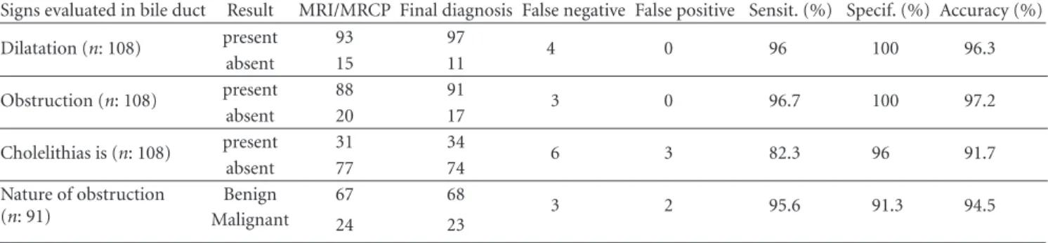
Signs evaluated in bile duct Result MRI/MRCP Final diagnosis False negative False positive Sensit. (%) Specif. (%) Accuracy (%) Dilatation (n: 108) present 93 97 4 0 96 100 96.3 absent 15 11 Obstruction (n: 108) present 88 91 3 0 96.7 100 97.2 absent 20 17 Cholelithias is (n: 108) present 31 34 6 3 82.3 96 91.7 absent 77 74 Nature of obstruction (n: 91) Benign 67 68 3 2 95.6 91.3 94.5 Malignant 24 23
malignant obstructions, in addition to axial images, when it was necessary coronal plan images were also obtained. If a tu-mor was present, the tutu-mor mass, size, the relationship with surrounding tissues, and contrast-enhancement pattern were evaluated.
3. Results
According to final diagnoses, in 34 of 108 patients (31.5%) bile duct obstruction was due to choledocholithiasis, in 23 of them (21.3%), bile duct obstruction was due to malignant obstruction, in 34 of them (31.5%), bile duct obstruction was due to various benign strictures; totally, in 91 patients (84%), it was found. In 17 patients (16%), obstruction was not seen. According to final diagnoses, in 97 of 108 patients (90%), there was bile duct dilatation. Dilatation of bile ducts was observed in 6 patients although any pathology had not been found to explain this. In 11 patients (10%), no dilatation was seen.
In 3 of 34 patients with choledocholithiasis, stones in intrahepatic bile ducts were also present at the same time. 36 of 108 patients had cholecystectomy operation, in 7 of them, stones were detected in common bile duct. In 37 of 72 patients, in whom the gallbladder were present, stone/stones were present in gallbladder, and in 27 of these 37 patients, choledocholithiasis was also observed at the same time. In 34 patients, the cause of the obstruction was determined as benign stricture. In 10 of these patients, iatrogenic bile duct stricture that developed due to cholecystectomy operation, in 8 of them papillary stenosis (fibrotic stenosis and Oddi sphincter dysfunction), in 7 strictures due to inflammatory conditions (e.g., sclerosing cholangitis), in 7 chronic pancre-atitis, in 2 Mirizzi syndrome, in 2 duodenal diverticula, and in 1 choleduct cyst were found (In 3 patients, more than one benign stricture were present). In 23 patients with malignant
obstruction, cholangiocarcinoma in 6 patients (Figure 1),
hepatocellular carcinoma in 2 patients, liver metastasis in 4
patients, metastasis to pancreas head in 2 patients, pancreas head adenocarcinoma in 4 patients, ampullary/periampulla-ry carcinoma in 4 patients, and gallbladder carcinoma in 1 patient were detected.
On the basis of reference results, in 93 of 97 patients with bile duct dilatation, dilatation could be accurately detected using MRI/MRCP; 4 patients had false negative diagnosis. In 88 of 91 patients in whom the obstruction were detected, the presence and location of obstruction were detected accu-rately; on the other hand, in 3 patients, the presence of obstruction and therefore the level of obstruction could not
be detected (Table1). Although in 34 patients with
choledo-cholithiasis calculi were detected accurately using MRCP in 28 patients, in 6 patients, false negative and in 3 patients false positive diagnosis were obtained. According to final diagno-ses, in 23 patients malignant strictures were found as the cause of obstruction, 21 of these patients had accurate dia-gnosis, but 2 had false negative diagnosis; in 68 patients with benign obstruction, the accurate diagnosis was obtained in 65 of them, but 3 patients were misdiagnosed as malignant obstruction.
When MRI/MRCP findings compared to final diagnoses, the sensitivity of MRI/MRCP in demonstrating bile duct dilatation was 96%, specificity was 100%, and accuracy was 96.3%, in the detection of the presence of obstruction, the sensitivity was 96.7%, specificity was 100%, and accuracy was 97.2%, in the diagnosis of choledocholithiasis, sensitivity was 82.3%, specificity was 96%, and accuracy was 91.7%, in the characterization of stenosis, sensitivity was 95.6%, specificity was 91.3%, and accuracy was 94.5%. Imaging findings that were obtained using MRI and MRCP, in comparison with
final diagnoses, are presented in Table2.
4. Discussion
In our study, intravenous contrast-enhanced MRI and MRCP images were evaluated together; in the determination
(a) (b)
(c) (d)
Figure 1: This figure demonstrates a mass lesion located in the left lobe of the liver close to hilus (a) hypointense on T1-weighted MR image, (b) heterogenous hyperintense on T2-weighted image, and (c) heterogenous contrast enhancement on contrast-enhanced fat-saturated T1-weighted image. (d) MRCP image demonstrates the mass that originated from left main bile duct and causes significant bile duct dilatation at the periphery of the liver (arrows). The lesion was considered to be a cholangiocarcinoma with MRI and MRCP findings and also confirmed surgically.
(a) (b)
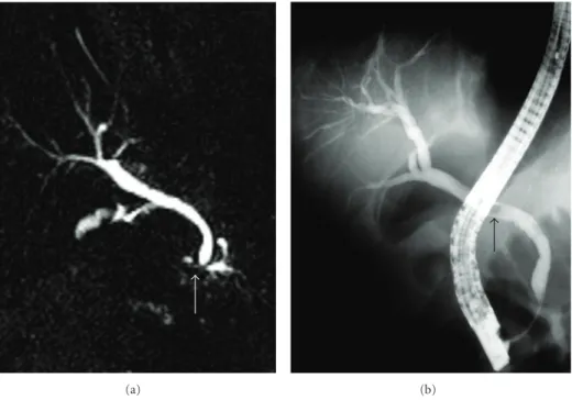
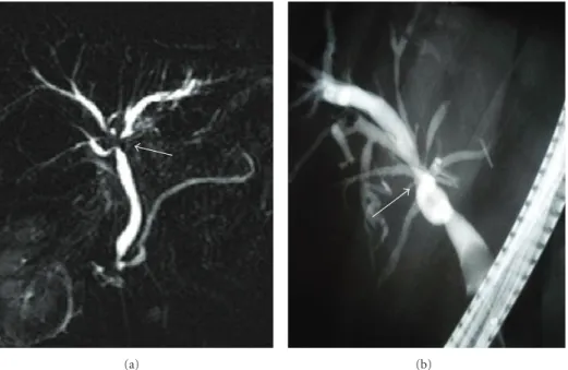
Figure 2: MRCP image demonstrates (a) a calculus (white arrow) located in the distal common bile that does not cause bile duct dilatation. This calculus was not detected during the MRI examination and received a false negative diagnosis. (b) ERCP image demonstrates the calculus (black arrow). This calculus was extracted during the procedure.
(a) (b)
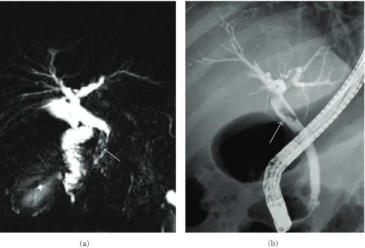
Figure 3: MRCP image demonstrates an irregular heterogenous hypointense region in the distal common bile duct (arrow). On MRI, this region evaluated as benign pathology such as calculus and sludge. ERCP image demonstrates a filling defect in this region (arrow). This filling defect was determined as Fasciola hepatica and extracted during the procedure.
of dilatation of bile ducts, choledocholithiasis, the presence of obstruction, location, and the nature of obstruction, high sensitivity and specificity were obtained.
MRCP can show the presence of bile duct obstruction
with 91%–100% accuracy [12,22,23]. In a study, in
deter-mining biliary obstruction with the combined use of MRI and MRCP, the sensitivity was found 82.3%, the specificity was found 93.8%, and accuracy was found 89% [14]. In our study which was similar to this study, in the determination of the presence of the obstruction, sensitivity was found 96.7%, specificity was found, 100% and accuracy was found 97.2%. In 3 patients, false negative results were obtained. In all of them, millimetric calculi in distal common bile duct and the obstruction were caused by them could not be revealed with MRI/MRCP, but with ERCP, these calculi could be detected.
MRCP can show the level of obstruction with a rate of
85%–100% [12, 14, 21–23]. In our study, the accuracy in
determining the level of stenosis was found 97.2%. False results came from 3 patients with choledocholithiasis men-tioned above. In these patients, with MRCP, the small calculi could not be shown thus the stenosis level could not be deter-mined; therefore, false negative results were obtained. Our results in detection of bile duct obstruction and in the deter-mination of the level of obstruction were quite compatible with the results of the literature.
MRCP can show the intra- and extrahepatic bile duct dilatation with high accuracy. Bile duct dilatation could have been determined with 94%–100% accuracy in recent studies
[16,18]. In our study, in the detection of the presence of
dilatation, the sensitivity of MRCP was found 96%, speci-ficity was found 100%, and accuracy was found 96.3%, and this was compatible with the results of literature. In our stud-y, in the detection of dilatation, in 4 patients false negative
results were obtained when MR images were compared to images based on the ERCP images. False negative results in these patients, as mentioned in many studies, was caused by the application of contrast material with high pressure to overcome the obstruction, and bile ducts were perceived wid-er than normal, and so, the false positiveness of ERCP played a role in this case. In these 4 patients, in the followup with USG, there was no dilatation in intra- and extrahepatic bile ducts.
Gallstones in common bile duct are the most common cause of the bile duct obstruction [16]. Gallstones on MRI, regardless of their chemical composition, commonly seen as signal-void areas in lumen, but sometimes they may be over-looked. In the determination of gallstone, the sensitivity of
MRCP depends on the size of the stone [5,16]. In the
litera-ture, 88%–100% sensitivity and 89%–100% specificity rates have been reported in diagnosis of choledocholithiasis with
MRCP [12, 20–25]. In our study, the sensitivity of MRI/
MRCP in the diagnosis of choledocholithiasis was found 82.3%, the specificity was found 92.2%, and accuracy was 91.7%; this was compatible with the results of the literature. Sensitivity was relatively low in our study, because in 6 of 34 patients with choledocholithiasis, the calculi could not be detected with MRCP. In 5 of these patients, the size of calculi detected and removed with ERCP was 1–5 mm. When MRCP images were retrospectively reexamined, it was understood that in 3 patients, any stone was seen, in 1 patient, 8 mm cal-culus in distal choleduct was interpreted as benign stricture, in 1 patient a few 1–3 millimetric calculi at papillary level and in 1 patient 5 millimeter calculus at papilla were overlooked
(Figure2). In the literature, authors have been reported that
∗ (a) ∗ (b) ∗ ∗ (c) (d)
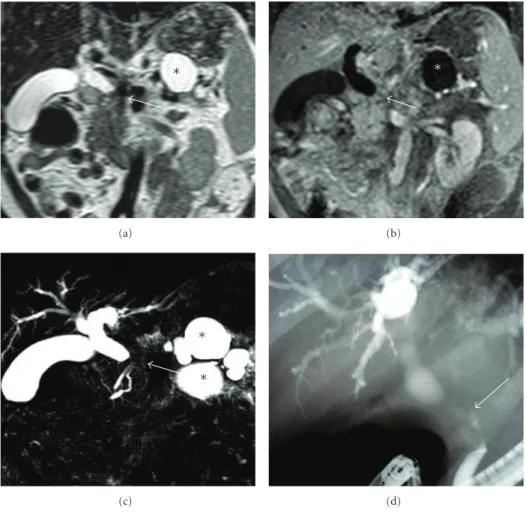
Figure 4: T2-weighted (a), contrast-enhanced T1-weighted MRI (b), and MRCP slices demonstrate significant stenosis in the common bile duct at the level of pancreas head (white arrow) and bile duct dilatation proximal to the stenosis. Any mass lesion is not seen at the stenosis level. On MRI and MRCP, the cause of stenosis considered as chronic pancreatitis because of pseudocysts located in corpus and tail of the pancreas (asterisk). (d) ERCP demonstrates the malignant character of stricture (black arrow), and the result of biopsy was adenocarcinoma of the pancreas.
the stone, stone may not be seen or small stones may be hid-den because of partial volume effect in hyperintense bile
[20,21]. The most difficult cases in diagnosis with MRCP
were the calculi impacted at papilla or millimetric calculi. To see the millimetric calculi, in addition to MIP reconstructed images, base images also must be investigated in detail. How-ever, if the calculi cannot be demonstrated with MRCP and the suspicion of calculi continues, invasive imaging
tech-niques may be needed for definitive diagnosis [24,25]. In our
study, false negative results were obtained in 3 patients. In one of these patients, benign strictures at the middle level of common bile duct, and in the other patient, benign stricture at distal common bile duct were evaluated as calculus. In the 3rd patient, the Fasciola hepatica parasite settled in the distal common bile duct which was extracted by papillotomy
dur-ing ERCP was interpreted in favor of calculus (Figure3). In
MRCP, strictures, intraductal air bubbles, protein plugs, blood clots, parasites, foreign bodies, surgical clips, and pul-sation artifacts caused by arteries near the neighborhood were seen as signal void areas and may receive the wrong di-agnosis as a calculus [20–24].
Stenoses occurred after cholecystectomies (mostly lap-aroscopic) constitute a significant part of benign bile duct stenoses. In addition, bile duct stenoses may be developed as a complication of surgical operations such as gastric and liver resection, liver transplantation, portocaval shunt oper-ation and interventional procedures such as ERCP. Primary sclerosing cholangitis, chronic pancreatitis, and papilla Vater stenosis may also cause bile duct stenosis. Factors such as papillary malformation, inflammation, and spasm secondary to irritation of stone may cause stenosis of papilla and may prevent the bile flow. Malignant strictures causing bile duct obstruction are caused by external compression and/or inva-sion secondary to pancreaticobiliary malignancies or
meta-stases of other malignancies [1–5,21]. In MRI/MRCP, benign
strictures manifest themselves as smooth gradual tapering of stenosis, regular border, and short segment involvement; in spite of this, pancreaticobiliary malignant lesions manifest themselves as abrupt termination of stenosis, irregular bor-der, and long segment involvement. In addition, hyperin-tense regions on T2w images around the stenosis or contrast enhancement on T1w images are other important criteria
(a) (b)
Figure 5: In a case who had undergone laparoscopic cholecystectomy operation 3 years ago, MRCP image demonstrates (a) stricture at hilar level (arrows) and mild dilatation of left main hepatic duct. Although this case had received false positive diagnosis as malignant stricture with MRI and MRCP findings, during ERCP, benign stricture developed secondary to cholecystectomy was diagnosed (arrows). Balloon dilatation and stent replacement was performed during ERCP procedure, and the result of biopsy was benign.
which show the tumoral lesions location and spread. Despite these criteria, MRCP may be insufficient in distinguishing the benign or malignant character of the stenosis from time
to time [2,3,14,18–26].
In the literature, in differentiation between malignant and benign obstruction, accuracy rates of MRCP ranging be-tween 30%–100% have been reported. Variability of rates may be caused by various sequences used in source images and evaluation of MRCP images without MRI slices. Some studies have shown that addition of T1w and T2w images into the examination increase the sensitivity in the evaluation
of malignant lesions [12,15,18,25,26]. In one study, when
only MRCP was used in the differentiation of benign and
malignant biliary obstruction, sensitivity 64.7%, specificity 81.2%, and accuracy 74.4% were found; however, when both MRI and MRCP were used, sensitivity 82.3%, specificity 93.8%, and accuracy 89% were found [14]. In our study, in
the differentiation of benign and malignant biliary
obstruc-tion, the sensitivity of MRCP/MRI method was found 95.6%, specificity was found 91.3%, and accuracy was found 94.5% and was compatible with the results of the literature. The reason why our results were higher than the literature might be caused by the positive contribution of intravenous con-trast-enhanced MRI to the diagnosis which all the patients underwent.
In our study, 3 patients with malignant stricture were misdiagnosed as benign. In one of these patients, there was a pancreas head carcinoma in which a mass could not be de-marcated on MR scans, no dilatation was seen in pancreatic ductus, and stenosis caused by pancreas head carcinoma was
evaluated as benign stricture (Figure 4). In the literature,
it has been reported that if there was no ductal dilatation,
with MRI and MRCP, the diagnosis of pancreas carcinoma
would be difficult [27,28]. In another patient, stenosis at the
papilla level was evaluated as inflammatory stenosis, but it was diagnosed as Ampulla Vateri tumor histopathologically. Obstruction at the level of ampulla can be caused by inflam-matory stenosis, Oddi sphincter dysfunction, edema, stone, ampullary carcinoma, or pancreatitis. In the evaluation of lesions in this region, MRI/MRCP has a low performance due to artifact caused by intraaluminal gas located close to duodenal wall [13]. Therefore, ampullary region is a region that diagnostic mistakes encountered frequently, and if clini-cal suspicion continues, ERCP method in which ampulla
visualized directly should be performed [28,29]. In the third
patient who had a wrong diagnosis, bilioenteric anastomosis had been performed because of chronic pancreatitis 10 years ago, and stenosis developed at anastomosis site was diag-nosed as fibrotic stenosis. In this patient, in biopsy specimens obtained during the bile duct revision operation performed for renewing anastomosis, carcinoma was demonstrated. In a wide research serial of 1003 patients with bilioenteric anas-tomosis, it has been reported that cholangiocarcinoma may develop in the late period; therefore, possibility of malig-nancy should be considered in these kinds of anastomosis stenoses [30]. In our study, two patients with benign stricture had wrong diagnosis. In a patient with Mirizzi syndrome who had wrong positive diagnosis, irregular stenosis in prox-imal common bile duct was defined as malignant stricture on MRI/MRCP. In the other patient, benign stricture at hilar region developed secondary to cholecystectomy operation
was interpreted as malignant (Figure 5). In some studies,
authors have reported that in the differential diagnosis of benign or malignant stenoses at hilar region or proximal bile
ducts experiencing difficulties [1,19,31]. On the other hand, in a recent study, the combination of MRI/MRCP in the eval-uation of cholangiocarcinomas at hilar region has been re-ported to be the most reliable imaging method [32].
5. Conclusion
In our study in which both contrast-enhanced MRI and MRCP were used together, high sensitivity and specificity values were obtained in the evaluation of biliary obstruction. In the detection of small stones, in the evaluation of patholo-gies in ampullary and periampullary region, and in the dif-ferential diagnosis of benign and malignant lesions at hilar region with MRI/MRCP, sometimes difficulties may be seen. For definitive diagnosis in this case, invasive imaging proce-dures in which direct visualization of ampulla and biopsies can be performed during the procedure may be needed. In general, MRI/MRCP is a noninvasive imaging method with high accuracy rates in the evaluation of obstructive biliary tract pathologies may replace the diagnostic ERCP and PTC.
Conflict of Interests
The authors do not have a direct financial relation with the commercial identity (e.g., “Magnevist, Schering”). They have no such conflict of interests.
References
[1] H. J. Schulz, “Indeterminate bile duct strictures,”
Gastroen-terologe, vol. 3, no. 1, pp. 6–13, 2008.
[2] J. A. Soto, E. K. Yucel, M. A. Barish, R. Chuttani, and J. T. Ferrucci, “MR cholangiopancreatography after unsuccessful or incomplete ERCP,” Radiology, vol. 199, no. 1, pp. 91–98, 1996.
[3] TJ Vogl, S. Zeuzem, S. Zangos, and R. Hammerstingl, “Mag-netresonanz-Cholangiopankreatikographie (MRCP) aus radi-ologischem und gastroenterradi-ologischem Blickwinkel. Magnetic resonance cholangiopancreatography (MRCP) from a radio-logical and gastroenteroradio-logical perspective,” Deutsche
Medi-zinische Wochenschrift, vol. 134, no. 19, pp. 1011–1014, 2009.
[4] A. Lubienski, M. Duex, K. Lubienski, J. Blietz, G. W. Kauff-mann, and T. Helmberger, “Interventions for benign biliary strictures,” Radiologe, vol. 45, no. 11, pp. 1012–1019, 2005. [5] F. Tse, J. S. Barkun, J. Romagnuolo, G. Friedman, J. D.
Born-stein, and A. N. Barkun, “Nonoperative imaging techniques in suspected biliary tract obstruction,” HPB (Oxford), vol. 8, no. 6, pp. 409–425, 2006.
[6] A. Filippone, R. Ambrosini, M. Fuschi, T. Marinelli, D. Pinto, and A. Maggialetti, “Clinical impact of MR cholangiopancre-atography in patients with biliary disease,” Radiologia Medica, vol. 105, no. 1-2, pp. 27–35, 2003.
[7] B. Glasbrenner, M. Ardan, W. Boeck, G. Preclik, P. M¨oller, and G. Adler, “Prospective evaluation of brush cytology of biliary strictures during endoscopic retrograde cholangiopancreatog-raphy,” Endoscopy, vol. 31, no. 9, pp. 712–717, 1999.
[8] M. Buset, F. Dunham, A. Gulbis, M. Boyazis, J. Jeanmart, and J. Toussaint, “Late complications of the diagnostic and operative endoscopy of the bile ducts and the pancreas. International study (author’s transl),” Acta Gastro-Enterologica Belgica, vol. 44, no. 9-10, pp. 396–403, 1981.
[9] M. G. Stiris, B. Tennoe, E. Aadland, and O. C. Lunde, “MR Cholangiopancreatography and endoscopic retrograde cho-langiopancreatography in patients with suspected common bile duct stones,” Acta Radiologica, vol. 41, pp. 269–272, 2000. [10] T. Rosch, A. Meining, S. Fruhmorgen et al., “A prospective comparison of the diagnostic accuracy of ERCP, MRCP, CT, and EUS in biliary strictures,” Gastrointestinal Endoscopy, vol. 55, pp. 870–876, 2002.
[11] B. Schumacher, “Diagnostik und Therapie von Gallenwegser-krankungen—ERCP und MRCP,” Gastroenterologie Up2date, vol. 2, no. 2, pp. 155–164, 2006.
[12] A. S. Fulcher and M. A. Turner, “MR cholangiopancreatogra-phy,” Radiologic Clinics of North America, vol. 40, no. 6, pp. 1363–1376, 2002.
[13] V. David, C. Reinhold, M. Hochman et al., “Pitfalls in the interpretation of MR cholangiopancreatography,” American
Journal of Roentgenology, vol. 170, no. 4, pp. 1055–1059, 1998.
[14] L. Zhong, Q. Y. Yao, L. Li, and J. R. Xu, “Imaging diagnosis of pancreato-biliary diseases: a control study,” World Journal of
Gastroenterology, vol. 9, no. 12, pp. 2824–2827, 2003.
[15] M. J. Kim, D. C. Mitchell, K. Ito, and E. K. Outwater, “Biliary dilatation: differentiation of benign from malignant causes— value of adding conventional MR imaging to MR cholan-giopancreatography,” Radiology, vol. 214, no. 1, pp. 173–181, 2000.
[16] J. E. W. Zajaczek and M. Keberle, “Value of radiological meth-ods in the diagnosis of biliary diseases,” Radiologe, vol. 45, no. 11, pp. 976–986, 2005.
[17] M. S. Park, T. K. Kim, K. W. Kim et al., “Differentiation of extrahepatic bile duct cholangiocarcinoma from benign stric-ture: findings at MRCP versus ERCP,” Radiology, vol. 233, no. 1, pp. 234–240, 2004.
[18] M. Baykara, N. Erdo˘gan, and N. ¨Ozcan, “Tıkanma tipi sarılı˘gı olan olgularda manyetik rezonans kolanjiyopankreatografi bulgularının perk¨utan transhepatik kolanjiyografi bulgularıyla kars¸ılas¸tırılması,” Erciyes Tıp Dergisi, vol. 27, no. 1, pp. 22–35, 2006.
[19] J. M. Dumonceau, C. Macias Gomez, C. Casco et al., “Grasp or brush for biliary sampling at endoscopic retrograde cholan-giography? A blinded randomized controlled trial,” American
Journal of Gastroenterology, vol. 103, no. 2, pp. 333–340, 2008.
[20] M. Bilgin, F. Shaikh, R. C. Semelka, S. S. Bilgin, N. C. Balci, and A. Erdogan, “Magnetic resonance imaging of gallbladder and biliary system,” Topics in Magnetic Resonance Imaging, vol. 20, no. 1, pp. 31–42, 2009.
[21] F. Kantarcı, D. Selc¸uk, S. Albayram, and G. ¨O˘g¨ut, “Pankreato-bilyer sistem patolojilerini de˘gerlendirmede manyetik rezo-nans kolanjiyopankreatografi (MRCP)’nin tanı de˘geri,”
Bil-gisayarlı Tomografi B¨ulteni, vol. 7, no. 4, pp. 177–182, 2003.
[22] A. M. Halefoglu, “Magnetic resonance cholangiopancreatog-raphy: a useful tool in the evaluation of pancreatic and biliary disorders,” World Journal of Gastroenterology, vol. 13, no. 18, pp. 2529–2534, 2007.
[23] P. Pavone, A. Laghi, V. Panebianco, C. Catalano, L. Lobina, and R. Passariello, “MR cholangiography: techniques and clinical applications,” European Radiology, vol. 8, no. 6, pp. 901–910, 1998.
[24] H. J. Brambs, M. Hoffmann, and S. Pauls, “Diagnosis and interventional therapy for ductal gallstones,” Radiologe, vol. 45, no. 11, pp. 1004–1011, 2005 (German).
[25] C. D. Becker, M. Grossholz, M. Becker, G. Mentha, R. De Peyer, and F. Terrier, “Choledocholithiasis and bile duct stenosis: diagnostic accuracy of MR cholangiopancreatography,”
[26] J. Romagnuolo, M. Bardou, E. Rahme, L. Joseph, C. Reinhold, and A. N. Barkun, “Magnetic resonance cholangiopancreatog-raphy: a meta-analysis of test performance in suspected biliary disease,” Annals of Internal Medicine, vol. 139, no. 7, pp. 547– I32, 2003.
[27] E. L. H¨anninen, H. Amthauer, N. Hosten et al., “Prospective evaluation of pancreatic tumors: accuracy of MR imaging with MR cholangiopancreatography and MR angiography,”
Radiol-ogy, vol. 224, no. 1, pp. 34–41, 2002.
[28] S. J. Diehl, K. J. Lehmann, J. Gaa, H. J. Meier-Willersen, K. Wendl, and M. Georgi, “The value of magnetic resonance to-mography (MRT), magnetic resonance cholangiopancreatog-raphy (MRCP) and endoscopic retrograde cholangiopancre-atography (ERCP) in the diagnosis of pancreatic tumors,”
Rofo, vol. 170, pp. 463–469, 1999.
[29] R. C. Semelka, N. L. Kelekis, G. John, S. M. Ascher, D. Burdeny, and E. S. Siegelman, “Ampullary carcinoma: demonstration by current MR techniques,” Journal of Magnetic Resonance
Imag-ing, vol. 7, no. 1, pp. 153–156, 1997.
[30] A. Tocchi, G. Mazzoni, G. Liotta, L. Lepre, D. Cassini, and M. Miccini, “Late development of bile duct cancer in patients who had biliary-enteric drainage for benign disease: a follow-up study of more than 1,000 patients,” Annals of Surgery, vol. 234, no. 2, pp. 210–214, 2001.
[31] S. S. Saluja, R. Sharma, S. Pal, P. Sahni, and T. K. Chattopad-hyay, “Differentiation between benign and malignant hilar ob-structions using laboratory and radiological investigations: a prospective study,” HPB (Oxford) Surgery, vol. 9, no. 5, pp. 373–382, 2007.
[32] G. Masselli, R. Manfredi, A. Vecchioli, and G. Gualdi, “MR imaging and MR cholangiopancreatography in the preoper-ative evaluation of hilar cholangiocarcinoma: correlation with surgical and pathologic findings,” European Radiology, vol. 18, no. 10, pp. 2213–2221, 2008.
Submit your manuscripts at
http://www.hindawi.com
Stem Cells
International
Hindawi Publishing Corporation
http://www.hindawi.com Volume 2014
Hindawi Publishing Corporation
http://www.hindawi.com Volume 2014
INFLAMMATION
Hindawi Publishing Corporation
http://www.hindawi.com Volume 2014
Behavioural
Neurology
Endocrinology
International Journal ofHindawi Publishing Corporation
http://www.hindawi.com Volume 2014
Hindawi Publishing Corporation
http://www.hindawi.com Volume 2014
Disease Markers
Hindawi Publishing Corporation
http://www.hindawi.com Volume 2014 BioMed
Research International
Oncology
Journal ofHindawi Publishing Corporation
http://www.hindawi.com Volume 2014
Hindawi Publishing Corporation
http://www.hindawi.com Volume 2014
Oxidative Medicine and Cellular Longevity
Hindawi Publishing Corporation
http://www.hindawi.com Volume 2014
PPAR Research
The Scientific
World Journal
Hindawi Publishing Corporationhttp://www.hindawi.com Volume 2014
Immunology Research Hindawi Publishing Corporation
http://www.hindawi.com Volume 2014
Journal of
Obesity
Journal ofHindawi Publishing Corporation
http://www.hindawi.com Volume 2014
Hindawi Publishing Corporation
http://www.hindawi.com Volume 2014
Computational and Mathematical Methods in Medicine
Ophthalmology
Journal of Hindawi Publishing Corporationhttp://www.hindawi.com Volume 2014
Diabetes Research
Journal ofHindawi Publishing Corporation
http://www.hindawi.com Volume 2014
Hindawi Publishing Corporation
http://www.hindawi.com Volume 2014 Research and Treatment
AIDS
Hindawi Publishing Corporation
http://www.hindawi.com Volume 2014 Gastroenterology Research and Practice
Hindawi Publishing Corporation
http://www.hindawi.com Volume 2014