DESIGN, SYNTHESIS and CHARACTERIZATION of
BIOINSPIRED NANOMATERIALS for ENGINEERING
and BIOMEDICINE
A DISSERTATION SUBMITTED TO
MATERIALS SCIENCE AND NANOTECHNOLOGY PROGRAM OF THE GRADUATE SCHOOL OF ENGINEERING AND SCIENCE
OF BILKENT UNIVERSITY
IN PARTIAL FULFILLMENT OF THE REQUIREMENTS FOR THE DEGREE OF
DOCTOR OF PHILOSOPHY
By
HAKAN CEYLAN August, 2014
I certify that I have read this thesis and that in my opinion it is fully adequate, in scope and in quality, as a thesis of the degree of Doctor of Philosophy.
……….
Asst. Prof. Dr. Ayşe Begüm Tekinay (Advisor)
I certify that I have read this thesis and that in my opinion it is fully adequate, in scope and in quality, as a thesis of the degree of Doctor of Philosophy.
……….
Assoc. Prof. Dr. Mustafa Özgür Güler (Co-Advisor)
I certify that I have read this thesis and that in my opinion it is fully adequate, in scope and in quality, as a thesis of the degree of Doctor of Philosophy.
………. Prof. Dr. Engin Umut Akkaya
I certify that I have read this thesis and that in my opinion it is fully adequate, in scope and in quality, as a thesis of the degree of Doctor of Philosophy.
………. Prof. Dr. Halil İbrahim Ünal
I certify that I have read this thesis and that in my opinion it is fully adequate, in scope and in quality, as a thesis of the degree of Doctor of Philosophy.
………. Asst. Prof. Dr. Ali Osmay Güre
I certify that I have read this thesis and that in my opinion it is fully adequate, in scope and in quality, as a thesis of the degree of Doctor of Philosophy.
………. Assoc. Prof. Dr. Hüseyin Özkan
Approved for the Graduate School of Engineering and Science:
………. Prof. Dr. Levent Onural
i
ABSTRACT
DESIGN, SYNTHESIS and CHARACTERIZATION of BIOINSPIRED
NANOMATERIALS for ENGINEERING and BIOMEDICINE
Hakan Ceylan
PhD in Materials Science and Nanotechnology Supervisor: Ayşe Begüm Tekinay, PhD Co-Supervisor: Mustafa Özgür Güler, PhD
August, 2014
Nature is an inspirational school for materials scientists. Natural selection process puts a massive pressure on biological organisms giving rise to effective strategies for fabricating materials, which generally outperform their man-made counterparts. Mimicking physical and chemical features of biological materials can greatly aid in overcoming existing design constraints of engineering and medicine. In this dissertation, a reductionist, bottom-up approach is demonstrated to recapitulate biological functionalities in fully-synthetic hybrid constructs. For material design, the potential of short, rationally-designed peptides for programmed organization into nanostructured materials is explored. The resulting nano-ordered materials exhibit multifunctional and adaptive properties, which can be tailored by the information within monomeric peptide sequences as well as the emerging properties upon their self-assembly. In light of these, design, synthesis and characterization of the prototypes of nanostructured functional materials are described in the context of regenerative medicine and biomineralization.
ii
Keywords: Bioinspired Materials, Functional Self-Assembly, Biomaterials, Surface
iii
ÖZET
BİYOLOJİK SİSTEMLERDEN ESİNLENEREK PROGRAMLANAN
NANO YAPILI MALZEMELERİN MÜHENDİSLİK ve
BİYOMEDİKAL UYGULAMALAR İÇİN TASARIMI, SENTEZİ ve
KARAKTERİZASYONU
Hakan Ceylan
Malzeme Bilimi ve Nanoteknoloji, Doktora Tez Danışmanı: Dr. Ayşe Begüm Tekinay
Eş Danışman: Dr. Mustafa Özgür Güler Ağustos, 2014
Malzeme bilimi araştırmacıları için doğa ilham verici bir okuldur. Canlı organizmalar, doğal seçilim eleklerinden başarıyla geçmelerini sağlayacak işlevsel olarak çok iyi organize olmuş malzemeler geliştirerek varlıklarını sürdürmektedir. Bu yönüyle biyolojik malzemeler benzer koşullar için yapay olarak üretilenlerden çok daha başarılı bir performans sergiler. Bu nedenle biyolojik malzemelerdeki fiziksel veya kimyasal etkenlerin yapay malzemelerde yeniden ortaya çıkartılması ile hâlihazırda endüstri ve tıp alanlarında karşılaşılan birçok zorluğun aşılabileceği değerlendirilmektedir. Bu tezde minimalist bir yaklaşım kullanarak, aşağıdan-yukarı fabrikasyon yöntemiyle geliştirilen hibrit yapılarda biyolojik malzemelerin işlevsel özellikleri taklit edilmektedir. Yapı malzemesi olarak, mantıksal olarak tasarlanan kısa sentetik peptit dizinlerinin programlı olarak organizasyonu (kendiliğinden toplanması) değerlendirilmiştir. Elde edilen nano-yapılı malzemelerin dinamik ve
iv
işlevsel özellikleri peptitleri oluşturan aminoasitlerin dizilimi ve toplanma sonrası ortaya çıkan yeni durum ile kontrol edilebilmektedir. Bu perspektifle, biyolojik malzemelerden esinlenerek geliştirilen, yenileyici tıp ve biyomineralizasyon alt başlıklarında uygulanan prototiplerin tasarımı, sentez ve karakterizasyonu incelenmektedir.
Anahtar kelimeler: Doğadan Esinlenerek Geliştirilen Malzemeler, İşlevsel Süpramoleküler Nanoyapılar, Biyomalzemeler, İşlevsel Yüzeyler,
Biyomineralizasyon
v
Acknowledgements
I would like to express my gratitude to my advisors, Prof. Tekinay and Prof. Güler for their guidance and support to my research. Their encouragement helped me develop a trans-disciplinary understanding, which I can carry with me throughout my research career.
I would like to acknowledge the PhD scholarship from TÜBİTAK (The Scientific and Research Council of Turkey) BIDEB 2211-A. I would also like to thank for two international conference supports (in the form of 2224-A) as well as the generous support for the 61st Lindau Nobel Laureate Meeting. TÜBİTAK has a very special place in my academic development.
This thesis simply would not have been possible by the endless devotion and exceptional support of Şeyma Özkan. She is truly a remarkable person. Her encouraging liveliness kept me motivated all the time. She is the unseen hero behind this work. Therefore, I would like to dedicate this thesis to her as my tribute.
I would like to express my most sincere thanks to Samet Kocabey, Seher Yaylacı and Hilal Ünal Gülsüner for their companionship in this long marathon. Their support has always kept me motivated. Their friendship deserves all compliments.
I would like to express my special thanks to Samet Kocabey, Hilal Ünal Gülsüner, Cağla-Özgit Akgün and Ruslan Garifullin for their fruitful collaboration. Together, we pursued interesting scientific questions, which we managed to publish in high quality journals. Another special thanks to Ayşegül Tombuloğlu who taught
vi
me many tools and techniques, which significantly eased my adaptation to the laboratory in my first year. I have never forgotten her kindness.
I would also like to thank Handan Acar, Melis Göktaş, Gülistan Tansık, Gülcihan Gülseren, Melike Sever, Berna Şentürk, Ceren Garip, Nuray Gündüz, Elif Arslan, Elif Ergül, Yasin Tümtaş, Öncay Yaşa, Ahmet Emin Topal, Alper Devrim Özkan and Mevhibe Yakut for creating such a warm working environment. My special thanks to Zeynep Ergül Ülger and Zeynep Erdoğan not only for their immense technical help, but also for their sincere friendship for all these years. It was wonderful to work with them.
I would like to thank Prof. Salim Çıracı for supporting my participation to a truly inspirational organization, the 3rd “Global Challenges- Opportunities for Nanotechnology Workshop”. It significantly contributed to my understanding of science and technology as productive means to address the growing complexity of global problems.
I would like to take this opportunity to give my tribute in memory of Prof. İhsan Doğramacı, the founder of Bilkent University. Bilkent University has been my home with all the unforgettable memories for the past nine years, including my undergraduate education. Prof. Doğramacı’s endless pursuit of reaching perfection opened wide avenues in my endeavors.
Finally, I would like to express my most sincere gratitude to my family who always supported me with their endless love to become who I am now. I am proud of them. I work for day and night to elevate their name and honor.
vii
CONTENTS
CONTENTS ... VII LIST OF FIGURES ... XIII LIST OF TABLES ... XVII ABBREVIATIONS ... XVIII CHAPTER 1
INTRODUCTION: CONCEPTS in BIOINSPIRED NANOMATERIAL
DESIGN ... 1
1.1 Sources of Inspiration ... 3
1.1.1 Underwater adhesion: Analogy between marine environment and mammalian tissues ... 3
1.1.2 The extracellular matrix: Learning from nature for biomaterial design 15 1.2 Bioinspired Design Strategies: From Supramolecular Chemistry to Hybrid Materials ... 28
CHAPTER 2 SELECTIVE ADHESION and GROWTH of VASCULAR ENDOTHELIAL CELLS on BIOACTIVE PEPTIDE NANOFIBER FUNCTIONALIZED STAINLESS STEEL SURFACE ... 34
2.1 Objective ... 34
2.2 Introduction ... 35
2.3 Results and Discussion ... 38
2.3.1 Synthesis of PA molecules and characterization of their self-assembly into nanofibers ... 38
2.3.2 Adsorption analysis and surface characterizations of the nanofibers on stainless steel surface ... 42
2.3.3 Characterization of the cellular responses on peptide nanofibers ... 46
2.3.4 Platelet adhesion on PA nanofibers ... 53
viii
2.5 Experimental Section ... 57
2.5.1 Materials ... 57
2.5.2 Synthesis and characterization of peptide amphiphiles (PA)... 58
2.5.3 Self-assembled nanofibrous network formation ... 59
2.5.4 Adsorption analysis and surface characterization of PA nanofibers on stainless steel ... 62
2.5.5 PA-coated surface preparation for in vitro characterizations ... 63
2.5.6 Cell culturing and maintenance... 64
2.5.7 Adhesion, spreading and cytoskeleton analyses of vascular cells on PA-coated surfaces ... 64
2.5.8 Viability and proliferation of vascular cells on PA nanofibers ... 66
2.5.9 Platelet adhesion on PA nanofibers ... 67
2.5.10 Statistical analysis ... 68
CHAPTER 3 SURFACE-ADHESIVE and OSTEOGENIC SELF-ASSEMBLED PEPTIDE NANOFIBERS for BIOINSPIRED FUNCTIONALIZATION of TITANIUM SURFACES ... 69
3.1 Objective ... 69
3.2 Introduction ... 70
3.3 Results and Discussion ... 73
3.3.1 Synthesis, characterization and self-assembly of peptide amphiphiles . 73 3.3.2 Surface adhesive osteoinductive nanofibers ... 74
3.3.3 In vitro biocompatibility of preosteoblasts on osteoinductive nanofibers……. ... 81
3.3.4 Osteoblastic maturation of preosteoblasts ... 85
3.4 Conclusion ... 88
3.5 Experimental Section ... 90
3.5.1 Materials ... 90
3.5.2 Synthesis and characterization of peptide-amphiphile building blocks . 90 3.5.3 Formation of peptide nanofibers and their characterizations ... 91
ix
3.5.5 In vitro cell culture tests ... 93
3.5.6 Statistical analyses ... 96
CHAPTER 4 BONE-LIKE APATITE NUCLEATING NANOFIBERS INDUCE DIFFERENTIATION of HUMAN MESENCHYMAL STEM CELLS into MATURE OSTEOBLASTS ... 97
4.1 Objective ... 97
4.2 Introduction ... 98
4.3 Results and Discussion ... 103
4.3.1 Design of the building blocks and self-assembly into multifunctional nanofibers ... 103
4.3.2 Surface stability of the nanofibrous peptide coatings ... 104
4.3.3 Surface mineralization with biological apatite... 106
4.3.4 Adhesion, spreading, migration, survival and proliferation of hMSCs 114 4.3.5 Osteogenic differentiation of hMSCs ... 115
4.4 Conclusion ... 124
4.5 Experimental Section ... 125
4.5.1 Synthesis and characterization of peptide amphiphiles ... 125
4.5.2 Formation of self-assembled peptide nanofibers ... 126
4.5.3 Stability of peptide nanonetworks on titanium substrate ... 128
4.5.4 Mineralization of peptide nanonetworks in simulated body fluid ... 129
4.5.5 Human mesenchymal stem cell culturing ... 130
4.5.6 Preparation of surfaces for in vitro assays ... 132
4.5.7 Cell adhesion, spreading and locomotion ... 132
4.5.8 Cell viability and proliferation ... 133
4.5.9 Osteogenic differentiation of hMSC ... 133
4.5.10 Alkaline phosphatase activity ... 134
4.5.11 Alizarin red staining ... 134
4.5.12 Quantitative reverse transcription polymerase chain reaction ... 135
4.5.13 Immunofluorescence and DMP-1 localization ... 136
x CHAPTER 5
MUSSEL INSPIRED DYNAMIC CROSS-LINKING of SELF-HEALING
PEPTIDE NANOFIBER NETWORK ... 137
5.1 Objective ... 137
5.2 Introduction ... 138
5.3 Results and Discussion ... 142
5.3.1 Self-Assembly of Mussel-Mimetic Peptide Building Blocks ... 142
5.3.2 Secondary Structure Characterization of the Peptide Nanofibers ... 150
5.3.3 Bulk Rheological Analyses of Cross-Linked Supramolecular Network…. ... 151
5.3.4 Influence of Temperature on the Curing of Mussel Inspired Cross-Linking in the Supramolecular Networks ... 157
5.3.5 pH Dependent Reversibility of the Mussel Inspired Supramolecular Network ... 158
5.3.6 Self-Healing Properties of the Networks ... 161
5.4 Conclusion ... 163
5.5 Experimental Section ... 164
5.5.1 Materials ... 164
5.5.2 Synthesis and Characterization of Peptide Amphiphiles ... 164
5.5.3 Cross-linked Gel Preparations ... 165
5.5.4 Scanning Electron Microscopy (SEM) ... 167
5.5.5 Circular Dichroism (CD) ... 167
5.5.6 Fourier Transform Infrared Spectroscopy (FT-IR) ... 168
5.5.7 Oscillatory Rheology ... 168
CHAPTER 6 SIZE-CONTROLLED CONFORMAL NANOFABRICATION of BIOTEMPLATED THREE-DIMENSIONAL TiO2 and ZnO NANONETWORKS ... 170
6.1 Objective ... 170
6.2 Introduction ... 171
xi
6.3.1 Synthesis and preparation of the peptide template for ALD ... 175
6.3.2 Characterization of TiO2 and ZnO nanonetworks ... 176
6.3.3 Atomic layer size control of the semiconductor nanotubes ... 182
6.3.4 Size-function relationship ... 186
6.3.5 Photoexcitation of surface-immobilized TiO2 and ZnO nanonetworks189 6.4 Conclusion ... 193
6.5 Experimental Section ... 194
6.5.1 Materials ... 194
6.5.2 Synthesis and characterization of peptide amphiphile molecule ... 194
6.5.3 Template preparation ... 195
6.5.4 Atomic layer deposition of TiO2 and ZnO ... 196
6.5.5 Characterization of TiO2 and ZnO nanonetwork ... 197
6.5.6 Photoexcitation reactions ... 198
CHAPTER 7 ... 200
MODULAR SHORT PEPTIDES for COUPLED SYNTHESIS and BIOFUNCTIONALIZATION of GOLD NANOPARTICLES for INTEGRIN-TARGETED CELL UPTAKE ... 200
7.1 Objective ... 200
7.2 Introduction ... 201
7.3 Results and Discussion ... 203
7.3.1 Design and Synthesis of Modular Peptides... 203
7.3.2 Development and Characterization of RGD peptide conjugated Gold Nanoparticles ... 204
7.3.3 Size Control on Synthesized Gold Nanoparticles ... 210
7.3.4 Biocompatibility and Cellular Uptake of Gold Nanoparticles ... 212
7.4 Conclusion ... 218
7.5 Experimental Section ... 219
7.5.1 Materials ... 219
7.5.2 Synthesis and Characterization of Modular Peptides ... 219
7.5.3 Synthesis of Gold Nanoparticles ... 220
xii
7.5.5 Colloidal Stability of Gold Nanoparticles ... 222
7.5.6 Ligand Density on Gold Nanoparticles ... 222
7.5.7 Cell Culture ... 222
7.5.8 Cytotoxicity of Gold Nanoparticles ... 222
7.5.9 Cellular Uptake of Gold Nanoparticles ... 223
7.5.10 Statistical Analysis ... 223
CHAPTER 8 CONCLUSION & FUTURE PROSPECTS ... 224
BIBLIOGRAPHY ... 227
xiii
LIST OF FIGURES
Figure 1.1 Interaction of water molecules and dissolved ions with surface.. ... 4 Figure 1.2 Mussel adhesion strategy inspires synthetic adhesives that can operate under water…………. ... 8 Figure 1.3 Mussel glue: Adhesion and curing mechanisms ... 10 Figure 1.4 ECM provides a physical and bioactive support to the cells forming a functional tissue and organ ... 16 Figure 1.5 Hierarchical assembly of collagen type I from nano- to meso-scales…….……... ... 19 Figure 1.6 Schematic domains in fibronectin and collagen type I alpha 1... 20 Figure 1.7 Adhesion of normal rat kidney cells to fibronectin-mimetic short peptides……… ... 21 Figure 1.8 Integrin activation and its time-based downstream signaling. ... 24 Figure 1.9 Steric conformation and functional groups are inspirational sites to mimic the biological function of GAGs. ... 25 Figure 1.10 Heparan sulfate proteoglycans carry out a number of biological functions………. ... 26 Figure 1.11 Sequence-specific information of DNA provides a vast source for programmable supramolecular materials. ... 29 Figure 1.12 Rationally-designed modular peptide amphiphiles can dynamically assemble into long one-dimensional nanofibers. ... 31 Figure 2.1 Design and self-assembly of REDV-PA and Dopa-PA molecules. ... 39 Figure 2.2 Liquid chromatography-mass spectrometry (LC-MS) analysis of the synthesized PAs ... 40 Figure 2.3 Zeta potentials of individual PA molecules and their mixtures at pH 7.4………. ... 41 Figure 2.4 Adsorption of REDV-PA/Dopa-PA nanofibers on stainless steel surface………….. ... 45 Figure 2.5 Representative calcein-AM stained fluorescent images of HUVECs attached on the stainless steel surfaces ... 47
xiv
Figure 2.6 Adhesion and viability of HUVECs and A7r5 cells on
REDV-PA/Dopa-PA-coated glass and tissue culture plate surfaces. ... 49
Figure 2.7 Spreading of vascular cells on REDV-PA/Dopa-PA, E-PA/Dopa-PA coated and bare stainless steel surfaces... 50
Figure 2.8 Cellular morphology, viability and proliferation at 24 h and 72 h……….….. ... 55
Figure 2.9 Relative attachment of platelets. ... 56
Figure 2.10 Synthesis route of a typical peptide amphiphile using solid phase peptide synthesis. ... 60
Figure 3.1 Schematic illustration of the immobilization strategy for osteogenic nanofibers on titanium surface based on the self-assembly of the KRSR-PA and Dopa-PA………. ... 75
Figure 3.2 Characterization of the self-assembled peptide nanofibers and the mechanism of assembly. ... 77
Figure 3.3 Specific immobilization of KRSR-PA/Dopa-PA nanofibers on titanium surface. ... 78
Figure 3.4 Dopa-mediated immobilization alters surface properties.. ... 80
Figure 3.5 Adhesion, viability and morphology of cells on functionalized titanium surfaces. ... 83
Figure 3.6 Adhesion and viability of MC3T3-E1 cells on functionalized titanium surfaces……… ... 86
Figure 3.7 High magnification confocal images of the cells at 24 h……… 87
Figure 3.8 Effect of immobilized PA nanofibers on osteogenic activity ... 89
Figure 4.1 Self-assembly of peptide amphiphiles into multifunctional osteoinductive nanofibers ... 102
Figure 4.2 Biophysical analysis of PA self-assembly ... 105
Figure 4.3 Dopa imparts surface stability to osteoinductive nanofibers ... 108
Figure 4.4 Hydroxyapatite formation on PA nanofibers in simulated body fluid... ... 109
Figure 4.5 Dopa-functionalization is critical for HAp formation in SBF. ... 110
Figure 4.6 Characterization of HAp formation ... 112
xv
Figure 4.8 Early-stage interactions of hMSCs with the nanofibers. ... 116
Figure 4.9 Osteoinductive effect of PA nanofibers on hMSCs ... 117
Figure 4.10 Morphology of hMSCs at day 0 and at day 28 ... 119
Figure 4.11 De novo calcium phosphate formation on E3-PA/Dopa-PA by differentiated osteoblast activity. ... 120
Figure 4.12 Differentiation of hMSCs into the osteoblast lineage cells by PA nanofibers……… ... 122
Figure 4.13 Liquid chromatography-mass spectrometry (LC-MS) analysis of the synthesized PAs. ... 127
Figure 4.14 Characterization of surface markers of hMSCs isolated from the donor bone marrow…….. ... 131
Figure 5.1 Mussel-inspired mechanical enhancement strategy for supramolecular peptide network. ... 143
Figure 5.2 Titration of mussel inspired peptide amphiphiles with NaOH. ... 145
Figure 5.3 pH dependent reactions of mussel inspired peptide nanofibers. ... 147
Figure 5.4 pH and Fe(III) dependent UV-Vis spectra of DopaK-PA... 148
Figure 5.5 SEM images of the mussel inspired, self-assembled peptide nanofibers……… ... 149
Figure 5.6 Secondary structure analyses of the mussel-inspired peptide nanofibers……… ... 152
Figure 5.7 Rheological characterizations of mussel inspired peptide gels at 1 wt% concentration ... 155
Figure 5.8 Relationship between equilibrium storage modulus and initial peptide amphiphile concentration. ... 156
Figure 5.9 Impact of temperature on the mechanical properties of peptide networks……… ... 159
Figure 5.10 pH dependent reversibility of the peptide networks. ... 160
Figure 5.11 Self-healing of the mussel inspired peptide gels. ... 162
Figure 5.12 Characterization of the purity and functionality of the mussel-inspired peptide amphiphiles. ... 166
Figure 6.1 Strategy for three-dimensional nanofabrication of TiO2 and ZnO nanonetworks on supramolecular nanofibers of peptide amphiphile nanonetwork..173
xvi
Figure 6.2 Characterization of as-synthesized TiO2 and ZnO nanonetworks
deposited with 350 and 100 ALD cycles, respectively. ... 178
Figure 6.3 Characterization of inorganic nanofabrication on peptide network..179
Figure 6.4 EDX mappings of Ti and Zn along transverse section. ... 180
Figure 6.5 Characterization of calcined TiO2 and ZnO nanonetworks prepared by the deposition of 350 and 100 ALD cycles, respectively ... 181
Figure 6.6 Size-controlled deposition of TiO2 and ZnO on peptide nanonetwork template through ALD cycle number... 183
Figure 6.7 Atomic layer deposition parameters. ... 184
Figure 6.8 Representative TEM images of TiO2 and ZnO nanonetworks after calcination at 450 °C. ... 185
Figure 6.9 UV-Vis spectra of anatase TiO2 and wurtzite ZnO nanonetworks (after calcination). ... 187
Figure 6.10 Photoexcitation of TiO2 and ZnO nanonetworks as a function of nanotube wall thicknesses. ... 190
Figure 6.11 UV-Vis spectra of methylene blue solutions to show degradation of methylene blue………. ... 191
Figure 6.12 Comparison of the photoexcitation activities of templated anatase TiO2 and non-templated, solution synthesized anatase. ... 192
Figure 7.1 Schematic illustration of the modular peptide design ... 205
Figure 7.2 One-pot synthesis, kinetics and colloidal stability of AuNPs.. ... 207
Figure 7.3 Energy-dispersive X-ray spectrum ... 208
Figure 7.4 Synthesis mechanism of AuNPs by a model RGD-functionalized modular peptide. ... 209
Figure 7.5 Size-controlled one-pot synthesis of RGD-functionalized AuNPs……… ... 213
Figure 7.6 TEM images of AuNPs synthesized-stabilized with the RGD peptide……….….. ... 215
Figure 7.7 Kinetics of AuNP formation ... 216
Figure 7.8 Cellular uptake of RGD-AuNPs. ... 217
Figure 7.9 Liquid chromatogram, mass spectrum and UV-Vis analyses of the modular peptide. ... 221
xvii
LIST OF TABLES
Table 1.1 Molecular weight and Dopa content of mussel adhesive proteins. ... 9 Table 4.1 Osteoinductive PA nanofiber compositions forming bone-mimetic cellular microenvironments ... 104 Table 4.2 Primer list used in the qRT-PCR... 135 Table 7.1 Size and surface characteristics of AuNPs: core size, estimated number of gold atoms per nanoparticles, and the number of RGD ligands on each nanoparticle. ... 214
xviii
ABBREVIATIONS
ALD : Atomic layer deposition
ALP : Alkaline phosphatase
ATR-FTIR : Attenuated total reflectance-Fourier transform infrared spectrometry BMP-2 : Bone morphogenetic protein-2
BSA : Bovine serum albumin
CD : Circular dichroism
DCM : Dichloromethane
DMF : N, N-Dimethylformamide
DMEM : Dulbecco’s modified Eagle’s medium
Dopa : 3,4-Dihydroxy-L-phenylalanine
ECM : The extracellular matrix
EdU : 5-ethynyl-2’-deoxyuridine
EDS : Energy-dispersive x-ray spectrometry
ESI : Electrospray ionization
FBS : Fetal bovine serum
FCS : Fetal calf serum
GAG : Glycosaminoglycan
HAp : Hydroxyapatite
HEPES : 2-[4-(2-hydroxyethyl)piperazin-1-yl] ethanesulfonic acid
HGF : Human gingival fibroblast
hMSC : Human mesenchymal stem cell
HPLC : High performance liquid chromatography EDTA : Ethylenediaminetetraacetic acid
xix
HUVEC : Human umbilical vein endothelial cell LC-MS : Liquid chromatography-mass spectrometry
MTT : 3-(4,5-dimethylthiazol-2-yl)-2,5-diphenyltetrazolium bromide
PA : Peptide amphiphile
PBS : Phosphate-buffered saline
Q-TOF : Quadrupole time of flight
SAED : Selected area electron diffraction
SBF : Simulated body fluid
SDS : Sodium dodecyl sulfate
SEM (Microscopy) : Scanning electron microscope SEM (Statistics) : Standard error of the mean
STEM : Scanning transmission electron microscope
TIS : Triisopropylsilane
TCP : Tissue culture plate
TEM : Transmission electron microscope
TFA : Trifluoroacetic acid
TRITC : Tetramethylrhodamine
XRD : X-ray diffraction
XPS : X-ray photoelectron spectrometry
1
CHAPTER 1
1
Introduction: Concepts in Bioinspired Nanomaterial
Design
This work is partially described in the following publication:
Ceylan H., Tekinay, A. B., Guler, M. O. Biological and Biomimetic Adhesives: Challenges and Opportunities, the Royal Society of Chemistry, 103-116, 2013.
Materials scientists are looking to nature in awe. Biological organisms develop highly sophisticated materials in tremendous diversity. Driven by the stringent natural selection process, physical or chemical demands occurring in a particular environment are met by elegant biological constructions having various components assembled following a distinct pattern.1 Hierarchical organization of material from nano- up-to- millimeter level is also highly economical in amount. These properties of biological materials inspire for novel technologies and more efficient engineering principles, as the performance of them outperform their man-made counterparts. This enterprise requires a multi-disciplinary approach, integrating chemistry, biology and materials science. This dissertation aims at developing new bioinspired design strategies for materials used in biomedical, environmental and industrial applications. However, one-to-one imitation of highly sophisticated biological materials is almost impossible and may even be inadequate depending on the type of the application. Therefore, here we followed a reductionist approach to reconstruct minimally
2
functional physical and/or chemical units of biological materials in a nanostructured backbone material. Such a strategy has an immediate advantage of larger scale production of the functional material through chemical synthesis. Besides, combination of different functionalities from different sources in one construct can enable design of hybrid materials that can be used at a wider scope with higher efficiency. Well-controlled chemistry and composition of the final material also provide robustness, i.e., less batch-to-batch variations, which are hardly controlled in dynamically evolving biological systems. Finally, nanostructured presentation of functional cues enables steric control over the molecular-level interactions. As the backbone material, we investigate the utility of rationally designed, short peptides, which can assemble into large macromolecular structures through noncovalent interactions. In the rest of this introductory chapter (Chapter 1), we mainly concentrate on the fundamentals of bioinspired nanomaterial design by first explaining sources of inspiration on the focus of this dissertation. Then, we provide an overview of supramolecular engineering concepts for material design. This design framework is then applied in the context of developing underwater adhesives as functional interfaces for cardiovascular and bone implants (Chapters 2, 3 and 4), selective microenvironments for competitive cell behavior in the context of guided tissue regeneration (Chapter 2 and 3), mechanically robust supramolecular hydrogels with self-healing and adaptive properties (Chapter 5), nano-templated atomic-layer mineralization semiconductor metal oxides (Chapter 6) and modular peptide design for one-step synthesis and functionalization of gold nanoparticles (Chapter 7). In Chapter 8, we provide an overview of the bio-inspired material design with a future perspective.
3
1.1 Sources of Inspiration
1.1.1 Underwater adhesion: Analogy between marine environment and mammalian
tissues
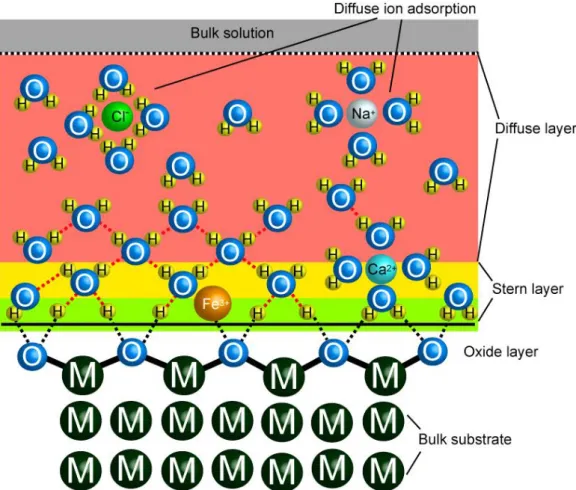
Underwater adhesion is a daunting task for organisms living in the seashores. Irregularities in salinity, abrasive wearing of the ocean waves, microbial invasions, and sharp thermal fluctuations create an environment of harsh extremes. On the other hand, continuous supply of nutrients moved from the ocean to the living zone of the organisms through the tides sustains an ecosystem inhabited by unique organisms, some of which exhibit exceptional adaptive characteristics for adhesion under highly unstable conditions. Adhesion strategies of different organisms living in these zones, such as sandcastle worm, adult barnacles and their larvae (the cyprid), and mussels have been under close examination by researchers in recent years with the purpose of translating their adhesive technology into synthetic platforms for industrial and medical applications.2-11 A natural underwater adhesive is usually synthesized and secreted onto the substrate with low initial viscosity followed by curing of the glue over the course of minutes to hours into its final hardened structure. An impressive feature of these adhesives is that they require little or no surface preparation prior to secretion.12 This capability is desirable in man-made adhesives which, so far, are incapable of functioning on highly solvated surfaces. Highly polar water molecules, dissolved ions and organic contaminants interact both with the surface and the adhesive molecules, interfering with the adhesion process (Figure 1.1).
4
Figure 1.1 Interaction of water molecules and dissolved ions with surface. Highly polar water molecules, dissolved ions and organic contaminants interact both with the surface and the adhesive molecules, interfering with the adhesion process.
5
The nature of biofunctional interface, separating an inert biomedical device from the native tissue while integrating the material into the body, is of utmost importance for the long-term efficiency of tissue regeneration. In order to achieve this, strong and biologically safe underwater synthetic adhesives, which can modulate cellular activities through biologically active signals, are required.13-15 The mainstream research in this field has largely concentrated on creating artificial cellular microenvironments by mimicking the architecture and biology of the native extracellular matrix. Toward this purpose, various polymers and self-assembled nanofibers of peptidic structures have been engineered to present desired biofunctional ligands to interfere with cellular signaling. On the other hand, immobilization of such materials onto a substrate has remained an unresolved issue. Physical and chemical conditions in the applications where these adhesives are normally used, both resemble the conditions in the intertidal zone. For example, the mechanical abrasiveness in load-bearing tissues, such as bone and cartilage, the high shear force experienced in blood vessels and the high ionic strength and polyionic environment of bodily fluids create a challenging environment for adhesives to operate efficiently. Therefore, natural underwater adhesives provide a plethora of inspiration towards developing biologically safe and reliable synthetic adhesives for medical applications.
Underwater adhesion of mussels, particularly Mytulis edulis and Mytulis californianus, has drawn escalating interest in biomimetics research.16, 17 In their natural habitat these sessile (non-motile) organisms cling themselves to underwater solid surfaces (e.g., rocks, wood, etc.) in the intertidal zones. The adhesion capacity of mussels encompasses virtually all types of surfaces, including metals, alloys,
6
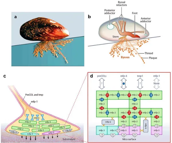
metal oxides, organic surfaces, and plastics, even polytetrafluoroethylene (TEFLON®).18, 19 Due to their extensive adhesion capacity, fouling of mussels on ship hulls and coastal infrastructure has been a growing economic concern. There is no man-made glue that can bind to such a broad variety of surfaces. The adhesion strength of mussels is one of the strongest known in natural underwater adhesives.16 In order to achieve this, mussels produce a special polyphenolic glue containing hierarchically organized proteins, known as mussel adhesive proteins (mfps), with varying content of 3,4-dihydroxy-L-phenylalanine (Dopa) residues (Figure 1.2 and Table 1.1). Spatial and temporal evolution of this phenolic residue within the wet glue precursor is believed to play an indispensible role in mussel adhesion and cohesion. The mussel adhesive unit is called a byssal thread, or byssus in plural, and contains three main functional and biochemical components: a stem embedded in the soft tissues of the mussel, a hard and flexible thread-like extension (byssal thread), and an adhesive plaque (Figure 1.2).17, 20 The whole structure is composed of different proteins called mfps. Up to now, six major proteins have been isolated and identified with different highly organized functions in adhesion process (mfp-1 to mfp-6).
The catechol side chain of Dopa can be involved in a variety of physical and chemical reaction mechanisms. For example, catechols can form exceptionally stable complexes with metals and metal oxides, thereby mediating adhesion to these surfaces. Lee et al. measured the dissociation force of a single tethered Dopa molecule from TiO2 using single-molecule atomic force microscopy (AFM).21 The dissociation force of the single Dopa-TiO2 bond (~800 pN) is about half of a covalent bond (~2000 pN) and much higher than the dissociation force of hydrogen
7
bonds that hold the DNA double helix intact (10-20 pN).21-23 In spite of the high bond strength, Dopa binding to TiO2 surface is completely reversible with thousands of break/reformation series.21 Catechol groups in mfps can also undergo covalent reactions that contribute significantly to the cohesive and water-resistant characters of the mussel glue.24, 25 Under the basic conditions of seawater (pH ~8.5), catechol is oxidized to highly reactive quinone and semiquinone species that further react with each other to covalently cross-link (cure) the mfps.19, 25 Further, Dopa-quinone was reported to covalently react with primary amine and sulfhydryl groups.21, 26
A byssal thread is composed of an extensible inner core and a hard outer shell (the cuticle).27 Typically, hardness of the cuticle is roughly five times higher than the hardness of inner core proteins.20 The inner core is formed by extracellular matrix proteins, with a central collagen core flanked by silk and elastin-like domains.28 Because of the spatial distribution of the silk- and elastin-collagen complexes in the thread, the distal portion (closer to the substrate) is typically an order of magnitude more rigid than the proximal (closer to the mussel) portion. However, the distal and proximal portions are extensible up to 109% and 200%, respectively, without breaking apart.29-31 The cuticle consists of densely packed granules, 0.8 µm in diameter, constituting ~50% of the cuticle volume.27 mfp-1 is a 108 kDa structural protein containing a unique repetitive decapeptide, and it coats the entire adhesion plaque and the distal portion of the byssus.32 Recent evidence suggests that the granules in the cuticle are formed by mfp-1 proteins densely cross-linked by ferric iron ions. mfp-1 can form reversible bis-Fe(Dopa)2 and tris-Fe(Dopa)3 complexes with iron ions in alkaline sea water.20
8
Figure 1.2 Mussel adhesion strategy inspires synthetic adhesives that can operate under water. (a) A typical mussel with its adhesive byssi. (b) A mussel adhesive organ consisting of a stem embedded in the soft tissues, a hard and flexible thread-like extension (byssal thread), and an adhesive plaque. (c, d) The thread and the adhesive pad consist of a number of proteins hierarchically assembled to maintain mechanical rigidity as well as facilitate surface adhesion (Reproduced from Ref. 17 with permission from Royal Society of Chemistry).
9
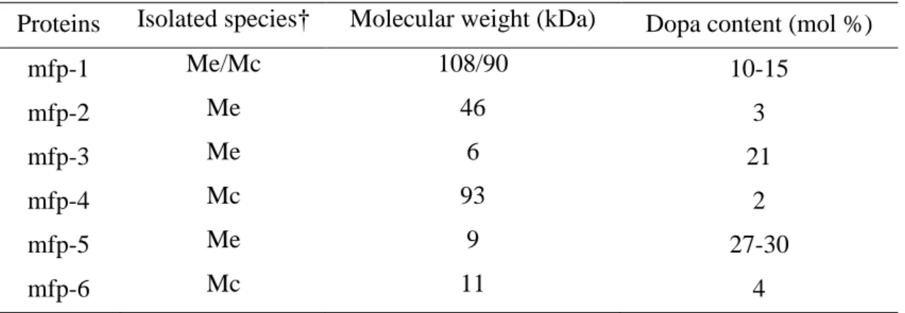
Table 1.1 Molecular weight and Dopa content of mussel adhesive proteins.8, 33-36
Proteins Isolated species† Molecular weight (kDa) Dopa content (mol %)
mfp-1 Me/Mc 108/90 10-15 mfp-2 Me 46 3 mfp-3 Me 6 21 mfp-4 Mc 93 2 mfp-5 Me 9 27-30 mfp-6 Mc 11 4
†Species are Mytilus edulis (Me) and Mytilis californianus (Mc).
These complexes have very high stability constants (log Ks ~37-40), implying that
iron ions are a critical element of mussel adhesion that endows the cuticle with both hardness and self-healing ability after fracture.37 Holten-Anderson et al. showed that removal of iron ions largely inhibits self-healing of Dopa-conjugated polymer and supramolecular networks.38 Despite its high stiffness, the cuticle is highly extensible, with ultimate tensile strain around 70%.39 Deformations up to this point are prevented from propagating by densely cross-linked granules, whereas micro tears that are formed during deformation are mended by a rapid healing process.20, 39 Adhesive plaques establish adhesion of the animal to the surface.8, 40 Proteins that are in direct contact with the substrate (mfp-3 and mfp-5) have the highest Dopa content, highlighting the significance of this phenolic residue for adhesion (Table 1.1). mfp-3 and mfp-5 both contain a high number of cationic arginine and lysine residues, respectively.17, 33 The ε-amine group of lysine is reactive with oxidized catechol (quinone), which may entail cross-linking in the mussel glue.26
10
Figure 1.3 Mussel glue: Adhesion and curing mechanisms (Reproduced from Ref. 41 with permission from Nature Publishing Group).
11
However, ex vivo studies of mussel proteins and Dopa-containing peptides have not yet confirmed such reactivity.42, 43 The current view is that the excess positive charge forms columbic interactions with surfaces that mussels bind to in their native environment, such as rocks that are rich in negatively charged silicates and aluminates.44-46 mfp-5 is also enriched in serine and phosphoserine residues, whose roles remain unknown.6 Although, mfp-3 and mfp-5 are more enriched with Dopa, the bulk of the adhesive plaque largely consists of mfp-1, mfp-2, and mfp-4. mfp-2 is known to be resistant to proteolysis and is thought to act as the stabilizer of byssus cement.40 This protein contains a large number of cysteine residues. mfp-4, on the other hand, is thought to serve as a bridge in the thread-plaque junction by linking the core collagen fibers of the distal byssus to plaque proteins, and has high levels of histidine, arginine, and lysine.47, 48 mfp-6, which was discovered in Mytilus californianus, has surprisingly low Dopa (4%) and high cysteine content (11%).35
Zhao and Waite reported the presence of 5-S-Cysteinyl Dopa, a cysteine-Dopa adduct. Therefore, this protein is believed to provide a cohesive link between the surface-coupling proteins (mfp-3 and mfp-5) and the bulk plaque proteins (mfp-2 and mfp-4).35 A very recent report, however, attributed a more fundamental role to this protein by suggesting that 6 prevents auto-oxidation of Dopa to quinone in mfp-3 before adhering onto substrate.49 Quinone formation dramatically reduces the adhesion capacity onto TiO2 and mica surfaces, by about 80%.21, 50 Thiol group on side chain of cysteine residues in mfp-6 successfully maintain Dopa by coupling oxidation of thiols to reduction of quinones.
Despite the high underwater adhesive performance of the mussel glue, the inimitable complexity and the hierarchical organization of its constituent proteins restricted the
12
practical use of this material. Even isolation of the individual mfps is a demanding task due to their labour-intensive and inefficient production yield. For instance, approximately 10,000 mussels are required to extract only 1 g of mfp-1; and the purity of this extract is not reliable due to high batch-to-batch variation.51 In order to produce this protein at large scale, E. coli and S. cerevisiae have been used,52-54 however, attempts to produce functional mfps failed mainly due to codon bias and small expression quantity.53-55 Although there has been partial success in the expression of mfp-1 repetitive sequences in S. cerevisiae53 and E. coli54, 55 using synthetic gene constructs, their adhesion profiles were found to be poor. In another study, Choi et al. took a recombinant approach by fusing domains of 1 and mfp-5 with functional groups of extracellular matrix proteins, such as RGD and YIGSR in order to create cell-friendly coatings.56
However, mfp production through recombinant production is still an unresolved challenge due to lack of the post-transcriptional modifications, including formation of Dopa by hydroxylation of Tyr residues. Limitations of obtaining high-purity and functional mfps led to alternative biomimetic approaches. Conjugation of Dopa, or the catechol group, to synthetic platforms has been the most widely recognized strategy. Dopa is chosen because not only can it adhere to a wide variety of substrates underwater, but also it has a very simple chemical structure, which can be easily grafted onto synthetic systems.25, 57, 58 Use of a synthetic backbone with well-defined chemistry, onto which Dopa can be attached, offers a more reliable platform with minimum batch-to-batch variation compared to natural adhesive proteins. Moreover, biomimetic reconstitution of mussel adhesives on synthetic platforms with additional functionalities provides a wider range of applications and development
13
strategies of novel hybrid materials with superior performance. Using this approach, catechol-conjugated poly(ethylene glycol) (PEG) was synthesized to obtain anti-fouling surfaces.59-61 Likewise, catechol-functionalized chitosan/pluronic thermo-responsive and injectable hydrogels were utilized as tissue adhesives.62 Using poly(dopamine methacrylamide-comethoxyethyl acrylate), a reversible dry/wet adhesive platform was developed by Lee et al. through combining mussel-mimetic Dopa adhesion with gecko-mimetic polydimethyl siloxane pillars.63 Dopa-modified polymers were used to functionalize not only bulk surfaces, but also surfaces of nanoparticles. For instance, binding of methoxy poly(ethylene glycol), which was grafted to hyper-branched polyethylenimine and polyDopa, onto hydrophobic nanoparticles provided stabilization in harsh biological environments.64 Oxidation-mediated grafting of catechols conferred redox activity to chitosan films.65 These films were characterized to be poor in direct electron transfer, whereas electrons can readily flow through soluble mediators. As a result of this interaction, catechol-modified chitosan films exhibited amplification, partial rectification, and switching capabilities, thereby holding promise for sensor development. At basic pH, dopamine undergoes an oxidation-triggered auto-polymerization reaction in water. Hong et al. revealed that both noncovalent assembly and covalent polymerization contribute to polydopamine formation.66 Virtually any type of surface, regardless of its chemistry, can be coated with polydopamine by simply dipping it into dopamine solution at pH ~8.5. Moreover, polydopamine coating thickness is proportional to the time of immersion and the chemical properties of this coating allow secondary modifications through coupling to nucleophilies.19, 26 Ryu et al. demonstrated that the polydopamine coating provides a general route for bone-like hydroxyapatite
14
crystallization on a surface.67 Wei et al. functionalized superparamagnetic iron oxide nanoparticles (SPIONs) with a dopamine sulfonate ligand to provide nanoparticles stability in water against pH and salinity changes in addition to enabling further functionalization with streptavidin or a maleimide dye.68 Very recently, Kang et al. demonstrated a one-step surface functionalization strategy by mixing dopamine with a diverse range of organic and inorganic species and dipping the substrate into this mixture.69
In summary, biomimetic materials field has emerged as a converging discipline to reconstitute adaptive characteristics of biological systems in synthetic platforms to solve structural and functional problems in engineering, materials science, and medicine. A high performance adhesive, stable in aqueous and saline environments but with surface versatility and environmental compatibility will find a broad range of applications in industry and medicine. Although natural underwater adhesives exhibit exceptional performance under highly abrasive conditions, the high cost of obtaining adhesives from their original natural sources promotes alternative biomimetic solutions. Because of the simplicity of catechol that allows easy grafting to synthetic materials and the versatility of substrates it could bind to, mussel-inspired surface functionalization has become a prevalent strategy. Due to its general biocompatibility and water-resistant character, the main application area of mussel-mimetic materials has been medical applications. Unique underwater adaptations of other aquatic organisms also offer potential opportunities and novel inspirations for the purpose of developing advanced functional materials. Improving their efficiency and conditions under which synthetic adhesives operate is a continuing venture to meet the ever-changing demands of industry. This process is highly analogous to the
15
evolution of natural adhesives under the force of natural selection in order to meet the ever-changing conditions of the environment.
1.1.2 The extracellular matrix: Learning from nature for biomaterial design
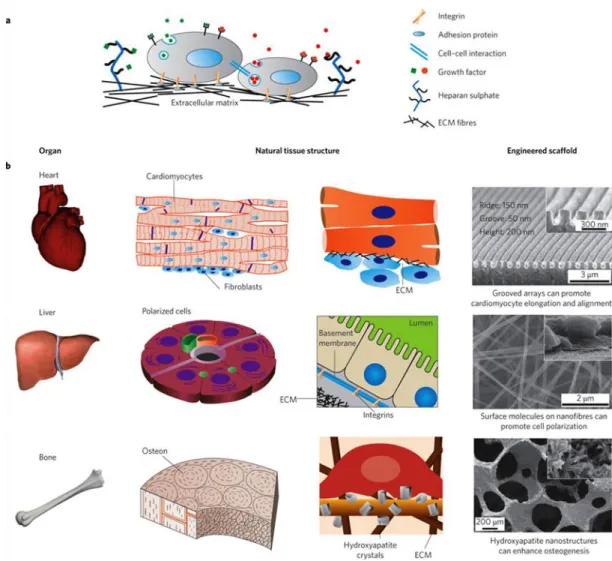
In most OECD countries, the population is ageing, which requires efficient treatment strategies for diseases related to tissue and organ malfunctions.70 Regenerative medicine is a translational research branch that aims at supporting tissue’s own healing process by providing them with appropriate physical and chemical cues. At the molecular level, these cues imitate the native extracellular matrix (ECM) by directly interacting with cell-surface receptors to guide adhesion, proliferation, migration and differentiation processes. In a native tissue, the ECM is a dense biomolecular network that surrounds cells, providing them with mechanical and bio-regulatory supports to form an organized, functional tissue (Figure 1.4). Almost every cell in the body is exposed to the ECM components.71 Osteocytes in bone, for example, are surrounded by a three-dimensional, mineralized composite matrix. Instead, the endothelial cells are exposed to two-dimensional basement membrane, which confers cell polarity. Even cells in the blood stream interact with soluble ECM proteins, such as fibronectin.72 The interaction between cells and the ECM is highly dynamic and reciprocal. Cells embedded in the ECM are able to remodel its microenvironment through enzymatic and non-enzymatic activities, depending on the type and the physiological state of the tissue. For example, the bone remodeling process, i.e., formation vs. resorption of inorganic bone composite, is under well-adjusted control of osteoblasts and osteoclasts.
16
Figure 1.4 ECM provides a physical and bioactive support to the cells forming a functional tissue and organ. (a) Cell-cell, cell-ECM and soluble factor-cell interactions collectively define the factor-cell fate. Growth factors can be sequestered in the ECM to regulate its bioactivity. (b) Hierarchically organized heart, liver and bone tissues show that ECM can take distinct nano- and micro-scale architectures that meet the physiological function. Consequently, a synthetic scaffold or cell support can be designed to mimic functionalities of the natural tissue to mimic the native microenvironment of the cells. Scanning electron micrographs of the synthetically engineered scaffolds are shown on the right hand side (Reproduced from Ref. 73 with permission from Nature Publishing Group).
17
Composition and structural organization of the ECM is also heterogeneous, varying within and between tissues (Figure 1.4).74 A typical example of the ECM of cardiac muscle tissue require elongated and aligned cell bundles that create an anisotropic syncytium.73 Therefore, aligned surface topography is a suitable biomimetic support for cardiac tissue engineering because they guide cardiomyocytes to align (Figure 1.4).
The ECM is mainly composed of two classes of biomacromolecules: fibrous proteins and glycosaminoglycans (GAGs).74 Fibrous ECM proteins, including collagen, elastin and fibronectin, constitute the structural backbone of the ECM. These proteins also contain a number of binding sites for cells and other ECM proteins. Collagens are the most abundant fibrous proteins in the ECM, forming up to 30% of the total protein mass of a multicellular animal.74 28 different types of collagens have been identified in vertebrates.75 The majority of the collagens form a triple helix, which then further assemble into fiber or network, thereby constituting the unique architecture of the ECM in a tissue.74 In the connective tissues, for example, fibrous collagens form the backbone of the interstitial tissue. On the other hand, network collagens are incorporated into the basal membrane structure. Collagen type I is the major fibrous collagen in bone, cornea, dermis, tendon and connective tissues. Synthesis and supramolecular organization of this collagen is well-understood from nano- to meso-scales, thereby providing a rich source of information for developing various biomimetic systems.76 Collagen type I is assembled into a triple helix via two α1 chains and one α2 chain. α chains are comprised from uniquely repetitive trimeric sequences. A glycine residue is required at the third position of each trimer in the form of X-Y-Gly where X is usually proline and Y is usually hydroxyproline.75 The
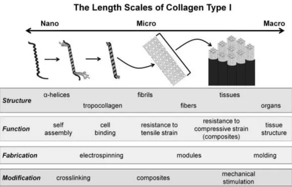
18
presence of glycine residue enables rotational freedom for helix formation. Proline and hydroxyproline residues cause kink formation, which confers steric stability for the formation of triple helix. At the flanking regions of each α chain, there are non-collagenous, i.e., non X-Y-G, regions which contain binding sites for cells and other ECM proteins (Figure 1.6).75 Preparation of functional helical collagen type I requires a number of post-translational modifications, such as hydroxylation of proline and lysine residues, glycosylation of lysine, as well as the cleavage of N- and C- terminal of the chains.75 For mechanical strength, the collagen fibers form bundles on the order of microns to centimeters.76 These bundles are then covalently cross-linked between lysine residues of the constituent collagen molecules by lysyl oxidase.77 This enzyme catalyzes aldehyde formation from amines, which further react with other aldehydes or with unmodified lysine residues to establish intermolecular covalent linkages. The degree of cross-linking can tune the mechanical properties of the ECM and hence the tissue. This provides one further level of regulation over the biological properties of the tissue.76 As such, mechanical properties have been shown to drastically regulate various cellular behaviors, such as adhesion and differentiation.78-80
Fibronectin is a multi-adhesive, fibrous ECM protein, which is secreted as a dimer linked by two disulfide bonds at the C-terminal of each chain (Figure 1.6). Each chain is 60-70 nm in length, 2-3 nm in thickness. Fibronectin mediates cell adhesion to the ECM proteins through its binding sites for collagen, heparin, and several cell-surface integrin receptors. There are over 20 isoforms of fibronectin that are generated by alternative splicing. A typical fibronectin contains repetitive sequences organized to form in three distinct domains (type I, II, or III domains).
19
Figure 1.5 Hierarchical assembly of collagen type I from nano- to meso-scales. The assembly mechanism of this protein is a source of inspiration for synthetic fibrous architectures, which finds a wide range of applications in regenerative medicine and biomineralization (Reproduced from Ref. 76 with permission from John Wiley & Sons, Inc.).
20
Figure 1.6 Schematic domains in fibronectin and collagen type I alpha 1. Both fibronectin (a) and collagen (b) contain a number of binding sites. Only one chain of the dimeric fibronectin is shown. Both chains have very similar sequences.81 Bioactive sequences on the ECM proteins inspire for developing biomimetic platforms to regulate cellular behaviors, including adhesion, proliferation, migration and differentiation.
21
Figure 1.7 Adhesion of normal rat kidney cells to fibronectin-mimetic short peptides. The synthetic peptides were investigated for their ability to promote the attachment cells by immobilizing peptides to polystyrene culture dish. The conservative substitutions of lysine for arginine, alanine for glycine, or glutamic acid for aspartic acid each resulted in abolition of the cell attachment-promoting activity characteristic of the natural sequence. In this sense, a minimal bioactive sequence can function to mimic the bioactivity of intact fibronectin protein (Reproduced from Ref. 82 with permission from the National Academy of Sciences of the U.S.A.).
22
Type III domain contains binding sites for integrins, such as the sequence Arg-Gly-Asp (RGD), which appears as a minimal sequence required for recognition by integrin receptors. In this sense, RGD can mimic the biological function of fibronectin protein despite the fact that its affinity for integrins is substantially less than that of intact fibronectin. Nonetheless, the bioactivity of this short peptide sequence and others found in both fibronectin and other ECM proteins is valuable in many dimensions. First, the ability of a short peptide sequence to mimic the function of the native ECM protein motivates for large scale production through chemical synthesis at considerably lower cost. Second, batch-to-batch variations in extracted natural ECM proteins can be eliminated with the synthetic, chemically well-defined materials. Besides, natural materials always have the risk of pathogen transmission and immunogenicity, all of which are dramatically reduced by the synthetic peptides. Presenting bioactive ligands on synthetic systems can also allow steric (geometric) control to understand of how cells recognize and respond to physical and chemical signals in their microenvironment. This will pave the way of designing more efficient synthetic supports, and hence well-defined platforms for next generation biomaterials.
The initial interaction between a cell and the ECM is towards establishing adhesion and spreading. A major route for cell-ECM interaction is mediated by integrins. Integrins are heterodimer transmembrane receptors containing α and β subunits. Following binding to an ECM ligand, integrins cluster into large focal adhesion complexes.83 Focal adhesion complex is connected to the actin cytoskeleton via vinculin proteins.84 Consequently, this complex can couple outside mechanical stress to the intracellular tension. This tension has a large impact on cell adhesion,
23
spreading and migration.72 On the other hand, a focal adhesion complex can transmit a signal from the ECM into the intracellular transduction machinery, which ultimately lead to changes in gene expression.85 The combination of integrin subunits determines which ECM components that cell can bind to.86 For example, major collagen receptors are integrin α1β1 and α2β1.87
Integrin α1β1 is particularly expressed by smooth muscle cells while α2β1 integrin is abundant in epithelial cells and platelets. Nonetheless, an integrin can bear affinities for the same ECM ligand in different tissue microenvironments. For example, α2β1 mediates cell adhesion to both fibrillar and basement membrane collagens. Many other cell types, including osteoblasts, chondrocytes and endothelial cells can express both of the receptors. Additionally, integrin-mediated signaling intersects with growth factor-mediated signaling through various levels of cross-talk.
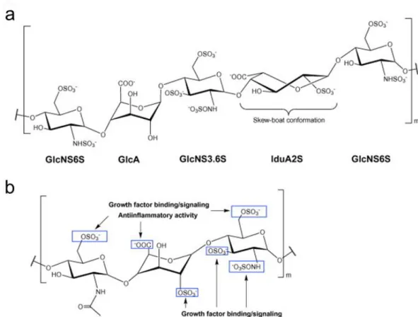
GAGs are highly polar, linear polysaccharides, which fill the extracellular space in the form of hydrogels. Depending on the molecular composition and the molecular weight, GAGs carry out wide variety of functions, including mechanical buffering, growth factor bioactivity, cell proliferation and differentiation. GAGs are found with distinct compositional and steric configurations.88 Each GAG consists of unique disaccharide repeats along with its chain length. GAGs, including chondroitin sulfate, heparin, heparan sulfate, dermatan sulfate and keratin sulfate bear dense sulfate and carboxylate groups, which can be attributed for the anionic character of these macromolecules. However, the negative charge density and position varies significantly within the disaccharide units. Hyaluronic acid is not sulfated and hence is the GAG with the least negative charge density.
24
Figure 1.8 Integrin activation and its time-based downstream signaling. In the first 0-10 min, lipid kinase activity is upregulated to elevate secondary messengers. Within several minutes, these immediate effects lead to the activation of signaling pathways for reorganization of the actin cytoskeleton. In the long term, integrin activation leads to activation of proliferation and differentiation pathways, which control the cell and tissue fate (Reproduced from Ref. 86 with permission from Cold Spring Harbor Laboratory Press).
25
Figure 1.9 Steric conformation and functional groups are inspirational sites to mimic the biological function of GAGs. (a) A specific pentasaccharide binding sequence on heparin for anti-thrombin III. (b) Important binding sites for growth factors to a heparin/heparan sulfate. Blue boxes label the position of the sulfate groups critical in growth factor binding (Reproduced from Ref. 88 with permission from Elsevier).
26
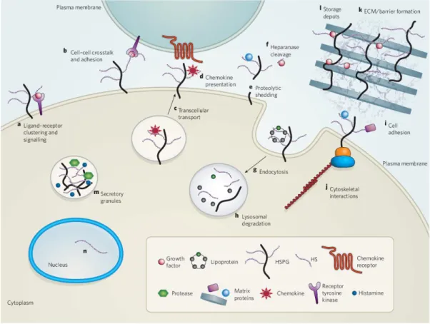
Figure 1.10 Heparan sulfate proteoglycans carry out a number of biological functions. (a, b) Heparan sulfate can act as co-receptors for growth factors and hence modulate their bioactivity. (c, d) They can also transport and modulate chemokine bioactivity by presenting them at the cell surface. (d-f) Proteolytic cleavage can liberate growth factors to modulate their bioactivity at longer distances. (g, h) Cell-surface heparan sulfates can also be internalized by endocytosis and recycled back to the cell surface. (i, j) Heparan sulfate proteoglycans can facilitate the cell adhesion to the extracellular matrix by forming a linkage between cytoskeleton and the ECM. (k, l) Heparan sulfate proteoglycans form growth factor depots and facilitate their later release following degradation (Reproduced from Ref. 89 with permission from Nature Publishing Group).
27
GAGs are found either as covalently attached to proteoglycans (e.g. heparan sulfate) or as independent macromolecules (e.g. hyaluronic acid). Many growth factors have been reported to bind to heparin and to heparan sulfate proteoglycans, such as fibroblast growth factors (FGFs) and vascular endothelial growth factors (VEGFs). Their interaction with the growth factors might be either sequence specific through certain binding domains or non-specific (Figure 1.9).88 Because these macromolecules are negatively-charged, positively-charged growth factors are attracted through electrostatic interactions.90 As a result, heparan sulfate proteoglycans act as a reservoir of growth factors by establishing stable gradients in the ECM. Such gradients of growth factors play important roles in developmental patterning and tissue heterogeneity. This binding can affect the growth factor bioactivity in two ways. First, heparan sulfate act as a cofactor of growth factor signaling, i.e., growth factor is simultaneously bind to heparan sulfate and its receptor (Figure 1.10). For example, binding of FGF to its receptor depends on its binding to heparan sulfate.89 In an alternative mechanism, growth factors can act after being released from the ECM through degradation of ECM proteins or GAGs. With this mechanism in action, GAGs can be seen as localized reservoirs for soluble growth factors that are released as soluble ligands (Figure 1.10).89
In conclusion, a complex variety of parameters create an array of interactions between cells and the ECM to orchestrate responses that have essential roles in tissue morphogenesis, homeostasis and repair.74 Such bio-regulatory parameters constitute a useful source of inspiration for guiding cellular behavior towards efficient tissue regeneration. Reconstitution of artificial microenvironments that direct cellular activities in a controlled way can provide cells with certain biofunctional cues that
28
guide cellular behaviors (e.g. adhesion, morphogenesis, viability, proliferation, migration and differentiation) for proper functioning of the regenerating tissue.
1.2 Bioinspired Design Strategies: From Supramolecular
Chemistry to Hybrid Materials
Living organisms are extremely sophisticated to be completely imitated. Organization of biological materials require both spatial (structural) and temporal (dynamic) control for the maximum functional efficiency. Despite the emergence of minimalist design principles that enables functional biomimetic material design, the ultimate efficiency of such a system is always limited by the lack a dynamic control over the system. To overcome that limitation, we need better strategies for complex material design and for structural mimicry of the biological materials. Supramolecular chemistry is a rapidly growing field of research studying complex molecular assemblies resulting from weak intermolecular forces.91 As opposed to traditional organic material synthesis, which requires covalent bond formation, synthesis of supramolecular species requires formation of noncovalent interactions, such as dispersion forces, hydrogen bond, and hydrophobic interactions. Biological systems rely heavily on such assemblies with highly stereospecific processes, such as multi-protein complexes, receptor-ligand interactions, complete viral assembly, and so forth.1, 91 Although individual strength of such forces are significantly below to that of a covalent bond, a number of them can together hold a supramolecular species intact with an exceptional stability, as nicely exemplified in the formation of amyloid plaques in Alzheimer’s disease.92, 93
29
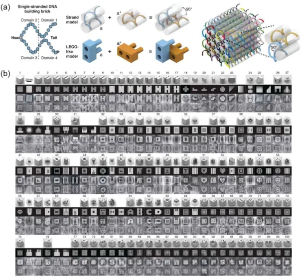
Figure 1.11 Sequence-specific information of DNA provides a vast source for programmable supramolecular materials. (a) DNA strands can be designed to share complementary domains which form larger LEGO-like bricks. (b) Bricks can be further used as the building blocks to form more complex three-dimensional structures with cavities (Reproduced from Ref. 94 with permission from American Association for the Advancement of Science).
30
In a typical supramolecular design, building blocks carry the stereospecific information, which drives the directional assembly of its components from a simple aggregation of a condensed matter to highly organized system with increasing complexity. Self-assembling building blocks that organize into well-defined supramolecular architectures can also be regarded as a programmed system. Therefore, understanding, inducing and directing the self-assembly process is key to building complex materials.95
DNA origami is a prime example of this endeavor. As the ultimate information carrier of life in the form of Watson-Crick pairing of nucleic acids, programmed assembly of DNA strands can be exploited to obtain intricate structures with remarkable spatial and temporal control (Figure 1.11).94, 96 Thus, the degree of information encrypted in the system determines the overall complexity of a material. Supramolecular polymers have recently opened a whole new avenue to introduce the supramolecular design principles into the polymer world. A supramolecular polymer can be synthesized by the directional, noncovalent assembly of monomers into one-dimensional high-aspect-ratio nanofibers.97 Therefore, an intrinsic advantage of a supramolecular polymer is their dynamic ability of depolymerization under mild conditions. This feature is commonly exploited by the living systems. For example, dynamic assembly-disassembly of long cytoskeletal structures is vital for a number of cell functions.97 Besides, noncovalent linkages can easily reform even at room temperature and atmospheric pressure, thereby imparting a self-healing character to supramolecular polymers. Aida et al. proposes two mechanisms for supramolecular polymerization: random-coil supramolecular polymers and ordered supramolecular polymers.97
31
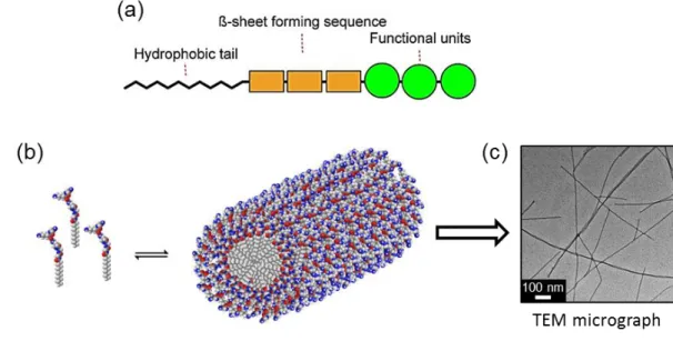
Figure 1.12 Rationally-designed modular peptide amphiphiles can dynamically assemble into long one-dimensional nanofibers. (a) Schematic of the modular design of a peptide amphiphile with basic domains of hydrophobic tail, β-sheet producing unit and the functional units. (b) (Left) Stimuli-responsive, reversible assembly of monomeric peptide amphiphiles into ultra-high-aspect-ratio nanofibers. (Right) TEM micrograph of peptide nanofibers stained with osmium tetroxide.