^ - V.;.. ·<* ί ' f ^ ■*“ ■·'■ - *'- 'İL ·* ί. ^ С 7 ‘ ·** ■ ■^' ■* ■ W · « · W ^ > <S· « м / ·> · 4 / .> w ' .'J Λ J t J •■•^ » ^ "Zf
Ğ ’Z
l O Z
Ijû S é
J B B 7
ESTABLISHMENT OF AN EXPERIMENTAL SYSTEM TO STUDY
p53 EFFECTS IN Saccharomyces cerevisiae
A THESIS
SUBMITTED TO THE DEPARTMENT OF MOLECULAR BIOLOGY
AND GENETICS AND THE INSTITUDE OF ENGINERING AND
SCIENCE OF BILKENT UNIVERSITY
IN PARTIAL FULFILLMENT OF THE REQUIREMENTS
FOR THE DEGREE OF
M ASTER OF SCIENCE
By
- ... TUBA DiNQER
J2oJi
't.'S 'ó 'I certify that I have read this thesis and that in my opinion it is fully adequate, in scope and in quality, as a thesis for the degree o f M aster o f Science.
I certify that I have read this thesis and that in my opinion it is fully adequate, in scope and in quality, as a thesis for the degree o f M aster o f Science.
P ro f Semra Kocabiyik
I certify that I have read this thesis and that in my opinion it is fully adequate, in scope and in quality, as a thesis for the degree o f M aster o f Science.
Assist. P ro f Marie Ricciardone
ABSTRACT
Establishment of an experimental system to study
p53 effects in Saccharomyces cerevisiae
Tuba Dinner
M. S. in Molecular Biology and Genetics Supervisor; Prof. Dr. Mehmet Öztürk
August 1997, 82 pages
The aim o f this vv^ork was to establish an experimental model to study human wild type and mutant p53 protein effects in yeast
Saccharomyces cerevisiae
cells.Wild type p53 was previously shown to be a DNA damage response gene that controls the genome stability in mammalian cells. We are interested in using yeast cells to study human p53-mediated cellular events after DNA damage. In this study we established an experimental model as an initial step. For construction o f this system pA K 31 plasmid expressing a human mutant p53-248W as well as control plasmid pL3 were used to obtain two different yeast cell populations, one expressing the mutant p53 248W protein and the other without p53 expression. Wild type p53 expression vector was avoided because o f its known grow th inhibitory effects in yeast cells.
In this experimental system initially p53 expression at pAK31 transformed yeast cells was shown. Then, the effect o f mutant p53-248W expression to growth rate o f yeast cells was analysed and no growth rate difference was detected between the cells expressing and non-expressing mutant p53-248W protein. To test the participation o f mutant p53-248W protein in DNA damage response in yeast cells, cells were exposed to DNA damaging agents; UVC and cisplatin that were reported to induce wild type p53 protein in mammalian cells. Codon 248 is a common site o f ‘hot spot’mutation and the arginine residue that corresponds to codon 248 encoded by the wild type p53 sequence is in the DNA interacting face o f the p53 protein. M utant p53-248 (arg->trp) is defective in specific DNA binding and it has lost the ability to act as a transcription factor. Although there are not many reports about mutant p53-248W response to DNA damaging agents, it was shown that mutant p53- 248W exhibit decreased repair o f active genes upon UV radiation. In this study cell survival was analysed in mutant p53-248W protein expressing yeast cells in parallel to mutant p53 protein levels following to DNA damage and no effect o f mutant p53 248W expression on cell survival upon DNA damage was detected. Also no difference was detected in mutant p53 protein levels following DNA damage.
ÖZET
p53 proteinin Saccharomyces cerevisiae mayasındaki etkilerini
incelemek için deney sisteminin oluşturulması
Tuba Dinçer
Yüksek Lisans Tezi,Moleküler Biyoloji ve Genetik Bölümü Tez yöneticisi: Prof. Dr. Mehmet Öztürk
Ağustos 1997, 82 Sayfa
Bu çalışmanın amacı,
Saccharomyces cerevisiae
maya hücrelerinde insan p53 proteininin ve mutant formunun etkilerini görmek için deneysel bir modelin oluşturulmasıdır.p53 proteininin DNA hasarına karşı tepki gösteren ve bu şekilde genom dengesini kontrol eden bir protein olduğu memeli hücrelerinde daha önce gösterilmişti. Biz DNA hasanna karşı insan p53 proteininin maya hücresinde gösterdiği etkilerle ilgilendik ve bunun için de ilk olarak mayada deneysel bir sistem kurduk. Bu deneysel sistemin oluşturulmasında, mutant p53-248W proteinini ekspres eden pA K 31 plasmidi ve kontrol olarakta bu proteini ekspres etmeyen pL3 plasmidi kullanıldı. Bu şekilde mutant p53 ekspres eden ve etmeyen iki farklı maya hücre populasyonu oluşturuldu. p53 proteininin hücre çoğalmasım engellediğini gösteren raporlardan dolayı çalışmalarda p53 proteini kullanılmadı. Bu sistemde ilk olarak pA K 31 vektörü ile transforme edilen maya hücrelerinde, p53 proteinininin ekspresyonu gösterildi. Daha sonra mutant p53-248W proteininin, maya hücrelerinin büyüme hızına etkisi analiz edildi ve mutant p53 ekspres eden ve etmeyen maya hücrelerinin büyüme hızında hiçbir fark görülmedi. M utant p53-248W proteininin maya hücrelerinde, DNA hasanna karşı bir tepki gösterip göstermediğini kontrol etmek için maya hücreleri DNA hasanna yol açan, UVC-ışınlanna ve cisplatine maruz bırakıldı. Memeli hücrelerinde p53 proteininin DNA zaranna tepkisi daha önceden UVC-ışınları ve cisplatinle gösterilmişti. p53 geninin aıjinin amino asidine denk gelen 248. kodonu en sık rastlanan mutasyon noktası olmasının yamsıra p53 proteininin D NA ile etkileşen kısmıdır. Mutant p53-248' in (arg->trp) DNA ile etkileşimindeki özgüllüğünden yoksun kalması dolayısıyla transkripsiyon faktörü olarak işlev görme özelliğini kaybeder. Mutant p53-248W proteininin DNA hasanna gösterdiği tepki hakkında çok fazla rapor bulunmamakla beraber, bir rapor da UVC-ışım karşısında mutant p53-248W proteininin aktif genlerin tamirini azalttığı gösterilmiştir. Bu çalışmada mutant p53-248 proteini ekspress eden maya hücrelerinin, DNA hasarı sonucunda hücrelerin canlı kalmasına olan etkisine paralel olarak, bu hücrelerde m utant p53 protein seviyeleri de incelenmiştir. DNA haşan karşısında mutant p53- 248W proteininin canlı kalan maya hücresi sayısına etkisi belirlenmediği gibi p53 protein seviyesinde de bir farklılık görülmemiştir.
ACKNOWLEDGEMENT
It is my pleasure to express my deepest gratitude to my supervisor Prof.
Dr.Mehmet Öztürk for his guidance, encouragement and invaluable efforts
throughout my thesis work. I have not only benefited from his knowledge but also
learned a lot from his superior academic character.
I would like to address my special thanks to Lütfiye Mesci, Birsen Cevher and
Esma Yolcu for sharing their experiences with me. I would also like to thank Dr.
Akbar Kuchkartayev for the plasmids pL3 and pA K 31, and yeast strain.
I would like to thank my housemate Hilal for her loveliness.
My dear friend Tolga, who shared many o f hard days with me throughout our
six years, thank you for your friendship, the life, is easier with you.
I would like to thank Burçak, Cemaliye and Reşat for making a joyful
environment and extending their helping hands whenever I needed.
My sincere thanks to my parents for their moral support and thanks to my
brother Captain Emre for his being that make the life easier and funnier. Last but not
TABLE OF CONTENTS Page
SIGNATURE PAGE
ABSTRACT
OZET
ACKNOWLEDGEMENT
TABLE OF CONTENTS
LIST OF TABLES
LIST OF FIGURES
ABBREVIATIONS
11 111 IV VI X XI Xlll1. INTODUCTION
2. GENERAL INFORMATION
2.1. p53 and cancer 2.2. Historic landmarks o f p53 2.3. Structure o f p532.3.1. p53 domains: structure-function relationship
1 2
2
3 4 42.3.2. Structural acpects o f the p53 protein in realtion to gene evolution 6
2.3.3. Highly conserved regions that coincide with the mutation clusters
o f p53 found in hum an cancers. 7
2.4. The cellular functions o f p53
2.4.1. M echanism leading to cell cycle arrest
2.4.2. Induction o f apoptosis
8 8 10
2.4.3. Transcriptional activation and repression by p53
2.4.4. Involvement o f p53 in cellular DNA replication and repair
2.4.5. The events in the p53 activation and its response
2.5. The p53 and yeast
12
14
15
1 1
3. Materials and Methods
3.1. M aterials20
20
3.1.1. Reagents 20
3.1.2. Enzymes and enzyme buffers 20
3.1.2.1. PC R enzymes 20
3.1.2.2. Restriction enzymes and buffers 20
3.1.3. M arkers 21
3.1.4. Commonly used buffers and enzymes 21
3.1.4.1. Buffers and solutions used in PCR 21
3.1.4.2. Stock solutions required for bacterial transformation 21
3.1.4.3. Stock solutions required for yeast transformation 21
3.1.4.4. Buffers and stock solutions for DNA isolation and Analysis 22
3.1.4.5. Buffers and stock solutions used for selection and testting for p53 expression
3.1.5. Commonly used media
З.1.5.1. M edia for bacteria
3.1.5.2. M edia for the yeast
24
27
27
27
3.2. Methods
3.2.1. Strains and M edia
3.2.1.1. Bacterial strains and media
28
28
28
3.2.1.2. Yeast strains and M edia 3.2.2. Plasmid constructs 3.2.3. Bacterial transformation 28 31 31 3.2.3.1. Plasmid purification 32
3.2.4. High efficiency transformation o f
S.cerevisiae,
AKY28 with LiAc 32 3.2.5. Analysis o f purified plasmids from bacteria and from genomicyeast DNA to test the state o f p53 with PC R and restriction
enzymes 33
3.2.5.1. Amplification o f 110 bp fragment o f exon 7 o f p53
cDNA with PCR 35
3.2.5.2. Purification o f PC R products 36
3.2.5.3. Analysis o f PCR amplified fragment in A garose/N uSieve agarose
gel electrophoresis 36
3.2.5.4. Restriction enzyme analysis o f PC R am plified 110-bp p53
cDNA fragment that contains mutant p53 sequence 36
3.2.6. Selection and testing for p53 expression 38
3.2.6.1. Cell lysis and protein extraction under denaturing conditions 38
3.2.6.2. Determination o f protein concentration 38
3.2.6.3. Analysis o f proteins in crude cell lysates by W estern blot 39
3.2.7. Application o fD N A damaging agents 40
3.2.7.1. UV-C treatment 40
3.2.7.2. Cisplatin treatment 41
3.2.7.3. Cell survival assay 42
4. RESULTS
4.2. W estern blot analysis o f mutant p53-248W protein expression in AKY28 cells
43
4.1. Verification o f mutant p53 sequence (codon 243) in pAK31 vector
and in pA K 31 transfected AKY28 yeast cells 46
4.3 Effect o f mutant p53-248W protein expression on growth rate o f the
yeast cells 54
4.4. Establishment o f an experimental model for p53 effects in yeast after
D NA damage 55
4.5. W estern blot analysis o f mutant p53 expression after UVC radiation o f
yeast cells 58
4.6. Effects o f mutant p53 on yeast survival upon another DNA damaging
agent cisplatin 59
4.7. W estern blot analysis o f mutant p53-248W expression in yeast cells that were grown in different concentrations o f cisplatin
containing medium 61
4.8. W estern blot analysis o f mutant p53 expression upon
UVC and cisplatin treatm ent among the AKY28 cells that were
grown in high Pi medium 62
5. D ISC U SSIO N 64
6. C O N C L U SIO N 69
7. P E R S P E C T IV E 70
8. R E F E R E N C E S 73
LIST OF TABLES
Table 1. Ingredients o f specific media
Page
29
Table 2. The sequence o f 110-bp fragment at exon 7 and localisation o f
primers on it 35
LIST OF FIGURES
Page
Figure 1. Schematic presentation o f human p53 7
Figure 2. The p53 pathway 14
Figure 3. M aps o f the plasmids, pL3 and pA K 31 45
Figure 4. Result o f the PCR amplification o f 110-bp fragment obtained from
pAK31 plasmid 46
Figure 5. M sp I and Hae III digestion o f 110-bp fragment amplified from
mutant p53 sequence (codon 248, R->W ) in PA K 31 vector 48
Figure 6. Results o f PCR amplification o f 1 10-bp fragment amplified from exon 7 o f p53 cDNA obtained from mutant p53 expressing yeast cells. 49
Figure 7. M sp I and Hae III digestion o f 110-bp fragment obtained from mutant
p53 expressing yeast cells. 50
Figure 8. Demonstration o f p53-248W protein expression in yeast using
different anti-p53 monoclonal antibodies 51
Figure 9. The results o f western blot analysis, mutant p53 expressed in low Pi
and high Pi medium 52
Fig. 10. Analysis o f p53 expression at 24“', 48“', and 72“' hr incubation in both
high Pi and Low Pi medium 53
Fig. 11. Effect o f mutant-p53 expression on grow th rate o f yeast cells 55
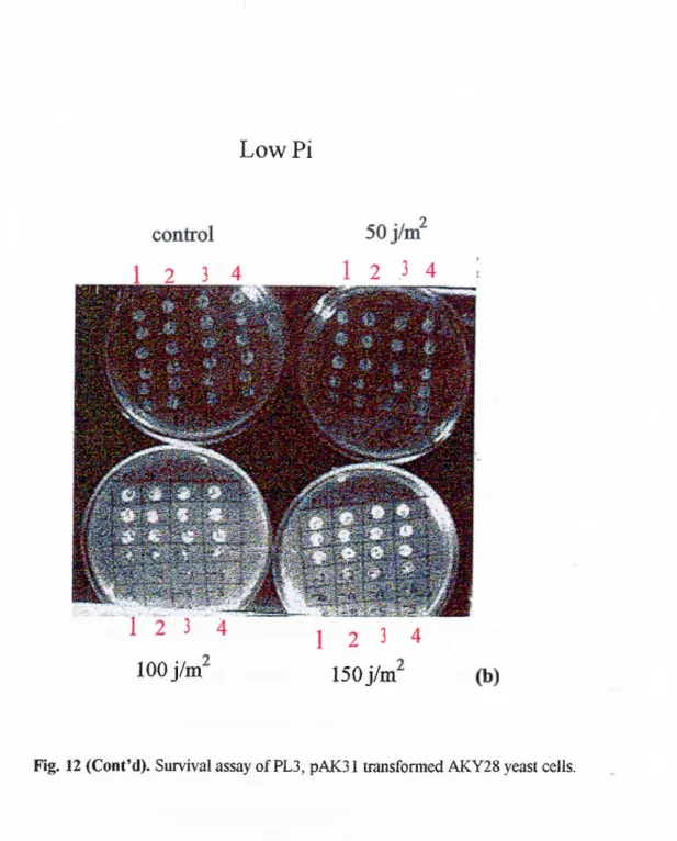
Fig. 12. Survival assay ofP L 3, pAK31 transformed AKY28 yeast cells 56
"Fig. 13. Survival curve o f mutant p53 expressing and non expressing yeast cells
in high Pi and low Pi medium upon UVC radiation 58
Fig. 14. W estern blot analysis o f m utant p53 protein at various time points (4 h,
Fig. 15. Survival assays o f mutant p53 expressing yeast cells upon different concentration o f cisplatin, 0.5 mM, 1 mM, 3 mM, and 5mM in high Pi and low
Pi medium 60
Fig. 16. Yeast cell survival curve for the mutant p53 expressing and non
expressing cells in cisplatin containing medium 61
Fig. 17. W estern blot analysis o f mutant p53 expression among the cells that
were grow n in different cisplatin concentration. 62
Fig. 18. W estern blot analysis o f the mutant p53 expression in yeast cells that
ABBREVIATIONS
bpBCIP
bisacrylamide cisplatin C-reminus cDNA dNTPs DNA ds DTT EDTA EtBr kDa LB LiAc min base pair5-bromo-4-chloro-3-indoyl phosphate disodium salt
N ’, N ’, methylene bis-acrylamide
cis-diamminechloroplatinium(II)
carboxy terminus
complementary deoxyribonucleic acid
deoxyribonucleoside triphosphate
deoxyribonucleic acid
double stranded
dithiothreitol
diaminoethane tetra-acetic acid
ethidium bromide kiloDaltons Luria-Bertani media lithium acetate minute XIII
M W molecular weight
N B T nitro blue tétrazolium chloride
N-terminus amino terminus
OD optical density
PAGE polyacrylamide gel electrophoresis
PE G polyethylene glycol
PBS phosphate buffered saline
PCI phenol: chloroform: isoamyl alcohol
PCR polymerase chain reaction
PM SF phenylmethylsulfonyl fluoride
PVDF polyvinylidene difluoride paper
Rpm revolutions per minute
SC synthetic complete media
SDS sodium dodecyl sulfate
SDS-PAGE SDS-polyacrylamide gel electrophoresis
TBE tris-boric acid-EDTA
TEM ED N, N, N ’, N ’-tetramethyl-l,2-diaminoethane
Tris Tris (hydroxymethyl-)methylamine
UV ultraviolet
1. INTRODUCTION
The p53 is a 53 kDa nuclear phosphoprotein that suppresses abnormal cell
proliferation and plays an important role for protection against cancer. p53 has been
named as “guardian o f the genome” by researchers to indicate its dictatory role in control
o f the genomic integrity. Recent studies indicate that the p53 protein is involved in gene
transcription, DNA synthesis and repair, senescence, genomic plasticity, and in
programmed cell death.
The p53 gene and protein product have become the center o f study ever since it
become clear that more than 50 % o f human cancers contain mutations in this gene.
These mutations were detected in more than 50 different cell lines and tissue types
(Hollstein
et a l,
1994). The nature o f these mutations in cancer cells is most commonly a missense mutation in one allele, producing a faulty protein that is than observed at highconcentrations in these cells. Deletions or chain termination mutations in the p53 gene
were obtained rarely. On the other hand, p53 “knockout” mice (no p53 expression or p53
-/-) were shown to be prone to different tumor development (Donehower et al., 1992).
p53 is a component o f biochemical pathways central to human carcinogenesis
(Harris, 1993). p53 protein alterations due to mostly missense mutations and rarely loss
o f p53 protein by nonsense or ffameshift mutations provide a selective advantage for
clonal expansion o f preneoplastic and neoplastic cells. There have been some suggestions
function” phenotype in addition to the abrogation o f p53 function (Dittmer et al., 1993).
p53 protein levels are mostly regulated by posttranslational mechanisms (Finlay et al.,
1988). Degradation o f p53 may involve ubiquitin-dependent proteolysis; most mutations
o f p53 disturb the degradation pathway, resulting in particularly high levels o f the mutant
protein (Scheffiier et al., 1990).
The mutational spectra at the p53 locus in different tissue types indicates a strong
role for diverse environmental mutagens with a set o f tissue preferences (Bressac et al.,
1991; H su et al., 1991; Brash et al., 1991). In addition there is strong selection for subset
mutations localised predominantly in the DNA binding domain o f p53 (Lin et al., 1995).
Thus both selection and a strong set o f environmental mutagens combine to produce
mutations in the p53 gene in human cancers.
2.GENERAL mFORMATION
2.1. p53 and cancer
Tum our suppressor genes maintain tissue homeostasis by controlling cellular
proliferation, terminal differentiation and apoptosis (Weinberg
e ta l,
1991). The p53 tum our suppressor gene has become a hot topic o f cancer research because it isprovide clues about the etiology and molecular pathogenesis o f cancer (Harris
et ah,
1993 ; Hollsteinet al.,
1991). Statistic value indicates that, o f the ~6.5 million cancer cases worldwide each year, 2.4 million tumours are estimated to contain a p53 mutation.About 50 % o f human cancers contain p53 mutations, including the cancers o f the breast,
cervix, colon, liver, prostate, bladder, and skin and these cancers are more aggressive,
more prone to metastasise, and more fatal (Harris
et al.,
1996).2.2, Historic landmarks of p53
The nuclear phosphoprotein p53 was originally discovered in extracts o f
transformed cells, reacting with antiserum from animals with tumours induced by Simian
Virus 40 (SV40) (Linzer and L evine, 1979; Lane and C raw ford, 1979). The protein was
found to form an oligomeric complex in SV40 transformed cells with the SV40 oncogene
product; the large T antigen. It was known that large T antigen is required for
maintenance o f transformed phenotype. p53 protein was also coprecipitated with E lB -
55 kDa protein that cause a transformed phenotype (Samow
et al.,
1982). At this time p53 was classified as a tum our antigen. The levels o f p53 in cells containing thosecomplexes were approximately 100 fold higher than in the non transformed cells and the
half life o f p53 is correspondingly extended from 20 min to 24 hr (Reich
et al.,
1983 ; Orenet al.,
1981). The results o f further studies have supported the hypothesis that increased levels o f p53 effect phenotype o f the normal cells; overexpression o f p53resulted in the immortalization o f rodent cells ( Jenkins et al., 1984 and Rovinski and
Benchimol,1988) and in conjunction with an activated
ras
oncogene in rodent cells, p53 3induced tumourogenic phenotype formation (Eliyahu et al., 1984, Parada e ta /., 1984).
However all o f these studies were performed with a expressing a mutant forms o f p53
(Hinds
et ah,
1989, Finlay et al., 1988, Eliyahu et al., 1988). When wild type p53 was introduced in to the cells, it suppressed the transformation o f cells in culture by otheroncogenes like
ras
(Finlayet al.,
1989). D NA screened from colon cancer patients revealed that p53 mutations occur with usually high frequency in tumour tissue (Bakeret
al.,
1989). It is frequently altered by somatic mutations in a wide variety o f human neoplasms (Hollsteinet al.,
1991). Beside spontaneous human cancers, germline p53 mutations are found at Li-Fraumeni syndrome in which affected family members developcancers at a young age (Malkin
et al.,
1990). High incidence o f tumours in p53 knockout mice is detected by six mounts o f age (Donehoweret al.,
1992). All these resultsconfirmed that p53 is a tum our suppressor gene that requires loss o f function mutations
for tum our formation but some mutant forms o f p53 exert a transdominant negative effect
on normal p53 that result in gain o f function for carcinogenesis (Milner et al., 1991,
Srivastava e /a /., 1993).
2.3. Structure of p53
2.3.1.
p53 domains: structure-function relationship
p53 is a phosphoprotein o f about 393 amino acids which can be divided
into five domains. p53 has been shown to possess a transcriptional activation function and
transcriptional activation domain has been mapped to the 1-42 amino acid residues at N-
sequence specific DNA binding domain o f p53 localised between amino acid residues
102 and 292, the central part o f the protein referred as core domain. The crystal structure
o f a complex containing the core domain and DNA has been determined at 2.2 A°
resolution (Cho
et ah,
1994). M ore than 90 % p53 o f mutations occur in this core domain and 40 % o f the missense mutations are localised to residues R175, G245, R248R249, R273, and R282 which play a role in the structural integrity o f this domain (Cho
et
al.,
1994). p53 is a nuclear p ro te in , a nuclear localisation sequence was found between amino acid residues 3 16-325.(Shaulskyet a l,
1990). The native p53 protein is a tetramer in solution. C terminal domain between amino acid residues 323 and 355 in human p53was mapped as oligomerization domain. Crystal structure o f the tetramerization domain
has been determined at 1.7 A° resolution (Jeffrey
et al.,
1995). The tetrameric form o f p53 protein that is a dimer o f a dimer, binds to four repeats o f consensus DNA sequence5’- PuPuPuC(A/T)-3’and this sequence is repeated in tw o pairs, each arranged in inverted
repeats such a s ^ <— > where is the sequence written above. This consensus
sequence was identified in the prom oter and/or an intronic regions o f the genes that are
transactivated by p53 (El- Deiry
et al.,
1992). The highly basic extreme C terminus which is connected to oligomerization domain with a flexible linker between the residues 368-387 can bind DNA sequence non-specifically and regulate non DNA binding-latent form
o f p53. This region negatively regulates p53 sequence specific DNA binding and deletion,
phosphorylation by protein kinase C or casein kinase II, binding o f antibody Pab421 to
this sequence all activate latent state o f p53 and provides its sequence specific DNA
binding (Hupp and Lane
et al.,
1994). The fifth domain defined approximately by amino acids 61-94 o f human p53 contains five repeats o f amino acid sequences PXXP (Pdesignate proline and X designate any amino acid). The fundamental importance o f
PXXP sequence, is supported by the fact that these motifs have been found in all proteins
known to bind directly to SH3 domain which have important roles in signal transduction.
A p53 cDNA deletion mutant (Apro AE) which lacks the entire fifth domain has been
shown to be completely dispensable for transcriptional activation. On the other hand a
deletion o f this p53 proline rich domain impairs p53 ability to suppress tum our cell
growth. In addition to transcriptional activation domain, prolin rich domain may play a
critical role in the transmission o f downstream signal (W alker and Levine, 1996).
Domains o f p53 protein are shmematically presented in Fig. 1.
2.3.2.
Structural aspects o f the p53 protein in relation to gene evolution
According to sequence analysis o f various p53 proteins five highly conserved
blocks were identified (Soussi
et al.,
1990). Four o f these five blocks (II to V) are found in the central domain and block I is located in the N-terminal.Starting with vertebrates, a large number o f p53 genes from different species have
been characterised. Interestingly p53 was also identified in invertebrates such as
squid
(Ishiokaet al.,
1995). Despite existence o f p53 in diverse organisms, up to now no protein homologous have been identified in lower eukaiyotes likeDrosophila
and yeast.2.3.3.
Highly conserved regions that coincide with the mutation clusters o f p53
found in human cancers.
Mutations in p53 have been shown to occur at nearly every position, with a
preferential occurrence in III, IV, V conserved blocks, like the mutations R175 found in
III*'’ block; R248, G 245 and R249 found in IV conserved block; R273 and R282 found
in V"* conserved domain (Fig 1). Those mutations are p53 somatic mutations that are
frequently seen in human tumours and cell lines (Hollstein
et al.,
1994).100 200 . m . 393
42 61 94 102
Activetion Protine rich domain Domain Mdm2 binding RMS G245 R248 R249 .R27:? R282 Oligomerization domain (323 -355) 292 316 323 355 368 387 I V
DNA binding domain
, NLS
Basic C Terminal domain (368 387) Ñon specific bNA binding DMA and RNA strand annealing Mismatch binding
NLS: Nuclear recognition sequence (3 1 6 -3 2 5 ) Evolutionary conserved regions I ~ V
Fig.1. Schematic presentation of human p53. Several hot spots for mutations R175, G245, R248, R249, R273, R282 are also Indicated
2.4. The cellular functions of p53
The high frequency o f p53 alterations in human cancers suggests that p53 play a
critical role in the control o f the growth o f normal cells. A number o f biological and
biochemical functions have been ascribed to wild type p53. Importantly mutant p53
proteins derived from human tumours are defective in some o f these functions, suggesting
that such functions like cell cycle arrest, apoptosis, transcriptional activation and
repression, D NA repair and replication are relevant for p53 mediated growth control and
tum our suppression
2.4.1. Mechanism leading to cell cycle arrest
All cells have mechanisms for coping with DNA damage. M ost eukaryotic cells
respond to DNA damage with transient delays in both G1 and G2 phases presumably to
allow for repair prior to DNA replication or division o f the cell. It was shown that tum our
suppressor p53 required for the G1 check point function in mammalian cells (Kastan
et
a l,
1992; Kuerbitzet a l,
1992). Fibroblasts from p53 nullizygous mice lack the G1 checkpoint function (Kastanet al.,
1992). Furthermore, normal human fibroblasts induce p53 and cause its accumulation in response to ionizing radiation (IR) whereas fibroblastsfrom the Ataxia talengiectesia (AT) patients are defective in this response (Kastan
et al.,
1992). This response o f p53 to DNA damage was also shown in many different cell types;cell lines having wt p53, exhibit G1 arrest following radiation, p53-null cells such as
Saos-2 cells continue to progress through S phase (Kuerbitz
et al.,
1992). Cells with mutant p53 enter S phase and amplify segments o f DNA, Li-Fraumeni syndrome cellscontaining w t p53 arrested in G1 and did not go gene amplification, the converse occur in
cells lacking w t p53 (Yin
et a l,
1995).The amount o f p53 in mouse cells increases 5-10 fold a few hours after UV
irradiation or treatm ent with the DNA damaging drug 4NQO (Maltzman and Czyzyk.
1984).
In normal conditions p53 is a labile protein. By pulse-chase experiments half life
o f p53 protein in untreated cells found to be 20 min. Agents which damage DNA induce
p53 to becom e very stable (M osner
et a l,
1995). The same group suggest that p53 is negatively auto-regulated by specifically inhibiting translation o f its own mRNA in vitro.According to studies in hepatocytes; nuclei o f untreated cells were always negative for
immune detectable p53. In contrast, nuclei o f UV damaged DNA stained positively for
p53 (Sun et al., 1995). Additionally a novel p53 promoter that contains 80 bp was
identified which is induced by genotoxic stress such as anti cancer drugs and UV (Sun
et
al.,
1995).The ability o f p53 to serve as a transcription activator suggests a mechanism for
the induction o f the damage responsive transcripts that might influence the cell cycle
progression. A strong candidate is (Nauba
et a l,
1995; El-Deiryet a l,
1993; Harperet a l,
1993), which is activated by p53 and likely to play an important role in the p53-induced D N A damage because it encodes a potent cyclin dependent kinase inhibitor.By this way p21CIPl /WAFlprevents phosphorylation o f the G1 cyclin/ CDK substrate pRB,
thereby preventing the transition from G1 phase to S phase (Slebos
e ta l,
1994).D N A damage is the triggering factor for the induction o f p53 activation. Types o f
dependent arrest mechanism can be activated by very few double strand breaks, and only
one may be sufficient. In addition to double strand break, nuclear injection o f linearized
plasmid DNA, circular DNA with a large gap or single stranded phagemid DNA is
sufficient to induce a p53 dependent cell cycle arrest (Huang
et al.,
1996).Beside G1 arrest, p53 has also been implicated in G2/M phase checkpoint. When
mitotic spindle inhibitors are added to the cells with wild type p53, the cells are blocked
in G2. In the absence o f wild type p53, the cells re-initiate DNA synthesis (Cross
et a l,
1995). I f the radiation applied before G1 checkpoint, the fibrosarcoma cells undergo G1arrest without subsequent G2 arrest and if the cells are irradiated after G1 checkpoint,
cells undergo G2 arrest without subsequent G1 arrest. Furthermore in those cell lines
which do not go radiation-induced apoptosis, the wild type p53 exhibit radioresistance in
terms o f clonogenic survival (Pellegata
et a l,
1996).2.4.2. Induction o f apoptosis
p53 initiate apoptosis if the damage to cell is severe. This protects the organism
from the growth o f damaged cells. p53 plays a role in triggering apoptosis under different
physiological conditions. Normal thymocytes will undergo apoptosis in response to DNA
damage whereas thymocytes from p53 (-/-) mice don’t undergo apoptosis in response to
same stimulus (Lowe et al., 1993). A number o f factors affect the decision o f a cell to
enter p53-mediated cell cycle arrest or apoptotic pathway; under conditions in which the
D NA is damaged, if survival factors are limiting or an activating oncogene is forcing the
cell in to a replicative cycle, p53 induced apoptosis prevails. Two o f the genes that are
and IGF-BP3 (Miyashita and Reed, 1995; Bucbinder
et ah,
1995). Bax regulate apoptosis by binding to BCL-2 that is a blocker o f apoptosis and IGF induce apoptosis by loweringthe mitogenic response o f cell. Sequence specific transcription capacity o f p53 is very
important for induction o f apoptosis like in the case o f induction o f Bax and IGF-BP3,
but beside this, it w as shown that a truncated p53 protein lacking sequence specific
transcription capacity is able to induce apoptosis in He-La cells. This result indicated the
existence o f another p53-dependent apoptotic pathway o f which occur without sequence
specific transcription function o f p53 (Haupt
et al.,
1995).2.4.3.
Transcriptional activation and repression by p53
p53 is a potential transcription factor (Fields and Jang
et a l,
1990) and once activated, it repress the transcription o f one set o f g e n e s, like c-fos, c-jun, IL-6, Rb andBcl-2. It was reported that only prom oters containing initiator elements and lacking p53
binding sites are inhibited by p53 (Mack
et al.,
1993). It stimulates the expression o f other genes that have p53 binding sites, some o f the target genes identified so far include;p21AVaf 1/CIPl which is a cyclin dependent kinase inhibitor that combine with cyclins
and PCNA and cause G1 arrest o f cell cycle (El-Deiry et al., 1993; Harper et al., 1993)
mdm-2 is a oncoprotein which is a cellular inhibitor o f the p53 in that it can bind to
transactivation domain and form an autoregulatory loop with p53 activity (Oliner
et al.,
1992), Gadd-45 is induced when cells are subjected to DNA damage leading to arrest inG1 (Kastan
et al.,
1992). It interacts with PCNA and inhibits DNA replication; cyclin G is a novel cyclin that is strongly activated in a p53-dependent manner in cells subject toDNA damage (Zauberman
et al.,
1995), bax is a member o f bcl-2 family that promotes 11apoptosis (Miyashita and Reed, 1995), IGF BPS is induced in cells after DNA damage. It
is anti-mitogenic factor (Bucbinder
et al.,
1995). Many effects o f p53, like growth arrest, apoptosis and inhibition o f carcinogenesis can be attributed to the biological functions o fits downstream genes.
2.4.4.
Involvement o f p53 in cellular DNA replication and repair
In addition to the damage-induced p53 functions, evidence for a direct
involvement o f p53 in cellular DNA replication and repair has accumulated. The products
o f two different p53 target genes, and Gadd45 has been shown to interact with
PCNA, a factor that is involved in DNA replication, both GADD45 and p21 discard
PCNA from DNA replication via binding to it (Waga
et al.,
1994; Smithet al.,
1994). These results suggest the idea that p53 involve in DNA replication regulation by inducingGADD45 and p21.
There are also evidences for a direct involvement o f p53 in cellular DNA repair.
Such an involvement can be deduced from several findings. In addition to sequence
specific DNA binding, p53 protein binds non-specifically to double stranded (ds) and
single stranded (sd) DNA (Kern
et al.,
1991). Furthermore it has been described to reanneal both RNA and D N A (Balkalkinet al.,
1994). These functions might beimportant in the processes associated with DNA repair like recognition o f DNA damage
in the form o f deletion insertion deletion mismatches (Lee
et al.,
1995) and prevention o f unscheduled recombination (Bertrandet al.,
1997). Another function o f p53 is its Mg^^ dependent 3 -5 exonuclease activity that could be mapped to the central core domain o fdependent polymerase Ô and s involved in DNA replication, recombination and
proofreading repair (Mummenbrauer
et al.,
1996).In addition, physical interaction between the human RAD 51 (homologues o f
bacterial Rec A) and w t p53 but not mt p53 was found in vivo. According to results; it
was shown that this interaction inhibit RAD 51 which catalyses three strand exchange and
ATPase activity. It was suggested that this physical interaction might play a major role in
controlling the extent and timing o f homologous recombination which is so important for
the genome stability (Sturzbecher
e ta l,
1996). Further investigations about p53 effect on homologous recombination in mammalian cells expressing a mutant p53 protein showedthat mt p53 increase the intrachrosomal homologous recombination (Bertrand
et al.,
1997).Also there are some clues about the p53 involvement in nucleotide excision repair
(NER) an important and versatile DNA repair mechanism, which is the major pathway for
repair o f UV-type lesions and damage by a variety o f important carcinogens and
mutagens. During the studies to determine the effects o f p53 on cellular sensitivity to UV
irradiation and on N ER in primary human skin fibroblasts from patients o f Li-Fraumeni
Syndrome, it was found that p53 mutant cell lines were resistant to UV-induced
cytoxicity and apoptosis but deficient in global repair (Ford
et al.,
1995). According to in vitro data, host cell reactivation experiments with a reporter plasmid that is damaged withUV, mutant p53 containing cell lines showed reduced repair o f U V induced DNA lesions
(Smith
et a l,
1995; Mckayet a l,
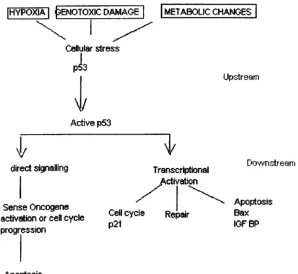
1997).The upstream events or signals flowing to p53 are mediated by several stressful
situations. In addition to DNA damage, hypoxia is also able to stimulate p53 levels and
activate p53 protein (Graeber
et al.,
1996). Another signal that activates p53 is generated when ribonucleoside triphosphate pools fall below the critical value (Linkeet al.,
1996). In the case o f p53 activation, activated p53 may induce either growth arrest or apoptosisvia transcriptional control or non-transcriptional control to maintain genomic integrity o f
cell. Figure 2 shows the major pathways leading to p53 activation and p53 mediated
cellular changes.
2.4.5. The events in the p53 activation and its response
[HYPOXIA I ^?ENOTOXIC DAMAGE | METABOLIC CHANGES
Cellular stress pia N/ Active p53
V
direct signaiiing U pstream Transcriptional Activation p21Figure 2. The p53 pathway Levine, A. J. 1997
Sense Oncogene _ ,, ,
activation or cell cycle progression Apoptosis D o w n s tre a m Apoptosis Bax IGF BP
2.5.The p53 and yeast cells
Although the term tumour suppressor connotes a function attributable only to
multicellular organism, the control o f DNA damage and cell cycle progression is critical
to all eukaryotes. Genes in which recessive mutations uncouple growth arrest signals
from cell cycle progression can be considered as yeast paradigms o f tum our suppressor
genes. For example, in
S.cerevisiae;
Rad9, R ad l7 , Rad24, Mec3, involve in recognition o f DNA damage and Pol2, Rfc5, Dpbl 1 involve in replication recognition o f replicationblocks; and the genes M ecl, Rad53 and D unl that transduce the growth arrest signals
(E lledgee/a/., 1996).
Although homologous o f p53 protein has not been identified in yeast, there are
several studies that demonstrate similarity between the effects o f wild type and mutant
p53 expression in yeast and human cells. Experiments in
S.cerevisiae
andS. pombe
with human p53 indicates that p53 can alter the growth o f lower eukaryotic cells. Accordingto studies in
S.cerevisiae,
the difference among colony growth between the cells expressing wild type human p53, mutant p53; mutant (Ala-143), mutant (His-273); andno p53 was shown. Colony growth difference was detected among the cells expressing
exogenous wild type p53, mutant p53, and no p53 (vector with or without insert).
Colonies expressing wild type p53 were considerably smaller than the control colonies.
Cells producing the Ala-143 mutant grew almost as well as the control cells, while cells
expressing the His 273 mutant exhibited an intermediate colony site. Colonies containing
vector alone (no p53) had a doubling time o f 4.4 hr. The doubling time o f cells containing
the wild type p53 was increased to 11.6 h and the doubling time o f the strains containing
the his-273 mutant was 6.5 hr. Also phase o f the cell cycle o f these arrested cells were
determined by flow cytometry. Strains containing wild type p53 exhibited G1 and G2/M
peaks. In those experiments, it was also observed that coexpression o f p53 with the
human cell cycle-regulated kinase, but not the yeast homologue CDC28, severely
inhibiting cell grow th and
S.cerevisiae
expressing both p53 and CDC2H accumulated largely in G1 phase. P53 was phosphorylated inS.cerevisiae
but differences inphosphorylation did not account for the apparent growth rate observed in various strains
containing p53 and CDC2H2 (Nigro
et al.,
1992). The same effect o f p53 to the growth o f fusion yeast;S.pombe
w as also observed (Biscoffet al.,
1992). Wild type and two mutant polypeptides His 273, His 175 were expressed to similar levels inS.pombe
and after 24 hr expression, although wild type p53 causedS.pombe
to stop dividing, tw o mutant forms continued to divide slowly compared to non p53 expressing cells. Inmammalian cells, p53 is predominantly a nuclear protein (Shaulsky et al., 1990). The p53
cellular localisation was also studied in
S.pombe
and it was shown that wild type protein was localised to the nucleus, the mutant polypeptides exhibited more extranuclearstaining than wild type p53 did. p53 is highly phosphorylated in mammalian cells. It was
shown that wild type p53, when expressed in
S.pombe,
was phosphorylated on serine 315 and serine 392 by casein kinase II and cdc2 kinase. It was shown that phosphorylation atSer-315 and Ser-392 doesn’t effect p53 growth inhibitory function in
S.pombe.
Also anS.pombe
strain was constructed to identfy dominant mutant alleles o f p53. Severaldifferent mutant alleles o f human p53 were introduced to wild type p53 expressing
His-175 that has been shown dominant to the wild type p53 in mammalian cells, were also
detected as dominant in this assay (Biscoff et al., 1992).
p53 is a transcription activator ( Fields and Jang
et al.,
1990). It was shown that mammalian wild type p53 expressed inS.cerevisiae
w as able to activate transcription o f a reporter gene under the control o f a C Y C l hybrid prom oter containing the 33 base pairp53 binding sequence whereas three mutants commonly found in human tumours, 175H,
248W, 273H were unable to activate transcription (Scharer
et al.,
1992). Based on this function continuity o f p53 in yeast, assays have been developed to detect p53 mutations.Dominant negative p53 mutations w ere selected by yeast transcriptional assay, using
URA3 as a reporter gene under the A D H l prom oter that contain p53 consensus DNA
binding sequence. According to this assay dominant-negative p53 mutations that suppress
wild type p53 transcription activity were screened according to their U ra' Foa^ phenotype
( Brachmann
et al.,
1996). Also the effect o f p53 mutations in the carboxy terminal to the transcription activity o f p53 was investigated using yeast transcriptional activity (Ishiokaet al.,
1995). For detection o f p53 mutations in blood samples, in tumours and in cell lines, yeast transcription assay was developed (Flamanet al.,
1995).During the studies to understand the mechanism o f p53 transcription activity
regulation in yeast
S. cerevisiae,
mutants were generated by EMS and UV mutagenesis that are defective for p53 transcription activity. To study sequence specific transcriptionactivity (SST),
S cerevisiae
was transformed by tw o vector, one o f them was p53 ' expressing plasmid and the other contained a reporter gene under the control o f URA3promoter that had specific p53 DNA binding site. Following cDNA screening to find a
gene that complements SST p53 activity in yeast, PA K l gene was identified. It was found
that all these mutants were complemented by PAKI gene, which is a ser/thr kinase.
Expression o f PAKI was associated with an increased specific activity o f p53 in DNA
binding assay and corresponding increased in transactivation (Thiagalingam
et a l,
1995). In an attempt to identify the p53 like a protein in the yeastS.cerevisiae,
a mutant yeast cell that require wild type p53 for its viability was isolated. The mutant, rft-1, isdefective in cell cycle progression and arrests before mitosis in the deficiency o f p53.
According to genetic and biochemical studies, it was shown that p53 complement the
activity o f rft-1 by forming a protein-protein complex (Koerte
et al.,
1995).Although there are homologues o f mammalian genes in yeast involved in DNA
damage and checkpoints like the
S.cerevisiae
genes; M E C l, T E L İ, POL2 which are the homologues o f human genes; ATR, ATM, Pole respectively, there is no homologue o fanother cell cycle control p53 gene (Elledge, 1996). But expression o f wild type p53
affects yeast cell growth (Nigro et al., 1992). Additionally p53 conserve its sequence
specific transcription activity in yeast (Shrarer et al., 1992) and a yeast gene that
complement this activity was found (Thiagalingam et al., 1995). All these results
encourage the further studies to investigate other p53 effects in yeast.
p53 response to DNA damage in yeast has not been studied yet. According to
studies in mammalian cells upon DNA damage, p53 is activated, it is stabilised,
accumulated in the nucleus and induce downstream genes to keep genomic stability.
In this study an experimental system was established to study p53 response in
yeast
S.cerevisiae
upon DNA damage. Considering the difficulties to use wild type p53 due to it’s growth inhibitory effect inS.cerevisiae
(Nigro et al., 1992), mutant p53 was preferred to establish this experimental system. As mutant, R248 (C C G -^TG G ) which isone o f the most common p53 mutation detected in human tumours (Cho et al., 1994) was
preferred. One o f the DNA damaging agents used in this study was UVC (254 nm); the
amount o f p53 in mouse cells increases 5-10 fold a few hour after U V radiation
(Maltzman and Czyzk, 1984). UV induced nuclear accumulation o f p53 was also
detected in primary human fibroblasts (Yamaizumi and Sugano, 1994). Beside p53
accumulation upon UV type DNA damage, it’s involvement in repair o f U V type DNA
damage was also shown (Smith et al., 1995).
In studies about p53. It was shown that p53 response might differ according the
status o f DNA damaging agents (Zhang
et al.,
1996, Vaseyet al.,
1996). To confirm the p53 effects upon DNA damage in yeast another DNA damaging agent was used. In thiscase as direct DNA damaging agent cis-diamminedichloroplatinium(II) (cisplatin) was
preferred. Cisplatin is an important anticancer drug which destroys tum our cells by
forming covalent adducts on DNA, blocking polymerases, and preventing replication,
transcription and cell division (Bruhn
et al.,
1990). It was shown that treatm ent o f mouse teratosarcoma cells with this chemotherapeutic agent led to an increase o f p53, 3 h afteraddition o f the drug (Küpper
et al.,
1994). InS. cerevisiae
cisplatin was also used as a DNA damaging agent to identify specific protein that bind cisplatin-DNA adducts (M cA’Nulty andLippard, 1996).
In this study an experimental system was developed to study the effects o f wild
type and mutant p53 protein following DNA damage.
3. M aterials and Methods
3.1. Materials
3.1.1. Reagents
All o f the chemicals obtained SIGMA except cisplatin that was obtained from
BELLON RHONE-POULENC RORER.
3.1.2 Enzymes and enzyme buffers:
S. 1.2.1. PCR enzyme
5u/|il Ampi-Taq DNA polymerase (PERKIN ELMER)
3.1.2. 2. Restriction Enzymes and buffers
25 u/|il M spl (STRATAGENE)lOx Assay Buffer #2 (For M sp l) (STRATAGENE)
10 u/pl Hae III (BOEHRINGER MANHEIM)
<1)х174/ H inf I digested 0.5 ng/ml (MBI Fermentas)
100 bp DNA ladder 0.13 pg/pl (PROMEGA)
3.1.4. Commonly used buffers and stock solutions
3.1.4. 1. Büßers and solutions used in PCR
lOx PCR bufFer-MgClz (PERKIN ELMER)
25 mM M gCb solution (PERKIN ELMER)
5 u/pl Ampi-Taq DNA polymerase (PERKIN ELMER)
10 mM dNTPs mix (MBI)
3.1.4.2. Stock solutions required fo r bacterial tranßormation
1 M CaClT^stock solution.54 g СаСЬ.бНзО was dissolved in 200 ml o f dH20. The solution was
filter sterilised using 0.22 pm filter sterilization unit (COSTAR).
3.1.4.3. Stock solutions required fo r yeast transformation
1.0 M LiAc stock solution. pH 8.4-8.910.2 g o f lithium acetate was dissolved in 100-ml water, pH is adjusted to
8.4-S.9.
The solution was filter sterilised with 0.22 pm filter sterilization unit. Solution was
stored at room temperature. 3 .1 . 3. Markers
50 g PEG 3350 was weighed out and transferred to graduated beaker, water
was added until the level ju st below the 100 ml mark, solution was stirred with
magnetic stirrer until PEG dissolved. Dissolved solution was transferred in to the
measuring cylinder and the volume w as completed to 100 ml. It was filter sterilised
with 0.22 pm filter unit and stored at R T in a tightly sealed container.
100 mM LiaC solution
500 pi o f 1.0-M stock solution was diluted in to 4.5 ml o f sterile water.
PEG/LiAc solution (40 % PEG, 100 mM Li Ac)
500 pi o f 1.0 M LiAc stock solution in 500 pi o f sterile water and 4.0
ml PEG 50 % (w/v) stock solution. Solution was vortexed vigorously for proper
mixing.
PEG stock solution. 50 % (w M
3.1.4.4. Buffers and stock solutionsfor DNA isolation and Analysis.
3.1.4.4.1. Buffer for isolation o f yeast genomic DNABreaking Buffer 2 % Triton X-100 1 % SDS 100 m M N aC l 10 mM Tris.Cl pH 8.0 1 mM EDTA.
3 M Sodium acetateCl Lt)
408-g sodium acetate.3 H2O dissolved in H2O, pH is adjusted to 5.2 with 3M
Acetic acid.
3.I.4.4.2. Buffers and solutions used for analysis o fD N A
TBE electrophoresis buffer lOx stock soln. (1 Lt)
108 g Tris Base
55 g Boric acid
40 ml 0.5 EDTA, pH 8.0
0 .5 M E D T A n Lt)
186.1 g Na2EDTA.2H20 was dissolved in 700 ml dH20, pH was
adjusted to 8.0 with 10 M NaOH (~50 ml), volume was completed to 1 Lt.
EtBr 10 mg/ml
0.2 g ethidium bromide was dissolved in 20 ml H2O and stored in dark.
Loading buffer 6x (stored at 4°C)
0.25 % Bromophenol Blue
0.25 % xylene cyanol
40 % sucrose in w ater
3.1.4.5.1. Buffers used for cell lysis and protein extraction under denaturing
conditions
Laemni Ivsis buffer
50 mM Tris.Cl pH: 6.8 (1.0 M Tris.Cl pH:6.8)
100 mM DTT (1 M stock solution)
2 % SDS ( 10 % SDS stock solution)
20 % Glycerol (v/v)
5 mM EDTA ( 0.5 M EDTA, pH; 6.8)
1 mM PMSF ( 1 0 mM PMSF ) Additionally added as protease inhibitor.
3.1.4.5.2. Solutions used for SDS-PAGE
10 % polvacrvamide resolving gel (40 ml)
15.9 ml deionized water
13.3 ml 30 % Acryamide mix
10 ml 1.5 M I r is (pH:8.8)
0.4 ml 10 % Ammonium per sulfate.
0.0016 ml TEMED
3.1.4.5. Buffers and stock solutions used fo r selection an d testing f o r p 5 3 expression
5 % stacking polvacrvamide gel (10 ml)
6.8 ml deionized water.
1.7 ml 30 % Acryamide mix
1.25 ml l.O M T ris. Cl pH: 6.8
0.1 ml 10% SDS
Tris-Glvcine electrophoresis buffer (1 Lt)
25 mM Tris
250 mM Glycine pH: 8.3
0 .1 % S D S
2x SDS gel-loading buffer (Store at 20° C)
100 mM Tris-Cl (pH 6.8)
200 mM DDT
4 % SDS
0.2 % bromophenol blue
20 % Glycerol
3.1.4.5.3. Buffers for W estern blot analysis.
Transfer buffer for W estern blot (1 Lt)
5.82 g Tris-base
3.75 ml 10 % SDS
200 ml methanol
2.93 g Glycine
0 .0 1 m l T E M E D
Ponceau S solution HOxl (10 ml)
0.5 g Poncue S was dissolved in 1-ml glacial acetic acid and volume was
adjusted to 10 ml
Blocking solution:
5%(w/v) non fat dry milk powder
0.2 % Tween-20 in PBS
Washing solution
0.3 % Tween 20 (v/v) in PBS
PBS lOx stock solution (1 Lt) pH 7.3
SO gN aCl
2 g K C l
l l .5 g N a2HPO4.7H2O
2 g KH2PO4
Chromogenic visualisation solution:
NBT stock solution (stored at 4°C)
0.5 g o f NBT dissolved in 10 ml o f 70 % dimethylforamide.
BCIP stock solution (stored at 4®C)
0.5 g BCIP dissolved in 10 ml 100 % dimethylformamide
Alkaline phosphatase buffer:
lOOmM Tris.Cl pH 9.5
100 m M N aCl
3.1.5.1. Media for bacteria
LB media (1 Lt) 10 gr Trptone 5 gr Bactoyeast extract lO g rN aC I LB-Amp media100 jig/ml Amphicillin in LB media
3.1.5.2 Media fo r yeast
YPD (rich medial (1 Lt)
10 g yeast extract
20 g peptone
20 g dextrose
20-g bactoagar was added for solid media.
SC media (1 Ltl pH: 5.6-6.0
6.7 g yeast nitrogen base (without amino acids)
20 g glucose
20 g bacto agar for solid media 3.1.5. Commonly used media
3.2 Methods
3.2.1. Strains and Media
3.2.1.1. Bacterial strains and media
E.coli
strain D H 5 a;F /endAl h sd R ll (r^mk) supE44 thi-1 recAl gyrA
(Ναι) relA l A(lacIZYA argF) U159 deopR (φ80άΙαοΔ91αοΖ)ΜΙ5)
was used forbacterial transformation assay. D H 5a were grown in standard LB media.
Transformants were selected and grown in LB-Amp (100 pg/ml amphicillin) media.
3.1.1.2. Yeast strains and media
S.cerevisiae
strain AKY28{Mata, leu 2-2,112, his 3-11, 15, pho3)
which is a derivative o f GRF18, was obtained from Dr. Akbar Kuchkartaev (Bilkent University,Department o f M olecular Biology and Genetics, Ankara, Turkey). M ata is the
mating type locus where a mating type genes, M ata l and M ata2 are expressed
(McKay and M anney 1974). leu 2-3, 112 is the non-reverting mutant allele o f LEU2
that express P-isopropylmalate dehydrogenase (Hsu et al., 1982). his3-l 1, 15 is the
non-reverting allele o f the HIS3 that express imidazoleglycerol-P dehydratase
(Struhl, 1982). pho3 is the mutant form o f the PH 03 that encodes acid phosphatase
that is involved in thiamine biosynthesis (Nosaka, 1990). AKY28 strain was grown
in rich YPD media.
Transformed AKY28 cells containing p53-expressing plasmids were selected
gene expression. p53 cD NA was located under the PH 05 inducible promoter.
Several genes encode acid phospatase in a repressible form but the primary gene
product is derived from the P H 0 5 gene. P H 0 5 gene encodes a secreted acid
phosphotase whose transcription is repressed when yeasts are grown in high
concentration o f inorganic phosphate (Bajwa et al., 1984). It is induced up to ~ 1000
fold in response to Pi starvation (Oshima
eta l.,
1982).The Pi o f normal synthetic SC media is 11.2 mM (1500 mg/liter) is sufficent
for complete repression . A Pi o f 0.22 mM (20 mg/ml) is the lowest initial Pi
consistent with a reasonable growth rate and yield (O’ Connell
et ah,
1992).High Pi and low Pi medium were prepared for repression and induction o f the
P H 0 5 promoter. SC media can also be used as high Pi media.
For high Pi and low Pi medium preparation, all the ingredients o f SC media
were used except bacto-yeast nitrogen base, which contains vitamins, trace elements
and salts. To obtain high Pi and low Pi medium vitamins, trace elements, and salts
were prepared separately and filter sterilized with 0.22 pm filter (Costar).
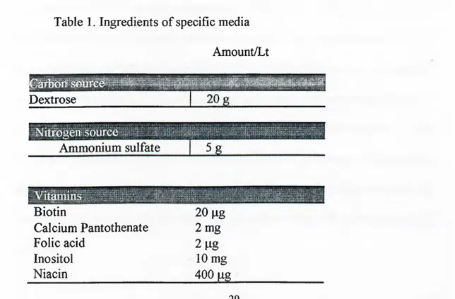
Table 1. Ingredients o f specific media
Amount/Lt Dextrose Biotin Calcium Pantothenate Folic acid Inositol Niacin 20 pg 2 mg 2 lig 10 mg 400 pg 29
p-Aminobenzoic acid 200 pg Pyridoxine hydrochloride 400 pg Riboflavin 200 pg Thiamine hydrochloride 400 pg 500 pg Copper sulfate 40 pg Potassium iodide 100 pg Ferric chloride 200 pg M anganese sulfate 400 pg Sodium molybdate 200 pg Zinc sulfate 400 pg
Potassium phosphate monobasic Magnesium sulfate Calcium chloride Sodium chloride r^Dow!iPi;s 1500 mg 20 mg 500 mg 500 mg 100 mg 100 mg 100 mg 100 mg Potassium chloride 1500 tng
This synthetic m edium is based on m edia described b y W ickersham and is marketed as ‘Y east nitrogen base w ithout am ino a cid s’ (Fink and Guthrie, 1991a).
Vitamins and trace elements were stored at 4" C. Salts were stored at room
temperature. AKY28 is a histine auxotroph. The specific medium also consists
histidine amino acid. lOOx stock solution o f histidine with a concentration o f 50
pg/ml was prepared and sterilised using 0.22 pm filter sterilisation unit and stock
solution was stored at 4 °C. During the medium preparation, ammonium sulfate,
distilled w ater and agar added for solid medium were autoclaved together at 122°C
for 30 Min. Vitamins, trace elements, glucose, salts and amino acid histidine were
added separately to the autoclaved ammonium sulfate and distilled water under the
hood. Final solutions named as either high Pi solution or low Pi solutions were also
All the plasmid constructs were obtained from Dr. Akbar Kuchcartaev
(Bilkent university. Department o f M olecular Biology and Genetics, Ankara,
Turkey). Two plasmid constructs were used. pL3 is the construct that was used as a
control; it doesn’t contain p53 cDNA, its size is 7.76 kb. pAK31 has human mutant
p53-248W cDNA and its size is 9.37 kb (Kuchkartayev, A. and Ozturk, M.,
unpublished)
pL3 is 2 |im circle based episomal plasm id that has 20-40 copy number per
cell. pAK 31 was derived from pL3 by insertion o f m utant p53-248W cDNA
between the P H 0 5 promoter and terminator. Plasmids contain an ampicillin
resistance gene as bacterial selectable m arker and LEU2 gene as yeast selectable
marker. LEU2 is one o f the most commonly used yeast selectable marker that
complement leu 2-3,112 double frameshift mutation . The maps o f the plasmids were
shown in section 4 (Results) figure 3.
3.2.3. Bacterial transformation
E. coli D H 5 a was transformed with plasm ids pL3 and pAK31, using calcium
chloride method (Sambrook et al., 1989a). To prepare the competent cells, a single
colony was obtained from plate that was freshly grow n o/n at 37°C. Then it was
inoculated to 20 ml
LB
and grown until itsODeoo
value became 0.3-0.4. The culture was kept on ice for 10 min. Cells were collected by centrifugation at 4800 rpm for 8min. The pellet was resuspended in 1 ml ice-cold 0.1 M CaCb and 200 pi o f 3.2.2. Plasmid constructs
competent cells were transferred in to eppendorf tubes. After plasmid DNA addition,
they were incubated on ice for 40 min. For heat shock, they were transferred to a
42°C
circulating water bath for 60 sec. They were then transferred to ice for 1-2 min. LB was then added and the cells were incubated for 1 hr in a 37°C w ater bath. 100 piwas taken from eppendorf tubes and spreaded on to LB-Amp plates to select cells
that were transformed with plasmids; pL3 and pA K 31. They were incubated at 37°C
for 18 hr in incubator until the colonies become visible.
3.2.3. IPlasmid purification
Wizard Plus midiprep DNA purification system ( PROMEGA)was used for
plasmids purification from pL3 and pA K 31 transformed D H 5 a cells.
3.2.4. H igh efficiency tra n sfo rm a tio n of
S.cerevisiae,
A K Y 28 w ith LiAc.AKY28 was transformed with pL3 and рА К ЗІ using highly efficient LiAc
transformation. (Gietz, D. R., and Woods, R. A. 1994). Strain AKY28 was grown
overnight at 30°C in a shaking rotary incubator and 20 pi o f this o/n culture was
added to 20 ml o f YPD (1/100 dilution) then grown in a shaking incubator until its
OD value at 600 nm became 0.3-0.4. OD values were detected by spectrophotometer
fixed wavelength program. (Du 640 spectrophotometer, BECKMAN). Cells were
harvested by centrifugation at 1500 g for 5 min. Collected cell pellets were washed in
20-ml sterile water. After second centrifugation, cells w ere resuspended in 1.0-ml
sterile dHjO water and transferred to eppendorf tubes and recentrifuged at top speed
for 10 sec. Collected cell pellets in eppendorf tubes were resuspended in 300 pi o f
vortexing briefly, 50-60 |al cell suspension were distributed to eppendorf tubes and
300 pi PEG/Li solution (40 % PEG, 100 mM Li Ac) were added to each tubes.
Incubation o f transformation eppendorf tubes in a waterbath at 30"C for 30 min was
followed by heat shock at 42°C for 20 min. PEG/LiAc solution was removed by
microcentrifugation at high speed for 10 sec. The cells w ere washed by 1.0 ml o f
sterile dH2 0 and diluted 100 times. 100 pi samples o f dilutions were spreaded on to
the SC selective medium without amino acids. Plates were incubated for 3-4 days at
30°C in incubator. After transformed yeast strain were obtained, immediately their
glycerol stocks were prepared. For glycerol stock preparation 1 ml o f transformed
yeast cells were grown in selective media (SC) until saturation or late log phase, and
1 ml 30 % glycerol were mixed with the saturated cell suspension. Stocks w ere
stored at -80" C.
3.2.5.
Analysis of purified plasmids from bacteria and from genomic
yeast DNA to test the state of p53 with PCR and restriction enzymes.
p53 mutant-248W containing plasmids purified from plasmid transform ed
bacteria D H 5a and AKY28 yeast cells that were transformed with those plasm ids
were analysed by PCR and restriction enzymes to confirm the state o f p53.
For analysis o f p53 state from pL3 (without p53 cDNA insert) and
pAK31(p53 mt-248W p53 cDNA in se rt) transformed AKY28 cells, total yeast
genomic DNA was isolated from transformed yeast cells (Ausubel et al., 1987b). For
total yeast genomic DNA isolation, 2-ml culture o f AKY28 cells carrying the
plasmids pL3 and pA K 31 were grown to stationary phase in SC media, the liquid
should be visibly turbid and pellet o f the cells should have accumulated at the bottom
o f the tube. 1.5 ml o f the saturated culture were transferred in to a microcentrifuge
tube and centrifuged 5 sec at room temperature. The supernatant was poured o ff and
cells w ere resuspended in 200 pi breaking buffer. The crystal beads were added just
below the meniscus o f the cell suspension and the mixture o f beads and cell
suspension were mixed by vortexing at max speed for 60 sec, tubes was placed on
ice for 1-2 min on ice. This step was repeated three times. Then 200-pl o f 25:24:1
phenol/chloroform/isoamyl alcohol (PCI) was added and tubes were mixed by
vortexing at max speed like previously done. The DNA containing mixture was
centrifuged at max speed for 5 min at room temperature. The upper aqueous phase
was transferred to another tube and one more 200 pi PCI was added and mixed by
vigorous vortexing for further purification from proteins and solute molecules. For
phase separation the tubes were centrifuged at max speed for 1 min at room
temperature. The top aqueous phase containing DNA was transferred to another
eppendorf tube containing 100 % ethanol that contain 1/10 vol o f 3 M sodium acetate
(If 200 pi D NA aqueous phase was collected, 20 pi from 3 M sodium acetate was
added). M ixture o f DNA aqueous phase, 100 % ethanol and sodium acetate were
mixed by vortexing and kept at -20° C for at least 30 min for precipitation o f DNA.
After precipitation step the eppendorf tubes were centrifuged at max speed for 10
min w hich was followed by aspiration o f 100 % ethanol, either with pipetting or
pouring off. For washing step 1-ml o f 70 % ethanol was added. Tubes were inverted
several tim es and spinned in microcentrifuge tubes. Supernatant was aspirated
carefully by pipetting. The pellet was dried under the lamp. After the ethanol
completely removed, the pellet was dissolved in 40-pl dd H2O. The concentration o f
obtained D N A was detected using the DNA/oligo quant, double strand DNA
To test the state o f p53 at D H 5a purified plasmids and plasmid transformed
AKY28 yeast cells, 110 bp fragment that coiresponds to exon 7 o f mutant p53-248W
sequence was amplified with PCR (PERKIN ELMER Gene Amp PCR system 2400).
3.2.5.1. Amplification o f 110 bp fragment o f exon 7 ofp53 cDNA with PCR
To analyse purified plasmids and transformed AKY28, 110-bp fragment o f
exon 7 o f the p53 cDNA sequence was amplified with PCR.
Table 2.The sequence o f 110-bp fragment at exon 7 and localisation o f
primers on it.
F3
ÏCATCCA CTACAACTAC
ATGTGTAACA GTTCCTGCAT GGGCGGCATG AACCGGAGGC
CCATCCTCAC cagg
R3
Primers F3 (5’ -GTTGGCTCTGACTGT ACC A C -3) and R3 (5’ -
CTGGAGTCTTCCAGTGTGAT- 3 ) were used. The primers were synthesised at
Department o f Molecular Biology and Genetics o f Bilkent university with
oligosynthesizer (OLIGO lOOOM BECKMAN). For 50 pi PCR solution; 5 pi 1 Ox
PCR buffer- MgCla, 1.5 mM MgCl2(25 mM stock solution), 200 pm dNTP mix (10
mM stock solution), 25 ng plasmid DNA, for amplification from yeast DNA, 100 ng
total yeast DNA, 10 pmole from each primer, 1 unit Taq polymerase (5 u/pl) were
used. PCR was performed by using Perkin Elmer Gene Amp PCR system 2400. PCR
was programmed as; 30 cycle including; 95**C 30 sec for dénaturation, 55 °C 30 sec