Original Article
Ozone (03)-oxygen mixture therapy
inhibits endometrial implant growth
Lebriz Hale Aktun1, Mustafa Acet1, Remzi Atilgan2, Nilay Karaca3, Betul Yorgunlar1, Behzat Can2, Adile Ferda Dagli2, Huseyin Fatih Gul2, Banu Kumbak1, Arzu İrban1
1Medipol University, Istanbul, Turkey; 2Firat University, Mezreh, Elazığ, Turkey; 3Bezmi Alem University, Istanbul,
Turkey
Received December 12, 2015; Accepted April 9, 2016; Epub June 15, 2016; Published June 30, 2016
Abstract: The aim of this study was to investigate potential therapeutic efficiency of ozone therapy in the treatment of experimental endometriosis in rats. Fifteen rats were divided into three groups, which were labeled as the (1) sham control, (2) the ozone (treated with intraperitoneal ozone-oxygen mixture) and (3) the GnRH-agonist (given single dose (1 mg) leuprolide acetate depot formulation) group. Endometrial implant activity of superoxide dis-mutase (SOD), malondialdehyde (MDA), interleukin-1β (IL-1β), IL-6, tumor necrosis factor-α (TNF-α), and vasculary endothelial growth factor (VEGF) were measured after ozone-therapy. Furthermore, peritoneal fluid activity of SOD, MDA, and TNF-α were also measured before and after ozone-therapy. Serum AMH levels of the rats those were given ozone-therapy and control groups were measured. The rats given ozone-therapy showed significantly reduced endometriotic implant volumes. After ozone-therapy, a significant increase in activity of SOD in peritoneal fluid was detected. Conversely, implant levels of SOD in rats given ozone therapy was found to be significantly decreased. Both peritoneal fluid and implant levels of MDA were significantly decreased after ozone-therapy. Implant levels of TNF-α, IL-1β, and IL-6 were significantly increased following ozone-therapy. VEGF levels of implant was found to be unchanged after ozone-therapy. Serum AMH levels of animals were given ozone-therapy and control groups were similar. The number of both primordial and preantral follicles were significantly decreased after ozone-therapy. However, the number of atretic follicles were similar in ozone-therapy and control groups. Repeated administration of ozone-oxygen therapy in non-toxic doses inhibits growth of endometrial implants.
Keywords: Endometriosis, ozone, oxidative stress, rat Introduction
Although, ozone therapy has found different fields of application in medical practise, poten-tial toxicity is a major factor preventing wide-spread use of it [1]. It has been reported that ozone is a pro-drug and does not need to recep-tor for its biological action [2].
Ozone therapy prevents oxidative stress-relat-ed cell damage by inducing adaptive mecha-nisms [3]. In agreement with studies reported that low dose ozone therapy supported an oxi-dative preconditioning by inhibiting cellular damage induced by reactive oxygen species (ROS) [4, 5]. In nontoxic doses, ozon leads to regulation of the biochemical pathways with the production of several critical precursors [3]. Accordingly, some antioxidant enzymes which
could be regulated by low dose ozone therapy support the possible impact of ozone in many pathological conditions including endometrio- sis.
Presence of an ozone-like mediator during inflammation has been reported [6]. By causing oxidative stress, excessive inflammation may lead to the pathologic condition to continue. Given that endometriosis is a disorder associ-ated with inflammation and oxidative stress [7, 8], we have postulated that intraperitoneal ozone treatment may protect antioxidant sys-tems and regulates the physiological concen-trations of inflammatuar substances inside the peritoneal implants. In this study, possible impacts of ozone therapy on rat peritneal endo-metriosis have been investigated by using his-tomorphological and biochemical markers.
Materials and methods
This study was carried out in the Experimental Research Laboratory of the Firat University Faculty of Medicine, complying with the approv-al of the ethic committee, the guidelines for care and use of experimental animals. Twenty one adult female Wistar rats each weighing between 200 and 240 g were supplied from Firat University Animal Laboratory. All rats were examined by a veterinarian and determined to be in good health conditions. The rats were housed in plastic cages and kept under stan-dard conditions: 12-h light and 12-h dark peri-ods, 20°C constant temperatures and a humid-ity ranging from 40 to 60%. The rats had free access to standard dry pellets ad libitum and tap water until the end of the study.
Considering the effect of menstrual cycle and estradiol that is known to be antioxidant, the two subcutaneous injections of estradiol that is known to be antioxidant all rats were hormon-ally synchronized before surgery in their 4 day estrus phase to exclude the differences in the steroid synthesis, cell adhesion and growth in implanted tissues. Synchronization was real-ized two subcutaneous injections of estradiol within 24 h intermission, followed by one injec-tion of progesterone 20 h after the last estra-diol injection. Daily vaginal smears of the rats were taken to establish the estrous cycle of each animal. Rats observed for at least two successive 4-day estrous cycles. Endometriosis was induced surgically by using the method described by previously [9, 10] during estrus. From the uterine horn, a 5 × 5 × 1 mm piece was excised by microscissors that was attached onto the peritoneum only on the right side of the ventral abdominal wall close to an artery by using the surgical auto transplantation tech-nique. After 3 weeks their daily vaginal smears were monitored and a second laparotomy was performed in their estrous phase to determine the attachment and viability of endometriotic implants. The pretreatment implant volumes in each group were calculated by measuring their dimensions (length, width and height, in milli-meters). For volume calculation the ellipsoid volume formula (π/6 × length × width × height) was used. Superficial peritoneal attachment of implants and non-rigid nature of the rat perito-neum allowed the measurment of implant’s length, width, and height without resection. Rats with volume calculation is not possible
due to heavy invasion of peritoneal implants were excluded from the study.
Of the 21 experimental rats, 2 rats died after operation and 4 rats developed abscess at implantation site and therefore these were excluded from the study. Afterwards, 15 rats were put into 3 groups of 5 rats by using ran-dom number tables as the sham control (group 1), the ozone-oxygen mixture (group 2), and the gonadotropin releasing hormone-agonist (GnRH-agonist, group 3). For the sham group, 4-0 nylon sutures, with or without fat tissues, were attached to the peritoneum except the auto transplantation of endometriotic implants. The rats in group 2 were treated with ozone-oxygen mixture (1 mg/kg body weight per rat, intra-peritoneally) every other day for a period of ten days. As a result, animals had received treatment for five days. The rate of ozone at the mixture is 3% percent. The rats in group 3 exposed to single dose leuprolide acetate depot formulation (1 mg/kg body weight per rat, s.c, Lucrin Depot-3M®; Abbott, Cedex, Istanbul, Turkey). This dose was determined based on a previous study in which 1 mg/kg leuprolide acetate was found to be optimal for female rats [11]. It has not been stated in any comments by the manufacturers whether oz- one has any effect on the estrous cycle. Therefore, daily vaginal smears were monitored during the treatment period and after the per-manence of the estrous cycle was confirmed, a third laparotomy was performed while the rats were fixed in the supine position. The volumes of the implants were measured again with the same method by the same researchers who were blinded to the groups. The cardiac blood samples were obtained to evaluate AMH levels. The endometrial implants and ovaries were then excised and processed for histological and biochemical studies. Peritoneal fluids of animals were collected before and after treat-ment for evaluating the biochemical markers. The endometriosis was diagnosed according to the histologic identification of endometrial glandular tissue and stroma.
Formalin-fixed endometriotic implants were embedded in paraffin, cut into 5-mm-thick sec-tions, and stained with hematoxylin and eosin. the endometriosis was diagnosed according to the histologic identification of endometrial grandular tissue and stroma. Likewise, forma-lin-fixed ovarian tissues were exhaustively
sec-tioned at a thickness of 5 mm using a micro-tome perpendicular to the long axis of the ovary. At least five slices were selected from the each ovary using systematic random sam-pling rules. Sections representing the largest two dimensional profile of each slice were selected for the follicle counting. The number of the follicles at each section were counted as previously described [12].
The follicles were classified into five types based on the classification of Mazaud [13]. In the light microscopic examination of ovary sec-tions belonging to all groups, follicle classifica-tion was carried out according to characteris-tics noted below:
Primordial follicle: Oocyte was surrounded either partially or completely by granulose pro-genitor cells.
Primary follicle: Follicle, in which a single layer of cubic granulose cells was observed around oocytes.
Antral (secondary) follicle: Follicle, in which oocyte was covered with more than two layers of granulosa cells and in which antrum forma-tion commenced.
Tertiary (Graafian) follicle: Follicle that possess-es a single and big space (antrum), in which a decreasing number of granulosa cells surround an antrum full of follicular fluid, and that oocyte surrounded by granulosa cells.
Atretic antral: Degenerated oocyte along with pycnosis in granulosa cells, low number of pyc-notic granulosa cells, and hypertrophic theca interna. For each rat, the total number of atretic follicles was calculated as decribed by Zhang [14]. A method by Pederson and Peders for determining the total follicle number was used to determine the possible impact of ozone ther-apy on follicular environment of the ovary [15]. Biochemical analysis was performed on blood samples, peritoneal fluid, and endrometrial implants. Samples were stored at -80°C until assay. The biochemist was blinded to the sam-ples. In order to evaluate the possible prooxi-dant-antioxidant activity of ozone gas, we mea-sured the endometrial implant levels of SOD, MDA, IL-1β, IL-6, TNF-α, and VEGF after ozone treatment. Furthemore, we have also measured the peritoneal fluid activity of SOD, MDA, and
TNF-α before and after ozone therapy. At the end of study in order to determine possible harmful effect of ozone therapy on ovarian fol-licles serum levels of AMH were also measured in all groups.
After centrifugation of both endometrial implant and peritoneal fluid with an equal volume of an ethanol/chloroform mixture (5:3, volume per volume [v/v]) at 5000 × g for 30 min, the clear upper layer (the ethanol phase) was taken and used in the SOD activity assay. All preparation procedures were performed at 4°C. Total (Cu-Zn and Mn) SOD (EC 1.15.1.1) activity was determined according to the method of Sun [16].
Malondialdehyde (MDA) levels in endometrial implants and peritoneal fluid were determined by the method of Ohkawa [17] which is based on the reaction with thiobarbituric acid (TBA) at 90-100°C. In the TBA test reaction, MDA or MDA-like substances and TBA react with the production of a pink pigment having an absorp-tion maximum at 532 nm. The results were expressed according to a standard graphic, which was prepared from a standard solution (1, 1, 3, 3-tetramethoxypropane).
Interleukin-1 beta (IL-1β) levels in endometriot-ic implant were determined with an enzyme-linked immunosorbent assay (ELISA) (Rat IL-1β ELISA-Catalog Number EKO393, USA); which could measure IL-1β in serum with a detection limit of 3 pg/ml. The standart curve range and lower detection limit for this kit is 3.12-2000 pg/ml and 1 pg/ml respectively. Both the intra- and inter-assay coefficients of variation (CV) were <10%.
Interleukin-6 levels in endometrial implant were determined with an enzyme-linked immu-nosorbent assay (ELISA) (Rat IL-6 ELISA-Catalog Number EKO412, USA); which could measure IL-6 in serum with a detection limit of 62 pg/ml. The standart curve range and lower detection limit for this kit is 62.5-4000 pg/ml and 5 pg/ ml respectively.
TNF-α ELISA analysis on the endometriotic tis-sue and peritoneal fluid was carried out in duplicate in a blinded fashion using commer-cially available ELISA kit (Rat TNF-α ELISA- Catalog number EKO526, Boster USA). The standart curve range and lower detection limit
for this kit is 7, 8-1000 pg/ml and 1 pg/ml respectively. The intra- and inter-assay CV were <5% and <10% respectively. IL-1β, IL-6 and, TNF-α concentration was measured from ab- sorbance of each well was read at 450 nm using with an auto-analyzer (ELX800). Back- ground absorbency of blank wells was subtract-ed from the standards and unknowns prior to determination of sample concentrations. VEGF levels in endometrial implant were deter-mined with an enzyme-linked immunosorbent assay (ELISA) (Rat IL-6 ELISA-Catalog Number EKO540, USA); which could measure VEGF in serum with a detection limit is 15 pg/ml. The standart curve range and lower detection limit for this kit is 15.6-1000 pg/ml and 1 pg/ml respectively. TNF-α concentration was mea-sured from absorbance of each well was re- ad at 450 nm using with an auto-analyzer (ELX800).
Serum levels of AMH were determined with an enzyme-linked immunosorbent assay (ELISA) (Rat IL-6 ELISA-Catalog Number E-EL-R0640, USA); which could measure VEGF in serum. The standart curve range and lower detection limit for this kit is 0.16-10 ng/ml and 0.1 ng/ml respectively. AMH concentration was measured from absorbance of each well was read at 450 nm using with an auto-analyzer (ELX800). Both the intra- and inter-assay CV were <10%. Statistical analysis
Data analysis was performed by using SPSS for Windows, version 11.5 (SPSS Inc., Chicago, IL, USA). Normality of distributions of continuous variables were determined by Shapiro Wilk test. Levene test was used for the evaluation of homogeneity of variances. In addition to pre- and post-treatment implant volumes SOD, MDA, IL-1β, IL-6, TNF-α, and VEGF levels were compared by Kruskal Wallis test. A p value smaller than 0.05 was considered statistically significant. Data are presented as mean ± SD. For all multiple comparisons, the Bonferroni Correction was applied for controlling Type I error.
Results
Rats given ozone therapy showed significantly reduced endometriotic implant volumes (P = 0.034) (Figure 1A, 1B). After ozone therapy, a significant increase in activity of SOD in perito-neal fluid was detected. Conversely, implant levels of SOD in rats given ozone therapy was found to be significantly decreased (P<0.028). Both peritoneal fluid and implant levels of MDA were significantly decreased in rats given ozone (Tables 1 and 2). Implant levels of TNF-α, IL-1β, and IL-6 were significantly increased follo- wing ozone therapy (P<0.009, P<0.009, and P<0.009 respectively). VEGF levels of implant was found to be unchanged after ozone therapy (P<0.175). Serum AMH levels of animals were given ozone therapy and control groups were similar. The number of both primordial and pre-antral follicles were significantly decreased after ozone therapy (Table 3; Figure 2A, 2B) however, the number of atretic follicles were similar in ozone therapy and control groups. When compared to lucrin group TNF-α, IL-1β, IL-6, and MDA levels of endometrial implants in rats given ozone therapy significantly de- Figure 1. A. Well defined endometriotic implant in
GnRH agonist group (arrow head). B. Endometriotic implant with defective epithelium after ozone treat-ment (arrowhead) (H&E × 200).
creased. Conversely, when compared to ozone group VEGF and SOD levels of endometrial implants in rats treated with lucrin significantly increased. Serum levels of AMH in both lucrin and ozone groups were similar (P<0.251). In the lucrin treated animals MDA levels of endometrial implants were significantly decre- ased (P<0.009). SOD levels of implants in lucrin group were found to be unchanged (P<0.754). In contrast to ozone group, implant levels of VEGF significantly increased after lucrin treatment (P<0.028). When compared to control group implant levels of TNF-α and IL-6 in lucrin group were similar (P<0.251, P<0.117 respectively). IL-1β levels in the endometrial implants of rats treated with lucrin were de- creased significantly (P<0.009). Similar to ozone group, serum AMH levels of rats given lucrin were not changed significantly (P<0.175).
In the lucrin group while peritoneal fluid levels of MDA decreased (P<0.029) SOD levels did not change (P<0.754). Peritoneal fluid levels of TNF-α in both ozone and lucrin groups increased significantly (P<0.016, P<0.009, respectively). The number of both primary and antral follicles were significantly decreased after lucrin treat-ment (P<0.034, P<0.036 respectively). Similar to ozone group, the number of atretic follicles were similar in rats given lucrin. There were no significant difference between ozone and lucrin groups in terms of follicle numbers.
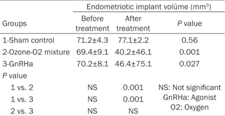
The mean pretreatment volume of endometri-otic implants in the sham control, ozone, and GnRH-agonist groups were found to be 71.2± 4.3, 69.4±9.1, 70.2±8.1 respectively. There were no statistically significant differences among the groups in regard to pretreatment volumes of endometriotic implants. The post-treatment volumes of endometriotic implants in the control, ozone and GnRH-agonist groups were noted to be 77.1±2.2, 40.2±46.1, 46.4± 75.1 respectively. There were significant differ-ences in the percentage of decline between pretreatment and post-treatment volumes of endometriotic implants in the ozone and lucrin groups (Table 4).
Discussion
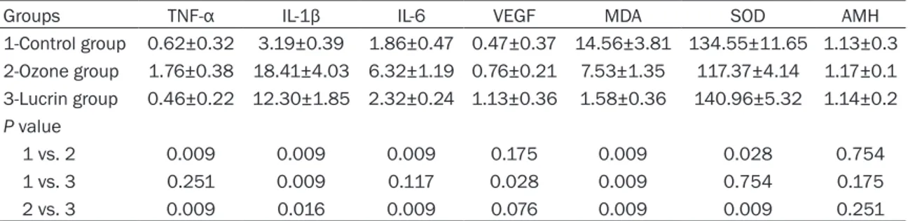
Preliminary results of our study have clearly demonstrated that ozone-O2 mixture therapy has a therapeutic potential in the treatment of peritoneal endometriosis. Concordantly, we detected a significant reduction in the volume of endometriotic implants after ozone-O2 treat-ment. Although this mixture leads to significant decline in the number of both primordial and preantral follicles the number of atretic follicles unchanged. Likewise, serum levels of AMH in Table 1. The concentration of TNF-α, IL-1β, IL-6, VEGF, MDA, and SOD in endometriotic implants after ozone therapy
Groups TNF-α IL-1β IL-6 VEGF MDA SOD AMH
1-Control group 0.62±0.32 3.19±0.39 1.86±0.47 0.47±0.37 14.56±3.81 134.55±11.65 1.13±0.3 2-Ozone group 1.76±0.38 18.41±4.03 6.32±1.19 0.76±0.21 7.53±1.35 117.37±4.14 1.17±0.1 3-Lucrin group 0.46±0.22 12.30±1.85 2.32±0.24 1.13±0.36 1.58±0.36 140.96±5.32 1.14±0.2 P value 1 vs. 2 0.009 0.009 0.009 0.175 0.009 0.028 0.754 1 vs. 3 0.251 0.009 0.117 0.028 0.009 0.754 0.175 2 vs. 3 0.009 0.016 0.009 0.076 0.009 0.009 0.251
The values are presented as mean and standard deviation. P value was significant at <0.05.
Table 2. Peritoneal fluid TNF-α, MDA and SOD levels before and after ozone therapy
Groups treatmentBefore After ozone therapy valueP Control group TNF-α 10.24±1.79 16.72±1.40 0.024 MDA 2.47±0.13 1.48±0.09 0.030 SOD 164.89±6.03 251.19±15.58 0.034 Ozone group TNF-α 17.05±4.58 38.87±2.56 0.016 MDA 2.46±0.25 1.46±0.13 0.028 SOD 257.98±19.46 327.23±15.57 0.009 Lucrin group TNF-α 17.64±3.68 36.29±4.30 0.009 MDA 2.59±0.27 1.50±0.91 0.029 SOD 277.60±17.46 255.85±26.59 0.754
The values are presented as mean and standard deviation. P value was significant at <0.05.
animals were given ozone therapy and control groups were similar. Collectively, we can strong-ly propose that inhibitor effect of ozon therapy on endometrial implant growth occured with minimal ovarian side effect.
ROS [18]. Elevated ROS levels may stimulate the expression of some gene and gene prod-ucts exert an oxidative activity in endometriotic implant cells. This might lead to increase in implant survival. Ozone mediated oxidative pre-conditioning might improve a moderate oxida-tive stress which, in turn, increases antioxidant enzymes protecting against further growth of endometrial implants. As a consequence, en- suring the redox homeostasis [18] inside the implant cells, ozone therapy may lead to in- crease in the expression of antioxidants en- zymes that may prevent ROS-mediated induc-tion of implant growing.
Imbalance in oxidant/antioxidant status in endometriotic implants has been noted [19]. It has been reported a significant suppression of peritoneal fluid antioxidant levels in women with endometriosis [20]. Concordantly, perito-neal fluid of women with endometriosis contain significantly lower levels of SOD [21] and in close aggrement, we also noted a significant increase in the activity of SOD in peritoneal fluid after ozon therapy. Conversely, implant lev-els of SOD in rats given ozone therapy was found to be significantly decreased. Different concentration of SOD in both peritoneal fluid and inside implant might seem paradox. De- creased SOD levels inside the endometrial implants after ozone therapy may be secondary to ozone-related oxidative preconditioning. Acc- ordingly, a great majority of SOD in endometrial implant might be used for removal or neutral-ization of oxidant product including MDA. In the present study both peritoneal fluid and implant levels of MDA were significantly de- creased after ozone therapy. Concordantly, it Table 3. Comparison of the mean follicle number of control,
ozone therapy and lucrin groups (mean ± SD)
Groups Primordialfollicle Primary follicle Preantral follicle follicleAntral Atretic follicle Corpus luteum 1-Control 11.8±2.4 6.4±1.8 9.2±1.4 7.8±2.5 3.4±1.1 14.0±3.8 2-Ozone 6.4±1.3 6.4±4.0 5.4±2.1 7.4±3.3 3.8±1.9 9.8±4.9 3-Lucrin 7.0±4.3 3.2±1.9 6.8±2.1 3.8±1.9 2.6±0.8 19.0±4.2 P value 1 vs. 2 0.008 0.916 0.025 0.916 0.914 0.173 1 vs. 3 0.171 0.034 0.055 0.036 0.228 0.093 2 vs. 3 0.916 0.207 0.454 0.085 0.228 0.024
The values are presented as mean and standard deviation. P value was signifi-cant at <0.05.
Figure 2. A. Decreased primordial (arrowhead) and preantral follicles (arrow) in ozone given animals. B. Unchanged corpus luteum number after ozone treat-ment (arrow heads) (H&E × 20).
Possible mechanisms of action of ozone therap on implant gr- owing is unclear. It is most likely that, ozone-O2 mixture therapy prevented oxidative stress, nor-malized levels of MDA and acti-vated the peritoneal fluid SOD levels. Another mechanism of action of ozone treatment on impant growing might be phar-macodynamic and superoxide scavenger properties of this pro drug. Although ROS can origi-nate from different sources a great number of intracellular im- pacts are mediated by different
has been reported that low doze ozone therapy improves an oxidative preconditioning by enhancing of antioxidant enzymes [4, 5].
For continued growth and survival of endome-triotic implant abnormal inflammatory reaction catalyzed by nuclear transcriptional factor kappa B (NF-kB) is required. Relatedly, the clas-sic NF-κB pathway is induced by TNF-α and IL-1β [22, 23]. In the present study ozone thera-py leads to increase in TNF-α, IL-1β, and IL-6 expression in the endometriotic implants. Mo- reover, peritoneal fluid TNF-α levels increased significantly in ozone given animals. Although the endometrial implants shrink, cytokine lev-els within the implants increase and that requires detailed explanation. Accordingly, mo- lecular mechanisms responsible for the in- creased cytokine levels after ozone therapy may be related to the administered dose of ozone. It is well known that high dose ozone-O2 mixture therapy induces severe oxidative stress by activation of NF-κB end up with an inflamma-tory response [24]. In contrast, administration of low ozone therapy stimulates mild oxidative stress which activates the nuclear factor ery-throid 2-related factor 2 as well as SOD [24]. Although we have not measured the endome-trial implant levels of NF kB, our results are more consistent with the last hypothesis. To- gether, our findings suggest that ozone therapy might inhibit the progression of endometriotic implants by regulating the oxidant-antioxidant balance within the implant cells.
Decline in the numbers of both primordial and preantral follicles after ozone therapy may be secondary to potential toxicity of ozone on
the possible impact of ozone on the ovarian fol-licles must be clarified.
Although some biological effects of ozone ther-apy in vivo are likely mediated by blood factors, a direct cellular impact cannot be ignored, especially when ozone is administered by insuf-flation in a tissue cavity. Relatedly, intraperito-neal insufflation of the ozone may prevent the growth of endometriotic implants. Furthermore, intrauterine ozone insufflation may be tried in order to restorate endometriosis-associated implantation defects.
This is the first study evaluating the effect of ozone therapy on implant growing in experi-mental endometriosis model. In this study, we have demonstrated that endometrial implants and peritoneal fluid of rats with endometriosis exhibit increased oxidative stress. By inducing the activation of endogenous antioxidant ca- pacity ozone-O2 mixture therapy have provid- ed a significant regression of endometriotic implants.
Acknowledgements
We acknowledge the help of Firat University Animal Laboratory.
Disclosure of conflict of interest None.
Address correspondence to: Lebriz Hale Aktun, Istanbul Medipol University, TEM Avrupa Otoyolu Göztepe Çıkışı No: 1, Bagcilar, Istanbul 34214, Turkey. Tel: +90-532-291 91 96; Fax: +90-212-460 70 70; E-mail: [email protected]
Table 4. Comparison of endometriotic implant volumes between the control and treatment groups
Endometriotic implant volüme (mm3)
Groups treatmentBefore treatmentAfter P value
1-Sham control 71.2±4.3 77.1±2.2 0.56 2-Ozone-O2 mixture 69.4±9.1 40.2±46.1 0.001 3-GnRHa 70.2±8.1 46.4±75.1 0.027 P value 1 vs. 2 NS 0.001 NS: Not significant GnRHa: Agonist O2: Oxygen 1 vs. 3 NS 0.001 2 vs. 3 NS NS
The values are presented as mean and standard deviation. P value was significant at <0.05.
ovary. Nevertheless, atretic follicle numbers in rats given ozone were not changed significantly. Furthermore, the number of both primary and antral follicles were significantly decreased after lucrin treatment. Taken together, decreased follicle number in ozone and lucrin given animals might be related with follicle counting methods. When reviewing the literature we did not find any information in terms of ozone gas and ovarian follicle number. Unchanged serum AMH levels after ozone therapy supports this idea. In order to recommend to use of ozone gas in the treatment of endometriosis
11597 Int J Clin Exp Med 2016;9(6):11590-11597 References
[1] Bocci V. Is it true that ozone is always toxic? The end of a dogma. Toxicol Appl Pharmacol 2006; 216: 493-504.
[2] Re L, Mawsouf MN, Menéndez S, León OS, Sánchez GM, Hernández F. Ozone therapy: clinical and basic evidence of its therapeutic potential. Arch Med Res 2008; 39: 17-26. [3] Bocci V. Does ozone therapy normalize the
cel-lular redox balance? Implications for therapy of human immunodeficiency virus infection and several other diseases. Med Hypotheses 1996; 46: 150-154.
[4] León OS, Menéndez S, Merino N, Castillo R, Sam S, Pérez L, Cruz E, Bocci V. Ozone oxida-tive preconditioning: a protection against cel-lular damage by free radicals. Mediat Inflamm 1998; 7: 289-294.
[5] Peralta C, León OS, Xaus C, Prats N, Jalil EC, Planell ES, Puig-Parellada P, Gelpí E, Roselló-Catafau J. Protective effect of ozone treatment on the injury associated with hepatic ischemia-reperfusion: antioxidant-prooxidant balance. Free Radic Res 1999; 31: 191-196.
[6] Wentworth P Jr, McDunn JE, Wentworth AD, Takeuchi C, Nieva J, Jones T. Evidence for anti-body-catalyzed ozone formation in bacterial killing and inflammation. Science 2002; 298: 2195-2199.
[7] Celik O, Celik E, Turkcuoglu I, Yilmaz E, Ulas M, Simsek Y, Karaer A, Celik N, Aydin NE, Ozerol I, Unlu C. Surgical removal of endometrioma de-creases the NF-κB1 (p50/105) and NF-κB p65 (Rel A) expression in the eutopic endometrium during the implantation window. Reprod Sci 2013; 20: 762-70.
[8] Celik O, Hascalik S, Elter K, Tagluk ME, Gurates B, Aydin NE. Combating endometriosis by blocking proteasome and nuclear factor-kap-paB pathways. Hum Reprod 2008; 23: 2458-65.
[9] Vernon MW, Wilson EA. Studies on the surgical induction of endometriosis in the rat. Fertil Steril 1985; 44: 684-94.
[10] Celik O, Hascalik S, Elter K, Tagluk ME, Gurates B, Aydin NE. Combating endometriosis by blocking proteasome and nuclear factor-kap-paB pathways. Hum Reprod 2008; 23: 2458-65.11
[11] Altintas D, Kokcu A, Tosun M, Cetinkaya MB, Kandemir B. Comparison of the effects of ce-trorelix, a GnRH antagonist, and leuprolide, a GnRH agonist, on experimental endometriosis. J Obstet Gynaecol Res 2008; 34: 1014-9.
[12] Smith BJ, Plowchalk DR, Sipes IG. Comparison of random and serial sections in assessment of ovarian toxicity. Reprod Toxicol 1991; 5: 379 [13] Mazaud S, Guigon CJ, Lozach A. Establishment
of the reproductive function and transient fer-tility of female rats lacking primordial follicle stock after fetal gamma-irradiation. Endocri- nology 2002; 143: 4775-87.
[14] Zhang H, Zhang X, Yuan Z. Germ cell loss in-duced by 12C6+ ion irradiation in young fe-male mice. J Radiat Res 2006; 47: 131-4. [15] Pedersen T, Peters H. Proposal for a
classifica-tion of oocytes and follicles in the mouse ovary. J Reprod Fertil 1968; 17: 555.
[16] Sun Y, Oberley LW, Li Y. A simple method for clinical assay of superoxide dismutase. Clin Chem 1988; 34: 497-500.
[17] Ohkawa H, Ohishi N, Yagi K. Assay for lipid per-oxides in animal tissues by thiobarbituric acid reaction. Anal Biochem 1979; 95: 351-358. [18] Droge W. Free radicals in the physiological
con-trol of cell function. Physiol Rev 2002; 82: 47-95.
[19] Wang Y, Sharma RK, Falcone T, Goldberg J, Agarwal A. Importance of reactive oxygen spe-cies in the peritoneal fluid of women with endo-metriosis or idiopathic infertility. Fertil Steril 1997; 68: 826-30.
[20] Liu Y, Luo L, Zhao H. Levels of lipid peroxides and superoxide dismutase in peritoneal fluid of patients with endometriosis. J Tongji Med Univ 2001; 21: 166-7.
[21] Szczepańska M, Koźlik J, Skrzypczak J, Mikołajczyk M. Oxidative stress may be a piece in the endometriosis puzzle. Fertil Steril 2003; 79: 1288-93.
[22] Laird SM, Tuckerman EM, Cork BA, Li TC. Expression of nuclear factor κB in human en-dometrium; role in the control of interleukin 6 and leukaemia inhibitory factor production. Mol Hum Reprod 2000; 6: 34-40.
[23] Lindström TM, Bennett PR. The role of nuclear factor kappa B in human labour. Reproduction 2005; 130: 569-81.
[24] Sagai M, Bocci V. Mechanisms of action in-volved in ozone therapy: is healing induced via a mild oxidative stress? Med Gas Res 2011; 1: 29.