Clinical Thyroidology / Original Paper
Eur Thyroid J 2018;7:44–50A Case Report of Syndromic Multinodular Goitre
in Adolescence: Exploring the Phenotype Overlap
between Cowden and DICER1 Syndromes
Dorothée Bouron-Dal Soglio a Leanne de Kock b, c Richard Gauci d Nelly Sabbaghian b Elizabeth Thomas d, e Helen C. Atkinson f Nicholas Pachter f, g Simon Ryan h John P. Walsh f, i M. Priyanthi Kumarasinghe j Karen Carpenter k Ayça Aydoğan l Colin J.R. Stewart g
William D. Foulkes b, c, m Catherine S. Choong e, f
a Department of Pathology, CHU Sainte-Justine, Montreal, QC, Canada; b The Lady Davis Institute, Segal Cancer Centre, Jewish General Hospital, Montreal, QC, Canada; c Department of Human Genetics, McGill University, Montreal, QC, Canada; d Nuclear Medicine, Sir Charles Gairdner Hospital, Nedlands, WA, Australia; e Department of Endocrinology and Diabetes, Princess Margaret Hospital for Children, Subiaco, WA, Australia; f Faculty of Health and Medical Sciences, School of
Medicine, The University of Western Australia, Crawley, WA, Australia; g King Edward Memorial Hospital, Perth, WA, Australia; h Department of General Surgery, Sir Charles Gairdner Hospital, Nedlands, WA, Australia; i Department of Endocrinology and Diabetes, Sir Charles Gairdner Hospital, Nedlands, WA, Australia; j PathWest, QEII Medical Centre, Perth, WA, Australia; k Department of Diagnostic Genomics, PathWest Laboratory Medicine, QEII Medical Centre, Nedlands, WA, Australia; l Department of Molecular Biology and Genetics, Bilkent University, Ankara, Turkey; m Department of Medical Genetics, Research Institute of the McGill University Health Centre, Montreal, QC, Canada
Received: February 14, 2017
Accepted after revision: September 16, 2017 Published online: November 21, 2017
What Is Known about This Topic?
• Clinicians should be suspicious of an underlying genetic aetiology when a child or adolescent presents with nodular thyroid disease and a history of familial malignancy or syndrome-related diseases. Tar-geted testing for a genetic predisposition syndrome is based on the phenotypic presentation of the pa-tient and relatives.
What Does This Case Report Add?
• We present a case report of an adolescent female who developed multiple tumours, the spectrum of which could be attributed to either Cowden or DICER1 syndromes. A PTEN mutation was discovered, highlighting rare manifestations of Cowden syndrome including ovarian germ cell tumour and lung cysts.
DOI: 10.1159/000481620
Keywords
Thyroid nodules · PTEN · DICER1 syndrome · Mutations · Cowden syndrome · Hereditary
Abstract
Background: Hereditary tumour predisposition syndromes may increase the risk for development of thyroid nodules at a young age. We present the case of an adolescent female with Cowden syndrome who had some atypical phenotypic
features which overlapped with the DICER1 syndrome. Ma-terial and Methods: A 17-year-old female presented with a 3-month history of progressive right neck swelling. Fine nee-dle cytology of the thyroid revealed a follicular neoplasm with features suggestive of follicular variant of papillary thy-roid carcinoma and she underwent a hemithythy-roidectomy. Enlarging nodules in the remaining thyroid led to a comple-tion thyroidectomy at 19 years of age. The patient’s past medical history included an ovarian mixed malignant germ cell tumour, pulmonary nodules and cysts, renal cysts, mu-cocutaneous lesions, an arachnoid cyst, and a fibrous breast lesion. Macrocephaly was noted on physical examination. Results: Based on the patient’s complex phenotype and young age, a hereditary predisposition syndrome was sus-pected and genetic testing of PTEN and DICER1 was under-taken. A heterozygous truncating germ-line PTEN mutation was identified, which combined with clinical findings, met criteria for the diagnosis of Cowden syndrome. Additional loss of heterozygosity of the wild-type PTEN allele was de-tected in the right thyroid lesion and ovarian tumour. No DICER1 mutations were identified. Conclusions: Genetic testing was crucial in elucidating this patient’s predisposi-tion to the early development of neoplastic and non-neo-plastic conditions. Our report also highlights the phenotypic overlap between the Cowden and DICER1 syndromes and illustrates the importance of recognising the variable phe-notypic features of hereditary syndromes in order to enable timely implementation of appropriate care.
© 2017 European Thyroid Association Published by S. Karger AG, Basel
Introduction
Screening for cancer predisposition syndromes is
be-coming increasingly common practice in modern
paedi-atric oncology and approximately 10% of all paedipaedi-atric
malignancies are attributable to a hereditary
predisposi-tion syndrome [1]. Clinically overt thyroid disease is
un-common in childhood and adolescence [2] and clinicians
should be suspicious of an underlying genetic aetiology
when a child or adolescent presenting with nodular
thy-roid disease has either a family history of relevant cancers
or has already been diagnosed with benign or malignant
disease. In these situations, the phenotypic presentation
of a patient and relatives must be taken into account to
determine the most appropriate genetic test. Patients
who harbour a germ-line mutation associated with a
can-cer predisposition syndrome are at increased risk of
de-veloping a specific range of tumours over their lifetime,
and as a result, identifying the correct genetic cause and
instituting appropriate counselling and follow-up are
necessary.
Here we report a case of an adolescent female with
multinodular goitre who had developed multiple
tu-mours, the spectrum of which could be attributed to
ei-ther DICER1 or PTEN germ-line mutations, highlighting
the potential for phenotypic overlap between the
condi-tions associated with these mutacondi-tions. Of note is that both
syndromes are associated with thyroid nodules in
young-er individuals, as is recognised in the Amyoung-erican Thyroid
Association’s recently published management guidelines
for thyroid nodules in children [2].
Material and Methods
Patient
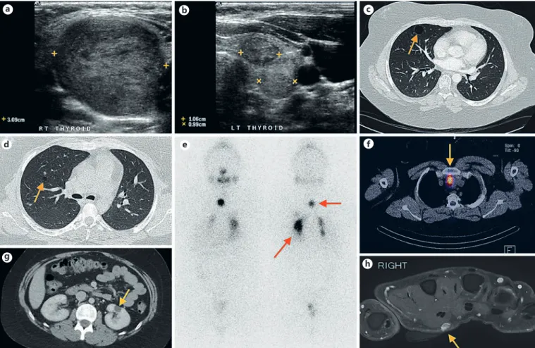
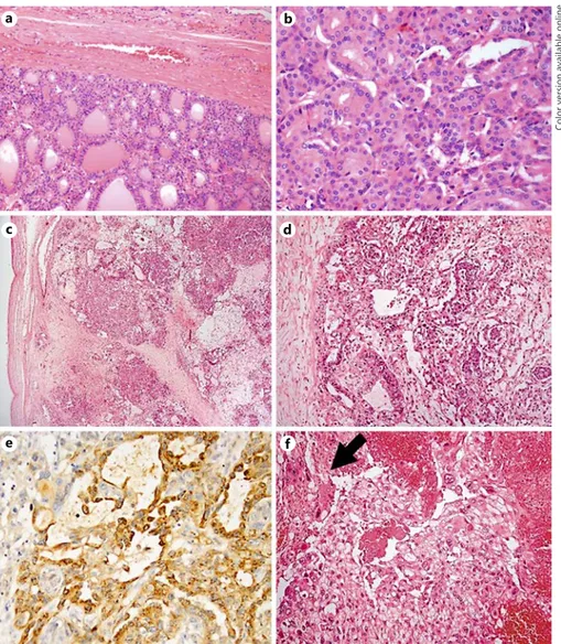
At 17 years of age, a female of Anglo-Saxon descent presented following 3 months of progressive right neck swelling. On exami-nation, there was a soft, non-tender right neck mass, which moved with swallowing. The patient was clinically and biochemically eu-thyroid. Ultrasound imaging demonstrated multiple nodules in both thyroid lobes, with the largest measuring 39 and 34 mm with-in the upper and lower poles of the right lobe, respectively (Fig. 1a). Ultrasound-guided fine needle aspiration (FNA) cytology revealed a follicular neoplasm and the patient underwent a right hemithy-roidectomy in October 2007. Histopathological examination re-vealed 2 thyroid nodules (Fig 2a, b). The first was a 35 mm circum-scribed nodule with a largely microfollicular, but also focally tra-becular and solid growth pattern. In a single section, there were focal areas where nuclei displayed overlapping, angulation, and grooving and some areas featured optically clear nuclei and rare nuclear pseudo-inclusions, in addition to abundant clear cyto-plasm. Thick colloid was noted focally. A CK19 immunostain showed patchy, weak immunoreactivity within tumour cells. The findings fell short of criteria needed to make a definitive diagnosis of follicular variant of papillary thyroid carcinoma, and the lesion was reported as a well-differentiated follicular thyroid neoplasm of uncertain malignant potential. In the current terminology of thy-roid neoplasms, this tumour would be called a non-invasive fol-licular neoplasm with papillary-like nuclear features (NIFTP). The second nodule measured 25 mm in diameter and had features of a follicular adenoma. After detailed multidisciplinary review and discussion with the patient, it was elected not to proceed to com-pletion thyroidectomy or radioactive iodine treatment, and close follow-up was instituted, including sonography of the remaining left lobe.
Ultrasound imaging demonstrated multiple enlarging nodules in the left lobe (Fig. 1b), the largest nodule measured 30 × 15 mm and was mixed solid and cystic in appearance. No cervical lymph-adenopathy was detected. FNA of the largest nodule was indeter-minate, as the sample was heavily bloodstained, limiting assess-ment. A repeat FNA was offered; however, the patient opted for surgical resection. She underwent a completion thyroidectomy, without prophylactic central neck dissection approximately 2 years following her right hemithyroidectomy.
Histopathology showed a multinodular architecture with mul-tiple hyperplastic nodules present throughout the entire lobe, as well as focal areas of lymphocytic thyroiditis and a single focus of follicular variant of papillary thyroid carcinoma measuring 4 mm in diameter. Chest computed tomography (CT) performed at the time identified sub-centimetre right pulmonary nodules and right lung cysts (Fig. 1c, d) which remained stable on subsequent serial imaging and were not fluorodeoxyglucose (18FDG) or iodine-131
avid.
The patient remained under surveillance with ultrasound and serial thyroglobulin (Tg) monitoring without radioiodine abla-tion. Unstimulated Tg 10 weeks following surgery was 5.0 μg/L (with TSH 6.8 mU/L). A nadir of 0.7 μg/L (with TSH 0.32 mU/L) was achieved 31 months after completion thyroidectomy. Tg re-mained relatively stable on thyroxine replacement for an addition-al 3 years (see online suppl. Fig. S1; for addition-all online suppl. materiaddition-al, see www.karger.com/doi/10.1159/000481620). At 24 years of age
(6.5 years after right hemithyroidectomy) Tg increased to 19 μg/L (with TSH 1.7 mU/L) suggesting recurrent or metastatic thyroid cancer (online suppl. Fig. S1). Ultrasound of the neck 3 months prior to this result had not detected disease in the thyroid bed nor in a cervical lymph node. She had CT of the neck and chest, which revealed a 7 × 16 mm lymph node in the superior mediastinum. She was staged and treated with 3 GBq of 131-I. Post-therapy im-aging demonstrated remnant uptake in the left thyroid bed and thyroglossal duct, inflammatory pulmonary activity as well as 2 iodine-avid lymph nodes in the left low cervical (5 mm) and supe-rior mediastinal (13 mm) regions. A repeat CT of her neck and chest 6 months following her 131-I showed a radiological response in the superior mediastinal lymph node, which now measured 9 × 6 mm. On the basis of persistent structural disease evident on CT and a modest dose of 3 GBq at initial therapy, a second dose of 3 GBq radioiodine was given 7 months following the first dose, with only the superior mediastinal lymph node remaining iodine
a b c
d e f
g
h
Fig. 1. Diagnostic imaging. a Right thyroid ultrasound showing a 39 mm nodule. b Two nodules seen on left thyroid ultrasound, measuring 1.06 and 0.99 cm, respectively. c, d Lung cysts (arrows). Pulmonary nodules were also present. e Post-therapy whole-body iodine scan (3 GBq): anterior view (left), posterior view (right). The bottom arrow highlights inflammatory lung activity. f The SPECT-CT image localises the focal midline iodine activity to a small superior mediastinal lymph node. g Left renal cyst indicated
by an arrow. h A 7 mm subcutaneous lobulated soft tissue mass detected on the right hand, immediately palmar to the second fin-ger lumbricals. The lesion is hyperintense on the fluid-sensitive sequences and following contrast, and demonstrates moderate ho-mogeneous enhancement. Immediately proximal to the lesion is a nest of mildly prominent vascular structures in keeping with veins. The appearances are consistent with a slow-flow vascular malfor-mation.
avid (Fig. 1e, f) on post-therapy imaging. The stimulated Tg was 4.4 μg/L (previously 13 ug/L) confirming a partial therapeutic re-sponse from the initial dose. Her unstimulated Tg 9 months fol-lowing the second dose is undetectable (<0.1 μg/L) with a TSH of 0.18 mU/L.
The patient’s past medical history included a mixed malignant germ cell tumour of the ovary diagnosed at 3 years of age that mainly exhibited yolk sac and choriocarcinomatous differentia-tion with more focal embryonal carcinoma and dysgerminoma el-ements (Fig. 2c–f). A left renal cyst (Fig. 1g), an arachnoid cyst, and mucocutaneous lesions consisting of a cutaneous venous angioma (Fig. 1h) and buccal fibroma were also discovered. A breast lump was detected at 23 years of age. Multiple FNAs and core biopsies were negative for malignancy and comprised predominantly fi-brous breast tissue. There was no history of developmental delay. The patient was noted to have macrocephaly with her head cir-cumference measuring 62 cm, 4 cm greater than the 98th percen-tile.
A cancer predisposition syndrome was not considered at her initial presentation and hemithyroidectomy; however, review of
the clinical phenotype of this young patient at a multidisciplinary meeting prior to radioiodine treatment raised the possibility of Cowden syndrome (CS) as macrocephaly was noted during a clin-ical review. DICER1 syndrome was also considered in the differ-ential diagnosis due to the presence of thyroid carcinoma in a background of MNG, lung cysts, and a renal cyst.
Genetic testing for germ-line DICER1 and PTEN mutations was undertaken. A heterozygous germ-line pathogenic PTEN mu-tation was identified (c.388C>T; p.Arg130Ter) (online suppl. Fig. S2). Additional loss of heterozygosity (LOH) of the wild-type
PTEN allele was detected in the ovarian tumour and the right
thy-roid lesion, consistent with a two-hit tumour suppressor gene in-activation. No LOH of PTEN was evident in the left thyroid lesion or breast tissue on Sanger sequencing (online suppl. Fig. S2). Be-cause of the previous identification of DICER1 RNase IIIb muta-tions in ovarian germ cell tumours [3, 4] and thyroid carcinomas [5, 6], we sequenced the RNase III domains in each lesion, but no such “hotspot” mutations were identified.
a b
c d
e f
Fig. 2.a, b Thyroid tumour. a Encapsulat-ed follicular pattern neoplasm. b Nuclear features suggestive of the follicular variant of papillary thyroid carcinoma. c–f Ovari-an tumour. c Low-magnification image mainly showing the yolk sac tumour. The ovary capsule (not involved by the tumour) is seen on the left. d Yolk sac tumour.
e Yolk sac tumour demonstrating AFP im-munoreactivity. f Choriocarcinoma (syn-cytiotrophoblast arrowed).
Consent and Sample Review
Consent for this case report was obtained according to institu-tional ethics procedures. This study was approved by the Institu-tional Review Board of the Faculty of Medicine of McGill Univer-sity, Montreal, QC, Canada, No. A12-M117-11A. The patient con-sented to genetic testing and provided consent to publish. All tumours were reviewed by pathologists (C.J.R.S. and M.P.K.) at the institution from which the samples were acquired and by our cen-tral reference pathologist (D.B.-D.S.).
PTEN Screening
Germ-line PTEN testing was performed in the standard fash-ion. The methods used in the genomic DNA (gDNA) and cDNA analyses are described in detail in the online supplementary Mate-rials and Methods. The full PTEN coding region and exon-intron boundaries were screened for somatic mutations in gDNA extract-ed from formalin-fixextract-ed paraffin embextract-eddextract-ed tumour samples. LOH analysis in tumour samples was performed by Sanger sequencing, according to previously published methods [7].
DICER1 Screening
We screened the full DICER1 coding region and exon-intron boundaries in gDNA extracted from peripheral blood lymphocytes using a custom Fluidigm Access Array followed by next-generation sequencing, as described previously [7]. Large deletions or duplica-tions were screened for using a Multiplex Ligation-Based Probe Am-plification assay [8]. Somatic “hotspot” mutations occurring within the regions encoding the DICER1 RNase IIIa and IIIb domains were screened for by Sanger sequencing, as previously described [5].
BRAF Screening
The region encompassing the BRAF c.1799T>A, p.V600E locus was PCR amplified and Sanger sequenced in DNA extracted from the left and right thyroid lesions. Neither of the lesions harboured the BRAF hotspot mutation.
Discussion
This case illustrates the complexity and variability of
phenotypes associated with tumour predisposition
syn-dromes and the importance of genetic testing in such
cas-es. The constellation of clinical features and tumours
observed in this adolescent female were unusual and
prompted consideration of an underlying genetic
suscep-tibility. The skin lesions and breast lump were suggestive
of CS, whereas the presence of lung and renal cysts led to
suspicion of DICER1 syndrome. The macrocephaly and
ovarian germ cell tumour favoured CS, but
macrocepha-ly has recentmacrocepha-ly been noted as a feature of the DICER1
syndrome [9] and, in general, non-epithelial ovarian
tu-mours, especially ovarian sex cord-stromal tutu-mours,
fa-vour DICER1 syndrome. Thyroid lesions are common
features of both DICER1 syndrome and CS. This
high-lights the overlap between the respective phenotypic
Table 1. Comparison of clinical features exhibited in an adolescent: Cowden versus DICER1 syndromes (designated as common, rare or not reported)
Clinical feature CS D1S Diagnosis (CS or D1S) favoured in this patient Malignant GCT of the
ovary Rare [15, 16] Not reported in germ-line mutation carriers; somatic DICER1 mutations described1 [4]
CS
Thyroid disease
Nodular hyperplasia Common [11] Common [19] Equally favoured Differentiated thyroid
carcinoma Reported, frequency uncertain (follicular and papillary variants) [11] Reported, frequency uncertain (mostly follicular variant of PTC) [5, 6, 19]
Equally favoured
Lung cysts Rare [12] Common (septated) [19] D1S Renal cysts Not reported (RCC rather than cysts)
[11] Common (particularly CN; septated) [19] D1S Mucocutaneous lesions Common [11] Not reported CS Breast lesions Common [11] Rare2 CS
Macrocephaly Common [11] Common [9] Slightly favours CS CN, cystic nephroma; CS, Cowden syndrome; D1S, DICER1 syndrome; GCT, germ cell tumour; PTC, papillary thyroid carcinoma; RCC, renal cell cancer. 1 It can be challenging to pathologically distinguish between some ovarian tumours; certain non-epithelial tumours
spectra observed in CS and the DICER1 syndrome (Table
1). Nevertheless, testing of the DICER1 RNase IIIb
do-mains identified no somatic mutations and, furthermore,
no germ-line DICER1 mutation was present in the
pa-tient.
CS is an autosomal dominant predisposition cancer
syndrome associated with germ-line mutations in the
PTEN tumour suppressor gene [10]. PTEN, or
phospha-tase and tensin homologue, is a dual-specificity
suppres-sor phosphatase, whose substrate is a
phosphatidylinosi-tol, a phospholipid in the phosphatidylinositol 3-kinase
(PI3K) pathway. PTEN shares homology with the
adhe-sion molecules tensin and auxilin. A heritable mutation
in PTEN has been found in 85% of CS patients. PTEN
mutations have also been detected in 65% of patients
with Bannayan-Riley-Ruvalcaba syndrome and 20% of
Proteus-like syndrome patients. Considering the
over-lapping clinical phenotype of these syndromes and their
common genetic origin, they are now combined under
the umbrella term PTEN hamartoma tumour syndrome
(PHTS).
The diagnosis of CS is based on several major and
mi-nor criteria (outlined in online suppl. Table S1), which
were first defined in 1996 by the International Cowden
Syndrome Consortium and later revised in 2008 [11]. A
clinical diagnosis of CS in an individual can be made
based on mucocutaneous lesions alone in sufficient
num-bers, because of the high prevalence of PTEN mutations
in adult-onset Lhermitte-Duclos disease (LDD). A
nosis of LDD alone is also sufficient for a clinical
diag-nosis of CS. Outside of these pathognomonic criteria, a
clinical diagnosis is established with major and minor
criteria: 2 or more major criteria of which 1 must be
macrocephaly or LDD, 1 major and 3 minor criteria, or 4
or more minor criteria. In the event an individual has a
first-degree relative for whom the diagnosis of CS has
been made, the criteria are relaxed. Although rare, there
are reports of CS patients with lung cysts [12, 13] and
ovarian tumours including dysgerminoma [14] and
gran-ulosa cell tumour [15, 16]. In the presented case, the
pa-tient had macrocephaly and was phenotypically
suspect-ed of having CS.
The recently described DICER1 syndrome is caused by
germ-line loss of function mutations in DICER1 [17] and
is inherited in an autosomal dominant fashion. DICER1
is a member of the ribonuclease III (RNase III) family of
proteins and cleaves non-coding small RNA precursors
to generate mature microRNAs (miRNAs), which, in
turn, post-transcriptionally regulate gene expression.
Carriers of DICER1 mutations are predisposed to
devel-oping a broad range of tumours of predominantly
paedi-atric or adolescent onset [17], as outlined in online
sup-plementary Table S2. The association between DICER1
syndrome and the majority of these tumours is well
estab-lished, but since the number of cases with DICER1
muta-tions is still limited, new tumoural associamuta-tions are
pos-sible. Notably, although macrocephaly is common and
severe in CS [11], it has recently been identified as a
sur-prisingly common feature of the DICER1 syndrome as
well (42% of mutation carriers are macrocephalic) [9].
The patient’s ovarian tumour was a malignant germ cell
tumour, which would be highly unusual in the DICER1
syndrome [4]. To our knowledge, no cases of ovarian
germ cell tumours have been reported in germ-line
DICER1 mutation carriers. However, there are reports of
somatic DICER1 RNase IIIb mutations in these tumours
[3, 4]. Given that the diagnosis of ovarian tumours is
of-ten challenging [18], it should be kept in mind that in
certain instances, DICER1 mutation analysis may assist in
distinguishing between the types of non-epithelial
ovar-ian cancers [19].
Ultimately, the identification of a heterozygous
patho-genic PTEN mutation (c.388C>T) in germ-line DNA,
combined with LOH of the wild-type PTEN allele in the
ovarian tumour and right thyroid lesion supported the
diagnosis of CS (or PHTS) over DICER1 syndrome in this
patient. This report emphasizes the importance of
recog-nising the variable cancer phenotype associated with
PTEN mutations, and the possible overlap with other
tu-mour predisposition syndromes, such as the DICER1
syndrome (and vice versa). The BRAF V600E mutation
was not detected in the neoplasms on the right or the left
thyroid lobe, as expected. Follicular patterned neoplasms
including NIFTPs are not known to harbour BRAF V600E
mutations.
Management of this patient was challenging, not least
because her original lesion was reported as a follicular
neoplasm of uncertain malignant potential. If a
heredi-tary predisposition syndrome had been recognised
earli-er, it is likely that total thyroidectomy and radioiodine
treatment would have been recommended at the age of 17
years. Annual thyroid ultrasound is recommended for
surveillance in patients with PTEN mutations, because of
the risk of thyroid cancer [20, 21], and it has been
sug-gested that prophylactic thyroidectomy may be justified
[22], although it is not recommended by the authors. It is
thus critical for clinicians to recognize the phenotypic
features associated with the different syndromes in order
to enable timely implementation of appropriate
surveil-lance. This necessitates the accurate diagnosis of tumours
and the determination of the specific tumour subtype
where indicated, as certain tumours/subtypes may be
more closely associated with one or other syndrome.
Acknowledgements
We thank Dr. A. Bateman and Southampton University Hos-pital, UK, for providing the ovarian tumour, Dr. John R. Priest for reviewing the diagnostic images, and Talia Boshari for assistance with sample acquisition and the associated administration work.
Disclosure Statement
The authors have no conflicts of interest to disclose.
Funding Sources
This work was supported by Alex’s Lemonade Stand Founda-tion (awarded to Dr. William D. Foulkes) and the Telethon Perth Children’s Hospital Research Fund (awarded to Dr. Catherine S. Choong). Leanne de Kock is a recipient of the Vanier Canada Graduate Scholarship.
References
1 Rao A, Rothman J, Nichols KE: Genetic test-ing and tumor surveillance for children with cancer predisposition syndromes. Curr Opin
Pediatr 2008;20:1–7.
2 Francis GL, Waguespack SG, Bauer AJ, Ange-los P, Benvenga S, Cerutti JM, Dinauer CA, Hamilton J, Hay ID, Luster M, Parisi MT, Rachmiel M, Thompson GB, Yamashita S: Management guidelines for children with thyroid nodules and differentiated thyroid
cancer. Thyroid 2015;25:716–759.
3 Heravi-Moussavi A, Anglesio MS, Cheng SW, Senz J, Yang W, Prentice L, Fejes AP, Chow C, Tone A, Kalloger SE, Hamel N, Roth A, Ha G, Wan AN, Maines-Bandiera S, Salamanca C, Pasini B, Clarke BA, Lee AF, Lee CH, Zhao C, Young RH, Aparicio SA, Sorensen PH, Woo MM, Boyd N, Jones SJ, Hirst M, Marra MA, Gilks B, Shah SP, Foulkes WD, Morin GB, Huntsman DG: Recurrent somatic DICER1 mutations in nonepithelial ovarian
cancers. N Engl J Med 2012;366:234–242.
4 Witkowski L, Mattina J, Schonberger S, Mur-ray MJ, Choong CS, Huntsman DG, Reis-Fil-ho JS, McCluggage WG, NicReis-Fil-holson JC, Cole-man N, Calaminus G, Schneider DT, Arse-neau J, Stewart CJ, Foulkes WD: DICER1 hotspot mutations in non-epithelial gonadal
tumours. Br J Cancer 2013;109:2744–2750.
5 de Kock L, Sabbaghian N, Soglio DB, Guiller-man RP, Park BK, Chami R, Deal CL, Priest JR, Foulkes WD: Exploring the association between DICER1 mutations and differenti-ated thyroid carcinoma. J Clin Endocrinol
Metab 2014;99:E1072–E1077.
6 Rutter MM, Jha P, Schultz KA, Sheil A, Harris AK, Bauer AJ, Field AL, Geller J, Hill DA: DICER1 mutations and differentiated thyroid carcinoma: evidence of a direct association. J
Clin Endocrinol Metab 2016;101:1–5.
7 de Kock L, Sabbaghian N, Plourde F, Srivas-tava A, Weber E, Bouron-Dal Soglio D, Hamel N, Choi JH, Park SH, Deal CL, Kelsey MM, Dishop MK, Esbenshade A, Kuttesch JF,
Jacques TS, Perry A, Leichter H, Maeder P, Brundler MA, Warner J, Neal J, Zacharin M, Korbonits M, Cole T, Traunecker H, McLean TW, Rotondo F, Lepage P, Albrecht S, Hor-vath E, Kovacs K, Priest JR, Foulkes WD: Pi-tuitary blastoma: a pathognomonic feature of germ-line DICER1 mutations. Acta
Neuro-pathologica 2014;128:111–122.
8 Sabbaghian N, Srivastava A, Hamel N, Plourde F, Gajtko-Metera M, Niedziela M, Foulkes WD: Germ-line deletion in DICER1 revealed by a novel MLPA assay using syn-thetic oligonucleotides. Eur J Hum Genet
2014;22:564–567.
9 Khan NE, Bauer AJ, Doros L, Schultz KA, De-castro RM, Harney LA, Kase RG, Carr AG, Harris AK, Williams GM, Dehner LP, Mess-inger YH, Stewart DR: Macrocephaly associ-ated with the DICER1 syndrome. Genet Med
2017;19:244–248.
10 Orloff MS, Eng C: Genetic and phenotypic heterogeneity in the PTEN hamartoma
tu-mour syndrome. Oncogene 2008;27:5387–
5397.
11 Hobert JA, Eng C: PTEN hamartoma tumor
syndrome: an overview. Genet Med 2009;11:
687–694.
12 Cottin V, Thomas L, Loire R, Chalabreysse L, Gindre D, Cordier JF: Mesenchymal cystic hamartoma of the lung in Cowden’s disease.
Respir Med 2003;97:188–191.
13 Lee EJ, Jung WS, Ko JM, Park HJ: Multiorgan involvements of Cowden disease in a 50-year-old woman: a case report and literature
over-view. J Korean Soc Radiol 2013;69:251–255.
14 Cho MY, Kim HS, Eng C, Kim DS, Kang SJ, Eom M, Yi SY, Bronner MP: First report of ovarian dysgerminoma in Cowden syndrome with germline PTEN mutation and PTEN-re-lated 10q loss of tumor heterozygosity. Am J
Surg Pathol 2008;32:1258–1264.
15 Neumann S: Cowden syndrome with an ovar-ian tumor (multiple hamartoma syndrome)
(in German). Chirurg 1991;62:629–630.
16 Smpokou P, Fox VL, Tan WH: PTEN hamar-toma tumour syndrome: early tumour
devel-opment in children. Arch Dis Child 2015;100:
34–37.
17 Foulkes WD, Priest JR, Duchaine TF: DICER1: mutations, microRNAs and
mecha-nisms. Nat Rev Cancer 2014;14:662–672.
18 Wu MK, de Kock L, Conwell LS, Stewart CJ, King BR, Choong CS, Hussain K, Sabbaghian N, MacRae IJ, Fabian MR, Foulkes WD: Functional characterization of multiple DICER1 mutations in an adolescent. Endocr
Relat Cancer 2016;23:L1–L5.
19 Foulkes WD, Gore M, McCluggage WG: Rare non-epithelial ovarian neoplasms: pathology, genetics and treatment. Gynecol Oncol 2016;
142:190–198.
20 Haugen BR, Alexander EK, Bible KC, Doherty GM, Mandel SJ, Nikiforov YE, Pa-cini F, Randolph GW, Sawka AM, Schlum-berger M, Schuff KG, Sherman SI, Sosa JA, Steward DL, Tuttle RM, Wartofsky L: 2015 American Thyroid Association management guidelines for adult patients with thyroid nodules and differentiated thyroid cancer: The American Thyroid Association Guide-lines Task Force on Thyroid Nodules and Differentiated Thyroid Cancer. Thyroid
2016;26:1–133.
21 Tan MH, Mester JL, Ngeow J, Rybicki LA, Or-loff MS, Eng C: Lifetime cancer risks in indi-viduals with germline PTEN mutations. Clin
Cancer Res 2012;18:400–407.
22 Milas M, Mester J, Metzger R, Shin J, Mitchell J, Berber E, Siperstein AE, Eng C: Should pa-tients with Cowden syndrome undergo
pro-phylactic thyroidectomy? Surgery 2012;152:
1201–1210.
23 de Kock L, Druker H, Weber E, Hamel N, Traubici J, Malkin D, Arseneau J, Stewart CJ, Bouron-Dal Soglio D, Priest JR, Foulkes WD: Ovarian embryonal rhabdomyosarcoma is a rare manifestation of the DICER1 syndrome.