THE IMPORTANCE OF RED CELL DISTRIBUTION WIDTH (RDW) IN PATIENT FOLLOW UP IN INTENSIVE CARE UNIT (ICU)
RABIAÖZDEMIR1, NEVZATMEHMETMUTLU2, METINÖZDEMIR3, MURATAKÇAY2, CIHATYEL4, IŞILÖZKOÇAKTURAN2
1Department of Anesthesiology and Reanimation, Medipol University Hospital, İstanbul - 2Department of Intensive Care, Ankara
Numune Education and Research Hospital, Ankara - 3Department of Intensive Care, Ankara Numune Education and Research
Hospital, Ankara - 4Department of Emergency, Antakya State Hospital, Antakya, Turkey
Introduction
Intensive care units (ICU) are units designed for the management of both acute disorders and life-threatening complications of chronic disorders, where necessary equipment and personnel are available for close monitoring and rapid manage-ment of patients(1). Patients admitted to ICU have an increased mortality. A study involving 12162 patients admitted to 24 ICUs found an in-hospital mortality rate of 0.91-1.34% whereas the corre-sponding rate for ICU was 24-41%(2).
Erythrocyte distribution width is a parameter measured in complete blood count that reflects the distribution of erythrocytes’ sizes in circulation(3). Studies have shown that RDW is not only correlat-ed to anemia, but a variety of other pathological conditions(4-6). RDW was found to be correlated to mortality in sepsis, trauma, cardiovascular disor-ders, and patients admitted to general ICUs(7-12). Although the exact mechanism of the association of RDW with mortality is yet to be fully explained, several studies, particularly those conducted in sep-sis, have implicated inflammatory cytokines in the
Received May 30, 2015; Accepted January 02, 2016
ABSTRACT
Objective: In this study we aimed to determine the effect of RDW on mortality by comparing the temporal changes in RDW
level in deceased and surviving ICU patients.
Materials and method: This study was retrospectively conducted in 776 patients admitted to intensive care unit between
01.01.2013 and 31.12.2013. Age, sex, clinical diagnosis, comorbid conditions, RDW levels at intensive care admission and dischar-ge, duration of intensive care stay, and mortality rate were evaluated. Kolmogorov Smirnov, student t-test, Mann Whithey u test, and Chi-square test were used for data analysis. The results were provided at a confidence interval of 95% and a significance level of p<0. 05.
Results: Conducted on 776 patients in Ankara Numune Hospital's general intensive care clinic 3, this study revealed a
morta-lity rate of 38.2%. The mean age of the study population was 65.4±18.3 years, with the deceased patients having a greater mean age (p<0.05). Four hundred and thirteen (53.2%) patients were male, and there was no significant difference between the ratios of both genders (p>0.05). The deceased patients had significantly increasing RDW levels whereas the surviving patients had significantly reducing RDW levels (p<0.05). There was a significant correlation between the magnitude of RDW increment and duration of hospi-tal stay in the deceased patients (p<0.05).
Conclusion: The results of this study suggest that RDW, with known association with mortality, can be used as a simple marker
for patient follow-up and monitorization of effectiveness of the administered therapies in ICU.
Key words: RDW, intensıve care unit, mortalite.
disease pathogenesis(7). Studies conducted on patients with sepsis have indicated that RDW may have prognostic implications(13). However, as these studies have only focused on patients with sepsis, no consensus has been reached as to whether it has a similar prognostic role in other conditions man-aged in ICU.
Herein, we aimed to analyze the temporal changes in RDW levels of patients deceased in or discharged from intensive care unit in an attempt to determine its prognostic value.
Materials and method
After being approved by the Local Ethics Committee of Ankara Numune Training and Research Hospital, our study retrospectively exam-ined patients managed in intensive care unit between 1.01.2013 and 31.12.2013. Patients' data were accessed from patient records and hospital's local automation system.
Age, sex, clinical diagnosis, comorbid condi-tions, RDW levels at intensive care admission and discharge, duration of intensive care stay, and mor-tality rate were evaluated.
Our study excluded patients who were below 18 years of age; who were pregnant; who had myelodysplastic syndrome, bone marrow metas-tases, or hematological disorders; who were trans-fused blood within 2 weeks; who were referred from another hospital; and patients who underwent a procedure.
Study data were analyzed in SPSS Windows 18 software package. Descriptive statistics included mean, standard deviation for parametric data; medi-an medi-and interquartile rmedi-ange (IQR) for non-parametric data; and number and percentile for categorical data. The distribution of the data was tested with Kolmogorov Smirnov test. Continuous parametric variables were compared with student t-test, and non-parametric variables were compared with Mann Whitney U test and Chi-square test. The cor-relation between the duration of ICU stay and RDW was tested with Pearson correlation analysis. The results were evaluated at a confidence level of 95% and a significance level of p<0.05.
Results
Conducted in 776 patients admitted to the gen-eral ICU 3 clinic of Ankara Numune Training and Research Hospital, this study revealed a mortality
rate of 38.2%. The mean age of the study popula-tion was 65.4±18.3 years, with the deceased patients having a significantly higher mean age (p<0.05). Four hundred and thirteen (53.2%) patients were male; the two groups had no signifi-cant difference with regard to the proportions of both genders (p>0.05) (Table 1).
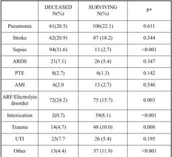
Sepsis was the most common diagnosis among the deceased patients while pneumonia was the most frequent diagnosis among the discharged patients. In the deceased patients the rates of sepsis and acute renal failure (ARF)/electrolyte disorder were significantly higher whereas intoxication, trauma, and other disease groups were significantly more common in the surviving patients (p<0.05) (Table 2).
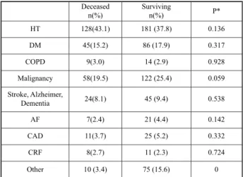
The analysis of the comorbidities of the study subjects revealed that hypertension (43.1%; 37.8%) and diabetes mellitus (15.2; 17.9%) were the
com-DECEASED Mean/ N(%) SURVIVING Mean/ N(%) Total Mean/ N(%) p Age 67.6±16.1 64.1±19.3 65.4±18.3 0.008* Sex Male 160 (54.1) 253(52.6) 413(53.2) 0.754** Female 136(45.9) 227(47.4) 363 (46.8) Total 296 (100.0) 480 (100.0) 776 (100.0)
Table 1: Demographic characteristics of the study
subjects.
* student-t test, ** X2test
DECEASED N(%) SURVIVINGN(%) P* Pneumonia 61(20.5) 106(22.1) 0.611 Stroke 62(20.9) 87 (18.2) 0.344 Sepsis 94(31.6) 13 (2.7) <0.001 ARDS 21(7.1) 26 (5.4) 0.347 PTE 8(2.7) 6(1.3) 0.142 AMI 6(2.0 13 (2.7) 0.546 ARF/Electrolyte disorder 72(24.2) 75 (15.7) 0.003 Intoxication 2(0.7) 39(8.1) <0.001 Trauma 14(4.7) 48 (10.0) 0.008 UTI 23(7.7 26 (5.4) 0.195 Other 13(4.4) 57 (11.9) <0.001
Table 2: Distribution of care unit diagnoses of the study
subjects.
* X2 test (ARDS:Acute Respiratory Distress Syndrome, PTE:Pulmonary Thromboembolism, AMI:Acute Myocardial Infarction, UTI: Urinary Tract Infection)
monest comorbidities. The rates of comorbid condi-tions were similar in the deceased and surviving patients (p>0.05) (Table 3).
The median RDW level was 15.2 (IQR 3.4) at admission and 15.5 (IQR 3.5) at discharge. While the median RDW level increased from 15.8 to 17.1, it remained constant at 14.8. RDW level was signif-icantly higher in the deceased patients at both admission and ICU discharge (p<0.05) (Table 4).
The analysis of changes in RDW levels during ICU stay revealed that it increased in 261 (87.9%) of the deceased patients and decreased in 31 (10.4%); it increased in 167 (34.8%) of the surviv-ing patients and decreased in 281 (58.5%) (p<0.05) (Table 4).
While the median duration of ICU stay was 5 (IQR=12) days for the deceased patients, it was 6 (IQR=11) days for those who were discharged. Whereas no significant correlation was found between the duration of ICU stay and the admis-sion, discharge RDW levels in the deceased patients
(p>0.05), there was a positive correlation between the RDW increment and duration of hospital stay (p<0.05).
No significant correlation was determined between duration of ICU stay and admission, dis-charge RDW levels and the amount of RDW change (p>0.05).
Discussion
Intensive care units are places where debilitat-ed patients with poor general status in nedebilitat-ed of con-tinuous monitoring and care are admitted and where the mortality rates are higher than that of other clin-ics(2). Studies have suggested that 5% of all beds of a hospital should meet ICU standards(14,15). Each institution flexes the criteria for ICU admission and discharge due to deficiencies in personnel and bed capacity of these units(15).
Studies have shown ICU mortality rates rang-ing between 24-43%(1,2,16). In our study the mortality rate of the patients was similarly found 38.2%. We are of the opinion that mortality rate is higher in this population since patients admitted to ICUs have more advanced age, worse overall status, and more severe forms of diseases; they are also exposed to nasocomial infections at a higher rate during ICU stay. Hence, a higher age and an infec-tion rate in the deceased patients support our hypothesis.
In a metaanalysis by Patel et al. involving 7 trials showed that the mean patient age ranged between 74 and 78 years, and that women predomi-nated ICU populations(16). In a domestic study by Ceylan et al. conducted on internal ICU patients, the mean age was found 63 years, and 62% of patients were male(17). In a study by Uysal et al. the reported mean age was 53 years, and 52% of sub-jects was male(1).
Our study population had a mean age and gen-der distribution consistent with the literature reports from our country, although these figures were lower than those reported by foreign studies. The main reason of this finding may be some international differences in living standards and average life expectancies.
Altun et al. reported that the most common ICU admission indications were stroke, COPD, and sepsis, and COPD and sepsis were associated with the highest mortality rate (18) Uysal et al, in a study from our country, reported that the most common indications of ICU admission were sepsis and neu-Deceased n(%) Surviving n(%) P* HT 128(43.1) 181 (37.8) 0.136 DM 45(15.2) 86 (17.9) 0.317 COPD 9(3.0) 14 (2.9) 0.928 Malignancy 58(19.5) 122 (25.4) 0.059 Stroke, Alzheimer, Dementia 24(8.1) 45 (9.4) 0.538 AF 7(2.4) 21 (4.4) 0.142 CAD 11(3.7) 25 (5.2) 0.332 CRF 8(2.7) 11 (2.3) 0.724 Other 10 (3.4) 75 (15.6) 0
Table 3: Comorbid conditions of the study subjects.
* X2 test (HT: Hypertension; DM: Diabetes Mellitus, COPD: Chronic Obstructive Pulmonary Disease, AF: Atrial fibrilla-tion, CAD: Coronary Artery Disease, CRF: Chronic Renal Failure). N Median (IQR) P* RDWadmission Deceased 296 15.8 (3.65) <0 .001 Surviving 480 14.8 (2.78) Total 776 15.2 (3.4) RDWfinal Deceased 296 17.1 (3.77) <0.001 Surviving 480 14.8 (2.8) Total 776 15.5 (3.5)
Table 4: RDW levels at admission and discharge.
rological disorders, with oncological diseases hav-ing the highest mortality rates. Increased rate of nasocomial infections is a common problem of ICUs(19). We believe that mortality is higher in these patients as a result of deteriorated general status due to infections and ARF/electrolyte disorders added on top of preexisting comorbidities. Moreover, increased nasocomial infection rate in hospitals probably led to an increase in overall infection rate. Although underlying malignancies modified the results of the study by Uysal et al, we opine that additional pathologies added onto malig-nancy actually led to death. Lower mortality rates found for intoxication and stroke cases support our hypothesis.
In a metaanalysis by Patel et al. comprising 11827 patients, the most common comorbidities in ICU populations were HT, DM, and malignancy(16). We found that HT, malignancy, and DM were the most common comorbid disorders in both groups. Since the subjects enrolled in our study had an advanced mean age, the rate of comorbidities was also high. Although Patel et al.(20) and Perlstein et al.(21)reported that RDW level predicted cardiovas-cular death and its level increased in cancer and chronic lower respiratory rate diseases, we found no significant differences between the RDW levels of both groups, possibly as a result of similar rates of comorbidities in deceased and surviving patients.
Wang et al. reported that mortality was corre-lated to increased RDW level(8). Anderson showed that RDW level was higher in patients who died as a result of cardiac pathologies(10). Lee et al. explored 30-day mortality in patients with pneumonia and found that RDW was correlated to mortality. Jo et al. studied patients in shock and demonstrated that RDW was correlated to sepsis severity and mortali-ty(22). Ku et al. reported that RDW was correlated to mortality in infectious diseases(23).
Fujita et al. suggested that RDW may be used as a predictor of mortality in patients admitted to ICU(24). Patel et al. stressed that, affected by many factors such as age, nutritional status, anemia, and inflammation, RDW independently predicted mor-tality(20). Braun et al. assessed 90-day mortality in patients with pneumonia and found that patients with increased RDW level had a higher mortality(5). The same study also found that RDW and blood urea nitrogen had a better ability to predict mortali-ty than glucose, hypernatremia, creatinine, and white blood cell count(5).
Hunziker et al. reported that RDW can be used as a simple physiological score for determining short- and long-term mortality(25).
In the metaanalysis by Patel et al. each 1 point increment in RDW level caused a 14% increase in mortality(16). Özdoğan et al. measured RDW serially for 7 days in patients with intraabdominal sepsis; they found that while RDW level was reduced in surviving patients, it was increased in the deceased ones(13). Kim et al, in a study where serial daily RDW measurements were done for 3 days, found that RDW level was correlated to mortality(26). In our study the RDW tended to increase in deceased patients whereas it had a tendency to decrease in those who were discharged. We think that enhanced inflammatory effect and impaired neurohumoral balance that result from a deterioration of the pre-existing clinical condition during ICU stay sudden-ly accelerates erythrocyte production in bone mar-row through excess levels of stimulants, with disor-ders in the maturation and/or morphogenesis of red blood cells, leading to increased RDW level. It is equally probable that RDW tended to decrease in patients with a tendency to convalesce thanks to an improvement of stress and neurohumoral mecha-nisms by administered therapies.
It is a common observation that RDW is increased independently of hemoglobin level by many pathologies, albeit by unknown mecha-nisms(5). Lippi et al. showed a direct relationship between RDW and CRP(25); Majercik et al.(9) report-ed that RDW increasreport-ed in response to increasreport-ed inflammatory milieu. Montana et al. reported that inflammatory cytokines such as TNF-α, IL-1 and IL-6 are released as a result of increased stress, and they both shorten cells’ life span and affect bone marrow to desensitize erythroid progenitors which disrupt erythrocyte maturation and cause anioscyto-sis(28). It was reported that increased stress causes neurohumoral changes to activate stress hormones, leading to significant changes in RDW level(29). Marinkoviç et al. showed that increased hemolysis resulting from oxidative stress causes RDW increase(30). Işik et al. reported that patients with gastrointestinal bleeding had increased RDW lev-els, and linked that increase to cytokines and bleed-ing-induced stress factors(31). Vashistha et al. report-ed that RDW was a powerful prreport-edictor of mortality in hemodialysis patients, an association which they linked to anemia(32). Jo et al. demonstrated that RDW may be elevated due to impaired blood flow in sepsis(7).
Several studies have indicated that mortality increases as the duration of hospital stay is pro-longed(33). Özay et al.(15) reported a duration of stay of 6.81 days; Uysal et al.(1)3.4 days; and Ayazoğlu et al.(34)4.7 days. The mean durations of ICU stay of both the deceased and surviving patients in our study were compatible with figures reported by other domestic studies.
Wang et al. reported that, in addition to RDW being a powerful predictor of mortality, the eleva-tion of its levels had a positive correlaeleva-tion to time to mortality, while it had a negative correlation to duration of hospital stay(8). We determined no rela-tionship between the magnitude of the decrements in RDW level and time to discharge but found a significant correlation between the magnitude of RDW elevation and time to death. As a result of the overcrowding of our hospital’s ICU, patients are transferred to regular wards as soon as they are deemed suitable for follow-up there. We are of the opinion that since time to transfer to a regular ward may sometimes be too short, RDW levels of patients discharged from ICU remain unchanged as there appears no opportunity for new erythrocytes to be produced. On the other hand, considering that 1% increments in RDW level increase mortality in deceased patients, the correlation between time to death and RDW level can be explained.
Our study concluded that RDW, which has a known association with mortality, may be used as a marker for disease monitoring and assessment of treatment efficacy.
Limitations
As our study has a retrospective design, clini-cal information including general condition, APACHE scores, and vital parameters could not be accessed. In addition, the criteria for transferring patients to regular ward was not clear. Exclusion criteria were based on medical records and were not confirmed by information obtained from patients or their relatives.
References
1) Uysal N, Gundoğdu N, Borekci Ş, Dikensoy O, Bayram N, Uyar M, et al. Prognosis of Patients in a Medical Intensive Care Unit of a Tertiary Care Centre. Yoğun Bakım Derg 2010; 1: 1-5.
2) Goldhill DR, Sumner A. Outcome of intensive care patients in a group of British intensive care units. Crit Care Med 1998; 26: 1337-45.
3) Isik T, Uyarel H, Tanboga IH, Kurt M, Ekinci M, Kaya A, et al: Rela-tion of red cell distribution width with the presence, severity, and complexity of coronary artery disease. Coron Artery Dis 2012; 23: 51-6.
4) Bekler A, Gazi E, Tenekecioglu E, Karaagac K, Altun B, Temiz A, et al. Assessment of the relationship between red cell distribution width and fragmented QRS in patients with non-ST elevated acute coronary syndrome. Med Sci Monit. 2014;13; 20: 413-9. 5) Braun E, Kheir J, Mashiach T, Naffaa M, Azzam ZS. Is
elevated Red cell distribution width a prognostic pre-dictor in adult patients with community acquired Pneumonia? BMC Infect Dis. 2014; 5; 14(1):129. doi: 10.1186/1471-2334-14-129.
6) Zhang Z, Xu X, Ni H, Deng H. Red cell distribution width is associated with hospital mortality in unselect-ed critically ill patients. J Thorac Dis. 2013; 5(6): 730-6.
7) Jo YH, Kim K, Lee JH, Kang C, Kim T, Park HM, et al. Red cell distribution width is a prognostic factor in severe sepsis and septic shock. Am J Emerg Med 2013; 31: 545-8.
8) Wang F, Pan W, Pan S, Ge J, Wang S, Chen M. Red cell distribution width as a novel predictor of mortality in ICU patients. Ann Med 2011; 43: 40-6.
9) Majercik S, Fox J, Knight S, Horne BD. Red cell distri-bution width is predictive of mortality in trauma patients. J Trauma Acute Care Surg 2013; 74: 1021-6. 10) Anderson JL, Ronnow BS, Horne BD, Carlquist JF,
May HT, Bair TL, et al. Usefulness of a complete blood count-derived risk score to predict incident mortality in patients with suspected cardiovascular disease. Am J Cardiol 2007; 99: 169-74.
11) Sadaka F, O’Brien J, Prakash S. Red cell distribution width and outcome in patients with septic shock. J Intensive Care Med 2013; 28: 307-13.
12) Wonnerth A, Krychtiuk KA, Mayer FJ, Minar E, Wojta J, Schillinger M, et al. Red cell distribution width and mortality in carotid atherosclerosis. Eur J Clin Invest. 2015 Dec 27. doi: 10.1111/eci.12584.
13) Özdoğan HK, Karateke F, Özyazıcı S, Özdoğan M, Özaltun P, Kuvvetli A, et al. The predictive value of red cell distribution width levels on mortality in intensive care patients with community-acquired intra-abdomi-nal sepsis. Ulus Travma Acil Cerrahi Derg. 2015; 21(5): 352-7.
14) Akpir K. Yoğun Bakım Seruveni: Dun, Bugun. Yoğun Bakım Derneği Dergisi, 2002;1: s 6 -7.
15) Özay HY, Bombacı E, Ercan GÇ, Çolakoğlu S. Retrospective Analysis of the Re-Admitted Cases in Intensive Care Unit; Reasons, Outcomes and Evaluation of the Factors That Affect Mortality. Turk Yo€un Bak›m Derneği Dergisi 2012;10: 91-6.
16) Patel KV, Semba RD, Ferrucci L, Newman AB, Fried LP, Wallace RB, et al. Red cell distribution width and mortality in older adults: a meta-analysis. J Gerontol A Biol Sci Med Sci. 2010; 65(3): 258-65.
17) Ceylan E, İtil O, Ellidokuz H,Uçan ES, Akkoçlu A. İç Hastalıkları Yoğun Bakım Ünitesinde İzlenmiş Hastalarda Mortalite ve Morbiditeyi Etkileyen Faktörler. Toraks Derg. 2001; 2(1): 6-12.
18) Altun Y, Demircan F, Mengeloğlu FZ, Namuslu M. The relationship between mean platelet volume and mortality in patients in intensive care unit. 2011; 2 (4): 408-11.
19) Spencer RC. Epidemiology of infection in ICUs. Intensive Care Med 1994; 20: 2-6.
20) Patel KV, Ferrucci L, Ershler WB, Longo DL, Guralnik JM. Red blood cell distribution width and the risk of death in middle-aged and older adults. Arch Intern Med. 2009; 169: 515-23.
21) Perlstein TS, Weuve J, Pfeffer MA, Beckman JA. Red blood cell distribution width and mortality risk in a community-based prospective cohort. Arch Intern Med. 2009; 169: 588-94.
22) Lee JH, Chung HJ, Kim K, Jo YH, Rhee JE, Kim YJ, et al. Red cell distribution width as a prognostic marker in patients with community-acquired pneumonia. Am J Emerg Med. 2013; 14(1): 72-9.
23) Ku NS, Kim HW, Oh HJ, Kim YC, Kim MH, Song JE, et al. Red blood cell distribution width is an indepen-dent predictor of mortality in patients with gram-nega-tive bacteremia. Shock. 2012; 14(2): 123-7.
24) Fujita B, Franz M, Figulla HR, Pfeifer R, Kabisch B, Fritzenwanger M, et al. Red cell distribution width and survival in patients hospitalized on a medical ICU. Clin Biochem. 2015 Nov; 48(16-17): 1048-52.
25) Hunziker S, Celi LA, Lee J, Howell MD. Red cell dis-tribution width improves the simplified acute physiolo-gy score for risk prediction in unselected critically ill patients. Crit Care. 2012; 14(3): R89.
26) Kim CH, Park JT, Kim EJ, Han JH, Han JS, Choi JY, et al. An increase in red blood cell distribution width from baseline predicts mortality in patients with severe sep-sis or septic shock. Crit Care 2013; 17: 282.
27) Lippi G, Targher G, Montagnana M, Salvagno GL, Zoppini G, Guidi GC. Relation between red blood cell distribution width and inflammatory biomarkers in a large cohort of unselected outpatients. ArchPathol Lab Med 2009; 133: 628-32.
28) Montagnana M, Cervellin G, Meschi T, Lippi G: The role of red blood cell distribution width in cardiovascu-lar and thrombotic disorders. Clin Chem Lab Med 2012; 50: 635-41.
29) Fukuta H, Ohte N, Mukai S, Saeki T, Asada K, Wakami K, Kimura G: Elevated plasma levels of B-typenatriu-retic Peptide but not C-reactive protein are associated with higher red cell distribution width in patients with coronary artery disease. Int Heart J 2009; 50: 301-12. 30) Marinkovic D, Zhang X, Yalcin S, Luciano JP,
Brugnara C, Huber T, Ghaffari S: Foxo3 is required for the regu-lation of oxidativestress in erythropoiesis. J ClinInvest. 2007; 117(8): 2133-44.
31) Isik B, Yilmaz MS, Yel C, Kavalci C, Solakoglu GA, Ozdemir M, et al. Importance of red blood cell distrib-ution width (RDW) in patients with upper gastrointesti-nal haemorrhage. J Pak Med Assoc. 2016 Feb; 66(2): 151-4.
32) Vashistha T, Streja E, Molnar MZ, Rhee CM, Moradi H, Soohoo M, et al.. Red Cell Distribution Width and Mortality in Hemodialysis Patients. Am J Kidney Dis. 2016 Jan 16. pii: S0272-6386(15)01494-8.
33) Bueno-Cavanillas A, Delgado-Rodriguez M, Lopez-Luque A, Schaffino-Cano S, Gálvez-Vargas R. Influence of nosocomial infection on mortality rate in an intensive care unit. Crit Care Med 1994; 22: 55-60.
34) Ayazoğlu TA, Candan A, Özkaynak İ, Eseoğlu İ. Efficacy of Intensive Care Unit for the Evaluation of Prognosis of the Patients APACHE IV model. Yeni Tıp Dergisi 2012; 29(2): 100-4.
_______
Corresponding author RABIAÖZDEMIR
Medipol Mega University Hospital
Anestesiology and Reanimation Department, Bağcilar İstanbul