DOI 10.24425/pjvs.2019.129216
Original article
Correspondence to: P. P. Akalin, e-mail: [email protected], [email protected], tel.: +90 326 2455845 ext: 1531, fax: +90 326 2455704
Glucose-6-phosphate dehydrogenase,
glutathione peroxidase, total glutatione
and reduced nicotinamide adenine
dinucleotide phosphate in milk cells
of subclinical mastitic cows
P.P. Akalin
1, Y. Ergün
2, N. Başpinar
3, G. Doğruer
2, A. Küçükgül
1, Z. Cantekin
4,
M. İşgör
1, M. Saribay
2, E. Koldaş
4, A. Baştan
5, S. Salar
5, S. Pehlivanlar
61 Hatay Mustafa Kemal University, Veterinary Faculty, Departments of Biochemisty, Alahan, 31060, Hatay, Turkey 2 Hatay Mustafa Kemal University, Veterinary Faculty, Department of Obstetrics and Gynecology, Alahan, 31060, Hatay, Turkey
3 Selcuk University, Veterinary Faculty, Departments of Biochemistry, Selçuklu, 42250, Konya, Turkey 4 Hatay Mustafa Kemal University, Veterinary Faculty, Departments of Microbiyology, Alahan, 31060, Hatay, Turkey 5Ankara University, Veterinary Faculty, Departments of Obstetrics and Gynecology, Dögol Street, 06100, Ankara, Turkey 6 Hatay Mustafa Kemal University, Veterinary Faculty, Department of Food Hygiene and Technology, Alahan, 31060, Hatay, Turkey
Abstract
This study aimed to determine the levels of milk cell total protein (TP), reduced nicotinamide adenine dinucleotide phosphate (NADPH), total glutathione (tGSH), activities of glucose-6-phos-phate dehydrogenase (G6PD) and glutathione peroxidase (GPx) in subclinical mastitic cows. Milk from each udder was collected and grouped by the California Mastitis Test. Then, a somatic cell count (SCC) was performed, and the groups were re-scored as control (5–87 × 103 cells), 1st
group (154–381 × 103 cells), 2nd group (418–851 × 103 cells), 3rd group (914–1958 × 103 cells),
and 4th group (2275–8528 × 103 cells). Milk cell TP, NADPH, tGSH levels, G6PD, and GPx
ac-tivities were assessed. Microbiological diagnosis and aerobic mesophyle general organism (AMG, cfu/g) were also conducted. In mastitic milk, TP, NADPH, and tGSH levels, and G6PD and GPx activities were significantly reduced per cell (in samples of 106 cells). In addition, milk
SCC was positively correlated with AMG (r=0.561, p<0.001), NADPH (r=0.380, p<0.01), TP (r=0.347, p<0.01) and G6PD (r=0.540, p<0.001). There was also positive correlation between NADPH (r=0.428, p<0.01), TP (r=0.638, p<0.001) and AMG. NADPH was positively correlated with TP (r=0.239, p<0.05), GPx (r=0.265, p<0.05) and G6PD (r=0.248, p=0.056). Total protein was positively correlated with tGSH (r=0.354, p<0.01) and G6PD (r=0.643, p<0.001). There was a negative correlation between tGSH and GPx activity (r=-0.306, p<0.05). The microbiological analysis showed the following ratio of pathogens: Coagulase-Negative Staphylococci 66.6%,
Streptococcus spp 9.5%, Bacillus spp 9.5%, yeast 4.8%, and mixed infections 9.5%.
As a conclusion, when evaluating the enzyme and oxidative stress parameters in milk, it is more suitable to assign values based on cell count rather than ml of milk. The linear correlation between the SCC and AMG, milk cell NADPH, TP and G6PD suggests that these parameters could be used as markers of mastitis.
Key words: subclinical mastitis, milk cell, glucose-6-phosphate dehydrogenase, glutathione
peroxidase, NADPHIntroduction
Mastitis symptoms depend on the state of the cow’s defense system, the characteristics of the responsible agents, and the type of intramammary infection (clini-cal or subclini(clini-cal). Somatic cell count (SCC) is the main tool for diagnosing subclinical mastitis. Milk somatic cells are comprised of leukocytes (lymphocytes, poly-morphonuclear leukocytes (PMN), and macrophages) and epithelial cells (Lee et al. 1980). When the mamma-ry tissue is infected, SCC in milk increase (Baştan 2010). Number of somatic cells in milk and their com-position depend on the type of infection and the type of causative agent (Leitner et al. 2000). For instance, the largest intramammary SCC increase in cows was found with Staphylococcus (Staph.) aureus infection, and the smallest increase was found with Corynebacterium
bovis infection (Schepers et al. 1997). A study (Leitner
et al. 2000) performed on the effects of different etio-logical agents on milk SCCs and composition reported that the majority of the cells in the somatic cell compo-sition of healthy milk are epithelial cells. The same study also found that, in milk from acutely infected mammary gland with Escherichia (E.) coli and Staph.
aureus, the SCC increased, and the majority of the cells
were PMNs. In addition, in case of acute infection with
Staph. aureus, mononuclear cell, lymphocyte, and
mac-rophage counts also increased to a certain level. In the case of chronic infection with Staph. aureus and Coa-
gulase-Negative Staphylococci (CNS), the majority of
cells in the somatic cell composition were T-lympho-cytes and macrophages. Epithelial cell counts decreased relative to neutrophil, PMN, and macrophage counts upon acute or chronic infection (Leitner et al. 2000).
Glucose-6-phosphate dehydrogenase (G6PD) is the first enzyme of the pentose phosphate metabolic pathway (Mehta et al. 2000). The pentose phosphate pathway has two primary functions: synthesizing the ribose-5-phosphate and the reduced nicotinamide ade-nine dinucleotide phosphate (NADPH) required for reductive reactions (Krebs and Eggleston 1978). NADPH protects the cell from the oxidative damage caused by free radicals (Ames et al. 1993, McCord 1993), and plays an important role in the regeneration of reduced glutathione (rGSH). Reduced GSH and GSH-dependent enzymes protect the cell from reactive oxygen species (Ayala et al. 1991).
Studies in humans have shown that insufficient ROS elimination in epithelial cells of individuals with G6PD deficiency, causes their cells to be more sensitive to Staph. aureus infection and apoptosis than healthy cells (Hsieh et al. 2013). The phagocytic activity of PMNs decreased in an individual with low G6PD levels, and the antioxidant capacity of these cells
is reduced (Cooper et al. 1972). G6PD is isolated from milk of sows, rats and rabbits (Grigor and Hartmann 1985), lactating mammary glands of rats (Shreve and Levy 1980) and bovine milk cells (Ritter et al. 1977) and is important for NADPH production, which is required to establish oxidative/antioxidative equilib-rium (Ritter et al. 1977, Shreve et al. 1980, Grigor and Hartmann 1985).
Pathogens that play a role in the development of subclinical mastitis are Staph. aureus, Staph. chromo-
genes, Streptococcus (S.) agalactia, S. bovis, S. uberis,
E. coli, Pseudomonas aeruginosa and other CNS
(Albenzio et al. 2002). In recent years, CNS has attrac- ted interest among the agents that cause subclinical mastitis (Yağcı et al. 2008).
When the biochemical parameter levels in milk will be investigated, many factors such as PMN, necrotic epithelial cells, and type of infectious agent will com-plicate the utilization of biochemical parameters (in ml milk) in mastitis diagnosis and prognosis. In pre-vious studies, (Dündar et al. 2000, Weiss et al. 2004, Erişir et al. 2011, Szczubial et al. 2012) biochemical parameters found in 1 ml of milk with subclinical mas-titis were evaluated. However, due to differences be-tween the changes in milk somatic cell composition (Leitner et al. 2000), we suggest to evaluate ‘parameter per somatic cell’.
No literature was found on changes in G6PD levels in the milk cells of cows with subclinical mastitis. Thus, this study was designed to (1) identify the effect of subclinical mastitis on milk cell G6PD and GPx activity and the levels of NADPH and total glutathione (tGSH) and (2) ascertain per cell parameter levels to illustrate the efficacy of using biochemical parameters to diagnose subclinical mastitis. This study also endeavored to identify the correlation between mastitis agents, SCCs, AMG, and biochemical parameters.
Materials and Methods
Materials
Three to five-year-old Holstein-Friesian cows (n=20) in the same period of lactation were included in the animal material of this study. Milk samples col-lected from the different mammary lobes (milk from one mammary lobe was accepted as one sample) of the cows were tested by California Mastitis Test (CMT) and grouped based on their CMT reactions (-negative, -/+ suspected, +1, +2, +3, n=15 for each, totally 75 milk samples from 20 cows). After the CMT, the teat was cleaned with 70% alcohol, and after the first 4–5 squirts of milk were discharged, 45–50 ml of milk was collec- ted from each quarter. Milk samples were stored
in a transfer box at +4°C and transported to the
labora-tories within 2 h of sampling. The experimental proto-cols were approved by the Local Animal Ethics Com-mettee of Mustafa Kemal University (No: 2014-04/1).
Method Somatic Cell Count
In the study, SCCs were performed on milk samples collected according to CMT scoring, which is a subjec-tive (Holtgrew-Bohling 2016) method, and after re-scoring, using a somatic cell counter, the samples were grouped as follows: control (1–87 × 103 cells),
mastitis 1st group (418–812 × 103 cells), 2nd group
(418–851 × 103 cells), 3rd group (914–1928 × 103 cells),
and 4th group (2614–8050 × 103 cells) n=15, each).
Sodium azide (0.05 ml, 24%) (Sigma-Aldrich) was added to the milk samples in order to preserve the cells. SCC was performed with a Bentley BactoCount IBCm (Bentley Instruments Inc., Chaska, MN, ABD), which is semi-automatic and works according to the principle of flow cytometry.
Biochemical Analysis
Milk brought to the biochemistry laboratory was centrifuged at 600 × g for 10 min at 4°C. After
centrifu-gation, the supernatant (milk serum) was eliminated by removing the upper layer of fat with a cotton pad; the remaining cell pellet was washed 2 times with cold phosphate-buffered saline (PBS) and centrifuged at 600 × g for 10 min at +4°C. Finally, the supernatant was
removed, and the remaining pellet was completed to 2 ml with PBS and sonicated (Bandelin Sonopuls HD 2070, Germany) (Akalın et al. 2016) for 5 repetitions of 10 sec each, with a 30-sec cooling period (on ice) between each repetition. After sonication, the homoge-nates were centrifuged at 13,000 × g for 15 min at +4°C. The supernatant was collected and stored at -86°C until further analysis. Then the total protein (TP), NADPH, and tGSH levels and the G6PD and GPx activities of these cell supernatants were determined.
Determination of G6PD activity
G6PD activity was determined by using the method developed by Beutler (1971) and calculated by the spectrophotometric measurement of the absorbance difference in optical density caused by the conversion of NADP+ to NADPH at 340 nm (UV 2100 UV–VIS
Recording Spectrophotometer Shimadzu, Japan). Results are presented as IU/ml, IU/mg protein, and IU/109 cells.
Determination of GPx activity
GPx catalyzes the conversion of H2O to H2O2 using GSH. Determination of GSH-Px activity in milk cells was done according to the method described by Beutler (1975). According to this method, GPx catalyzes the conversion of GSH to oxidized glutathione (GSSG) in the presence of H2O2. GSSG, formed by GSH-Px in an environment where H2O2 is present, is converted back to GSH with the help of glutathione reductase and NADPH. GPx activity was calculated by the spectro-photometric measurement of the absorbance difference in optical density caused by the conversion of NADPH to NADP+ at 340 nm. Results are presented as IU/ml,
IU/mg protein, and IU/106 cells.
Determination of Total Glutathione Levels
Total glutathione (GSSG + rGSH) levels in cell su-pernatants and milk serum were calculated using a com-mercial kit (Sigma CS0260). It is a kinetic method based on the principle of the reduction of 5.5’-dithiobis (2-nitrobenzoic) acid to trinitrobenzoat (TNB) by gluta-thione. When oxidized glutathione is regenerated by glutathione reductase and NADPH, TNB absorbance at 412 nm can be measured by spectrophotometry. Results are presented in nmol/ml, nmol/mg protein, and nmol/106 cells.
Determination of NADPH Levels
NADPH levels in cell supernatants were deter-mined spectrophotometrically using a commercial kit (Sigma MAK038). The reaction principle is based on the spectrophotometric (ELISA reader) analysis of the reduction of formazan dye by NADPH at 565 nm, which is synthesized enzymatically in the pentose phosphate pathway. Results are presented in nmol/ml, nmol/mg protein, and nmol/106 cells.
Determination of Total Protein Levels
Total protein levels in milk cell supernatants were determined by the Bradford (1976) method (Coomassie Brilliant Blue G, Sigma 27815-100 G). Protein concen-tration was determined spectrophotometrically by determining the absorbance at 595 nm. Bovine serum albumin (Merck 112018) was used for a standard. The results are given as mg/ml and mg/106 cells.
Microbiological Examination
Milk samples brought to the laboratory were plated on Blood agar, MacConkey agar and Sabouraud Dextrose agar. For bacterial isolation Blood agar and MacConkey agar plates were incubated in aerobic
me-dium for 2–3 days at 37°C; then the growth colonies were identified using by biochemical tests (Gram stain-ing, catalase, oxidase, coagulase, sugar fermentations, haemolysis, motility). In addition, for yeast and fungi isolation Sabouraud Dextrose agar plates were incubat-ed at 25°C for 7–10 days under aerobic conditions. Species-level identification of Aspergillus spp. was per-formed by microscopic examination after lactophenol cotton blue staining and Candida spp. species-level identification performed by microscopic examination after Gram staining (Quinn et al. 1994, Schultz et al. 2004).
Aerobic Mesophyle General Organism Count
Milk samples were brought to the laboratory in ster-ile containers and under cold chain. Each milk sample weighed 10 g and was homogenized with 90 ml 0.1% of Peptone Water. The required decimal dilutions were prepared, and Plate Count Agar cultivation was per-formed. The media were left to incubate for 48 h at 30°C (Swanson et al. 1992). The results are presented as cfu/g according to base log10.
Statistical Evaluation
The values obtained were evaluated by ANOVA in SPSS 15.0 program and group differences were deter-mined by Duncan test. G6PD levels do not show a normal distribution, thus, median values were assig- ned by using Man Whitney U test. Pearson correlation was performed for correlation analysis and p<0.05 indi-cated as important.
Results
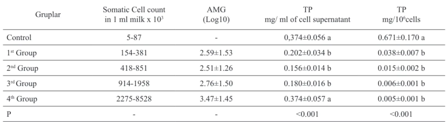
The SCC and AMG in 1 ml of collected milk sam-ples and TP levels in 1 ml of somatic cell homogenate supernatants are presented in Table 1. Total protein levels were found to be inconsistant when assessed as ml and were lower in the 1st, 2nd, and 3rd groups than
in the control and 4th groups. When the TP levels in one
million cells (106) were evaluated, a significant
differ-ence between the control group and all the mastitis groups was found (p<0.001).
The NADPH levels of the somatic cell supernatants are presented in Table 2. There was a significant
decre-Table 1. Somatic cell count, aerobic mesophyle general organism (AMG) count in milk and total protein (TP) levels in milk cells (Mean±SE) (n=15)
Gruplar Somatic Cell count in 1 ml milk x 103 (Log10)AMG mg/ ml of cell supernatantTP mg/10TP 6cells
Control 5-87 - 0,374±0.056 a 0.671±0.170 a 1st Group 154-381 2.59±1.53 0.202±0.034 b 0.038±0.007 b 2nd Group 418-851 2.51±1.26 0.156±0.014 b 0.015±0.002 b 3rd Group 914-1958 2.76±1.50 0.180±0.016 b 0.006±0.001 b 4th Group 2275-8528 3.47±1.45 0.374±0.057 a 0.005±0.001 b P - - <0.001 <0.001
a–b Means within a column with different superscripts differ.
Table 2. Reduced nicotinamide adenine dinucleotide phosphate (NADPH) and total glutathione (tGSH) levels in milk cells (Mean±SE) (n=15).
NADPH
nmol/ml nmol/mg proteinNADPH nmol/10NADPH6 cells
tGSH
nmol/ml nmol/mg proteintGSH nmol/10tGSH6 cells Control 1.233±0.175 4.24± 0.73 b 2.623±0.780 a 42.47±4.07 a 122.88±13.08 c 86.77±23.60 a 1st Group 0.895±0.120 6.35±1.50 ab 0.159±0.021 b 29.76±3.46 b 204.27±38.76 abc 5.33±0.67 b 2nd Group 1.156±0.176 9.03±2.30 a 0.116±0.022 b 33.23±3.05 ab 226.95±22.40 a 3.23±0.33 b 3rd Group 0.995±0.141 5.00±0.70 ab 0.035±0.005 b 36.09±2.36 ab 214.64±18.30 ab 1.28±0.15 b 4th Group 1.525±0.451 3.31±1.12 b 0.020±0.005 b 36.65±3.59 ab 139.82±34.33 bc 0.54±0.05 b P - <0.05 <0.001 <0.05 <0.05 <0.001
ase in the mastitis groups compared to the control group as regards nmol/106 cells (p<0.001).
tGSH levels in the somatic cell supernatants are also presented in Table 2. When assessed as nmol/mg protein, tGSH levels were suspicious; 2nd group levels
were higher than those in the control and 4th groups.
However, when assessed as nmol/106 cells, the level of
the control group was higher than that in all the mastitis groups (p<0.001).
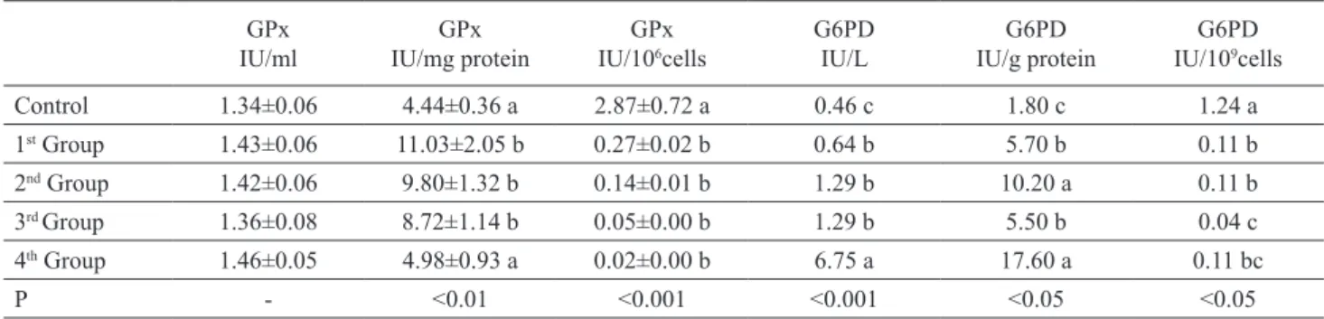
G6PD and GPx activities in the somatic cell super-natants are presented in Table 3. When assessed as IU/L and IU/g protein, the G6PD levels of the 4th group were
higher than those in the other groups, whereas when assessed as U/109 cells, the control group’s G6PD levels
were higher than those in the mastitis groups (p < 0.05). As regards GPx activity, when assessed as IU/mg protein, 1st, 2nd and 3rd group GPx activities were higher
(p<0.01) from that in the control and 4th groups,
where-as a significant decrewhere-ase wwhere-as determined in the mwhere-astitic groups compared to the control group as regards IU/106
cells (p<0.001).
The correlations between the biochemical parame-ters and SCC and AMG are presented in Table 4. Milk SCC was positively correlated with AMG (r=0.561,
Table 3. Glutathione peroxidase (GPx, Mean±SE) and glucose-6-phosphate dehydrogenase (G6PD, Median) activities in milk cells (n=15).
GPx
IU/ml IU/mg proteinGPx IU/10GPx6cells G6PDIU/L IU/g proteinG6PD IU/10G6PD9cells
Control 1.34±0.06 4.44±0.36 a 2.87±0.72 a 0.46 c 1.80 c 1.24 a 1st Group 1.43±0.06 11.03±2.05 b 0.27±0.02 b 0.64 b 5.70 b 0.11 b 2nd Group 1.42±0.06 9.80±1.32 b 0.14±0.01 b 1.29 b 10.20 a 0.11 b 3rd Group 1.36±0.08 8.72±1.14 b 0.05±0.00 b 1.29 b 5.50 b 0.04 c 4th Group 1.46±0.05 4.98±0.93 a 0.02±0.00 b 6.75 a 17.60 a 0.11 bc P - <0.01 <0.001 <0.001 <0.05 <0.05
a–c Means within a column with different superscripts differ.
Table 4. Correlations between the parameters (n=75).
AMG NADPH (nmol/ml) TP (mg/ ml) tGSH (nmol/ml) GPx (IU/ml) G6PD (IU/L)
Somatic Cell Count 0.561 * 0.380** 0.347** 0.174 0.136 0.540*
AMG 0.428** 0.638* 0.164 0.155 0.171 NADPH (nmol/ml) 0.239*** 0.155 0.265*** 0.248**** TP (mg/ml) 0.354** 0.048 0.643* tGSH (nmol/ml) -0.306*** 0.198 GPx (U/ml) 0.007 * p<0.001, ** p<0.01, *** p<0.05, **** p=0.056
Table 5. Pathogens isolated from mastitic milks (n=15, totally 60 milk samples).
Patogens Number- (%)
CNS (Coagulase Negative Staphylococcus) 28 (66.6)
Streptococcus spp. 4 (9.5)
Bacillus spp. 4 (9.5)
Yeast 2 (4.8)
Mixed Infection 4 (9.5)
Total Infection with pathogens 42 (100)
Total Infection with non-pathogens 18
No Growth 60
p<0.001), NADPH (r=0.380, p<0.01), TP (r=0.347, p<0.01) and G6PD (r=0.540, p<0.001). There was also positive correlation between NADPH (r=0.428, p<0.01), TP (r=0,638, p<0.001) and AMG. NADPH was positively correlated with TP (r=0.239, p<0.05), GPx (r=0.265, p<0.05) and G6PD (r=0.248, p=0.056). Total protein was positively correlated with tGSH (r=0.354, p<0.01) and G6PD (r=0.643, p<0.001). There was a negative correlation between tGSH and GPx activity (r=-0.306, p<0.05).
Pathogens isolated from the milk with mastitis are presented in Table 5. The ratio of the pathogens was as follows: CNS 66.6%, Streptococcus spp 9.5%, Bacillus
spp 9.5%, yeast 4.8%, and mixed infections 9.5%.
Because insufficient pathogens were found for each group, a correlation calculation between biochemical parameters and pathogens could not be conducted.
Discussion
Mastitis causes physical, chemical, and bacteriolog-ical changes in milk and pathologbacteriolog-ical changes in mam-mary glandular tissue (Sharma 2007). In this study, SCC was performed on milk samples which were col-lected according to CMT scoring and it was found that CMT scoring was not fully coherent with the SCC, which can be explained by the subjectivity of CMT scoring (Holtgrew-Bohling 2016). Correlations between the changes in the biochemical parameters in the super-natant obtained by concentration and homogenization of milk cells and SCC and AMG were determined, and the significant differences between the values of healthy milk and mastitic milk were identified.
When examining the TP levels in 1 ml of cell homogenate supernatant, we found that healthy milk had higher TP levels than the milk with mastitis (1st, 2nd,
and 3rd groups) (p<0.001). In the 4th group, the value
was closer to that of the healthy milk; this outcome can be explained by the elevated cell count and subsequent relative increase in TP levels. On the other hand, if the TP levels in 106 cells are considered, the TP levels per
cell decreased as mastitis intensity increased. The cor-relation between the TP levels of all samples and SCC (r=0.347, p<0.01, n=75) echoed the results reported by Ritter et al. (1987).
In the present study, as the intensity of mastitis increased, G6PD activity in the supernatant of 1 ml of milk cell homogenate increased, but the difference was only statistically significant in the 4th group (Table 3).
Milk somatic cells are comprised of leukocytes (PMN), macrophages, and epithelial cells (Baştan 2010). When an infection develops in mammary tissue, the ratio of leukocyte and epithelial cell counts in milk changes
(Miller et al. 1993, Zhao and Lacasse 2008, Baştan 2010). Due to the chemotactic agents induced by the infectious agents, the PMNs in blood leak into milk, and PMNs start to play a major role in somatic cell composition (increasing from 5–25% to 90%) (Leitner et al. 2000, Riollet et al. 2000).
G6PD has been found in milk, breast tissue epithe-lial cells, and milk leukocytes (Ritter et al.1977, Shreve and Levy 1980, Grigor and Hartmann 1985). Grigor and Hartmann (1985) reported that, in rats, G6PD activ-ity was substantially low in milk, while the mammary gland epithelial cell G6PD activity was rather high. They also concluded that G6PD found in milk was mainly derived from epithelial cell necrosis. In our study, the higher G6PD activity per cell in healthy milk than in mastitic milk can be attributed to the fact that healthy epithelial cell ratio was found to be higher in healthy milk than in mastitic milk.
The chemotactic effects of the infectious agents cause PMNs in the blood to seep into the milk through diapedesis, and PMNs play a major role in somatic cell composition (they can increase from 5–25% to 90%) (Leitner et al. 2000, Riollet et al. 2000). Milk epithelial cells in SCC also decrease proportionally. Meanwhile, some fluid also leaks into the mammary gland from the veins and alters the content of the milk. The function of PMNs is to phagocytize the pathogen, but they damage mammary tissue during this process. The destructive effects of PMNs on mammary epithelial cells are not yet clearly understood (Zhao and Lacesse 2008). Some researchers (Boulanger et al. 2002, Paape et al. 2002) suggest that PMNs exert their effects by releasing reac-tive oxygen species (ROS) outside the cell and secret-ing proteolytic enzymes (degranulation). Free radicals produced by leukocytes during inflammation are one of the body’s defenses against microorganisms. Research has shown that, depending on the degree of mastitis and amount of PMN in the milk, the number of free radicals released into the milk increases, leading to damage to the mammary gland epithelial cells (Zhao and Lacasse 2008). Meanwhile, parallel to increased ROS and pro-teolytic enzymes, mammary epithelial tissue starts to be damaged (Leitner et al. 2000, Zhao and Lacasse 2008). Since PMNs with low G6PD activity will have lower resistance against ROS (Cooper et al. 1972), they can be degraded after phagocytosis of the infectious agents; the ROS they contain will leak into the milk. A PMN cell with insufficient capacity for antioxidant enzymes can be degraded after bacteria phagocytosis, and its contents (ROS and proteolytic enzymes), which are released outside, can cause necrosis in mammary epi-thelial tissues. Thus, udder epiepi-thelial cells undergo necrosis, and their contents mix with milk. Thus, a higher number of epithelial cells in healthy milk than
in mastitic milk (Leitner et al. 2000) and a high level of G6PD in the epithelial cells will cause higher levels of G6PD per cell in healthy milk. On the other hand, in mastitic milk, high leukocyte levels and low epitheli-al cell colony numbers will reduce the amount of G6PD per cell. A positive correlation between G6PD and SCC was found in all samples, and these results cohere with those of Ritter et al. (1987). In addition, the positive correlation identified between NADPH levels and SCC and TP levels shows compliance with G6PD.
The microbiological results of this study showed that infections with CNS were much more frequent than those with other pathogens. It has been also reported by Yağcı (2008) that CNS in subclinical mastitis are more dominant than the other agents.
When the biochemical parameter levels in milk were investigated, many factors such as PMN, necrotic epithelial cells, fluid leak to milk from extracellular flu-id, and type of infectious agent complicated the utiliza-tion of biochemical parameters (in ml milk) in mastitis diagnosis and prognosis. In previous studies, (Dündar et al. 2000, Weiss et al. 2004, Erişir et al. 2011, Szczubial et al. 2012) biochemical parameters found in 1 ml of milk with subclinical mastitis were evaluated. However, due to differences between the changes in milk somatic cell composition (Leitner et al. 2000), the authors suggest that evaluating the parameter per somatic cell would be more appropriate. In future studies, separating epithelium, leukocytes, and patho-gens in milk with mastitis and identifying G6PD activity in each cell type is recommended.
Conclusion
Considering the parameters analyzed in this study in terms of ml and mg/protein, while different changes among groups were detected, the parameter levels per somatic cell were significantly lower than those with healthy milk. When evaluating the enzyme and oxida-tive stress parameters in milk, it is more suitable to as-sign values based on cell count rather than ml of milk. The linear correlation between the SCC and AMG, milk cell NADPH, TP and G6PD suggests that these param-eters could be used as markers of mastitis. The study also found that CNS was the dominant pathogens in subclinical mastitis cases.
Acknowledgements
This study was supported by Scientific Research Coordinating Office (BAP) of Mustafa Kemal Univer-sity (Project No. 12161).
References
Akalın PP, Başpınar N, Çoyan K, Bucak MN, Güngör Ş, Öztürk C (2016) Effects of ultrasonication on damaged spermatozoa and mitochondrial activity rate. Turk J Vet Anim Sci 40: 195-199.
Albenzio M, Taibi L, Muscio A, Sevi A (2002) Prevalence and etiology of subclinical mastitis in intensively managed flocks and related changes in the yield and quality of ewe milk. Small Rum Res 43: 219-226.
Ames BN, Shigenaga MK, Hagen TM (1993) Oxidants, anti-oxidants, and the degenerative diseases of aging. Proc Natl Acad Sci USA 90: 7915-7922.
Ayala A, Fabregat I, Machado A (1991) The role of NADPH in the regulation of glucose-6-phosphate and 6-phospho-gluconate dehydrogenases in rat adipose tissue. Mol Cell Biochem 105: 1-5.
Baştan A (2010) Breast health and problems in cows. 1st ed., Kardelen Ofset Press, Ankara, pp 64-73.
Bergonier D, Bertholet X (2003) New advances in epizootiol-ogy and control of ewe mastitis. Livestock Prod Sci 79: 1-16.
Beutler E (1971) Red cell metabolism. Manual of biochemical methods. Academic Press, London, pp 68-70.
Beutler E (1975) Red cell metabolism. In: Beutler E (ed) A manual of biochemical methods. 2nd ed., Grunef and Strottan Press, New York, pp 67-69.
Boulanger V, Zhao X, Lacasse P (2002) Protective effect of melatonin and catalase in bovine neutrophil-induced model of mammary cell damage. J Dairy Sci 85: 562-569. Bradford MM (1976) A rapid and sensitive method for the
quantitation of microgram quantities of protein utilizing the principle of protein-dye binding. Anal Biochem 72: 248-254.
Cappellini MD, Forelli G(2008) Glucose-6-phosphate dehy-drogenase deficiency. Lancet 371: 64-74.
Cooper MR, DeChatelet LR, McCall CE, LaVia MF, Spurr CL, Baehner RL (1972) Complete deficiency of leukocyte glucose-6-phosphate dehydrogenase with defective bac-tericidal activity. J Clin Invest 51: 769-778.
Dündar Y, Eryavuz A, Aslan R, Uçar M (2000) Malondialde-hyde and glucose-6-phosphate dehydrogenase levels in healthy and subclinical mastitic cows. Van Health Sciences Journal 6: 84-86.
Erişir M, Kandemir F M, Yüksel M (2011) The effect of sub-clinical mastitis on MDA, GSH levels and GSH-Px, CAT activities in milk of cows. Fırat Uni Sagl Bil Vet Derg 25: 67-70.
Grigor MR, Hartman PE (1985) NADP-linked dehydrogenases in secreted milk. J Dairy Res 52: 501-506.
Holtgrew-Bohling K (2016) Large animal clinical procedures for veterinary technicians, 3rd ed., Elsevier Inc Press, US, pp 445.
Hsieh YT, Lin MH, Ho MY, Chen LC, Chen CC, Shu JC
(2013) Glucose-6-phosphate dehydrogenase (G6PD)-
-deficient epithelial cells are less tolerant to infection by Staphylococcus aureus. PLOS One 8: e79566. Krebs HA, Eggleston LV (1974) The regulation of the
pentose phosphate cycle in rat liver. Adv Enzyme Regul 12: 421-434.
Leitner G, Shoshani E, Krifucks O, Chaffer M, Saran A (2000) Milk leucocyte population patterns in bovine udder infec-tion of different aetiology. J Vet Med B 47: 581-589.
Lee CS, Wooding FB, Kemp P (1980) Identification, proper-ties, and differential counts of cell-populations using electron-microscopy of dry cows secretions, colostrum and milk from normal cows. J Dairy Res 47: 39-50. McCord JM (1993) Human disease, free radicals, and the
oxidant/antioxidant balance. Clin Biochem 26: 351-357. Mehta A, Mason PJ, Vulliamy TJ (2000) Glucose-6-phos-phate dehydrogenase deficiency. Baillieres Best Pract Res Clin Haematol 13: 21-38.
Miller JK, Brzezinska-Slebodzinska E, Madsen FC (1993) Oxidative stress, antioxidants, and animal function. J Dairy Sci 76: 2812-2823.
Paape M, Mehrzad J, Zhao X, Detilleux J, Burvenich C (2002) Defense of the bovine mammary gland by polymorpho-nuclear neutrophil leukocytes. J Mammary Gland Biol Neoplasia 7: 109–121.
Pyorala S, Taponen S (2009) Coagulase-negative staphylo-cocci - emerging mastitis pathogens. Vet Microbiol 134: 3-8.
Quinn PJ, Carter ME, Markey BK, Carter GR (1994) Clinical Veterinary Microbiology. Mosby-Year Book Europe Limited, England, pp 209-236.
Rajala-Schultz PJ, Smith KL, Hogan JS, Love BC (2004) Antimicrobial susceptibility of mastitis pathogens from first lactation and older cows. Vet Microbiol 102: 33-42. Riollet C, Rainard P, Poutrel B (2000) Cells and cytokines in
inflammatory secretions of bovine mammary gland. Adv Exp Med Biol 480: 247-258.
Ritter C, Conti A, Morse GE (1977) Hexose shunt dehydroge-nase activity in leukocytes isolated from bovine milk, J Dairy Sci 60: 1987-1990.
Schepers AJ, Lam TJ, Schukken YH, Wilmink JB, Hanekamp WJ (1997) Estimation of variance components for somatic cell counts to determine thresholds for unin-fected quarters. J Dairy Sci 80: 1833-1840.
Sharma N (2007) Alternative approach to control intramam-mary infection in dairy cows- A review. Asian J Anim Vet Adv 2: 50-62.
Shreve DS, Levy HR (1980) Kinetic mechanism of glu-cose-6-phosphate dehydrogenase from the lactating rat mammary gland. Implications for regulation. J Biol Chem 255: 2670-2677.
Swanson KMJ, Busta FF, Peterson EH, Johnson MG (1992) Colony count methods. In: Vanderzant C, Splittstoesser DF (ed) Compendium of methods for the microbiological examination of foods. American Public Health Associa-tion, Washington DC, pp 75-95.
Szczubial M, Dabrowski R, Kankofer M, Bochniarz M, Komar MM (2012) Concentration of serum amyloid A and ceruloplasmin activity in milk from cows with sub-clinical mastitis caused by different pathogens. Pol J Vet Sci 15: 291-296.
Weiss WP, Hogan JS, Smith KL (2004) Changes in vitamin C concentrations in plasma and milk from dairy cows after an intramammary infusion of Escherichia coli. J Dairy Sci 87: 32-37.
Yağcı İP (2008) Subclinical mastitis in sheeps; etiology, epidemiology and diagnosis methods. Kafkas Univ Vet Fak Derg 14: 117-122.
Zhao X, Lacasse P (2008) Mammary tissue damage during bovine mastitis: causes and control. J Anim Sci 86(13): 57-65.