Review Paper
What does the broken brain say to the neuroscientist? Oscillations and
connectivity in schizophrenia, Alzheimer's disease, and bipolar disorder
E. Ba
şar
a,⁎
, C. Schmiedt-Fehr
b, B. Mathes
b, B. Femir
a, D.D. Emek-Sava
ş
c,d, E. Tülay
a, D. Tan
f, A. Düzgün
a,
B. Güntekin
a, A. Özerdem
c,d,e, G. Yener
a,c,d,e, C. Ba
şar-Eroğlu
ba
Istanbul Kultur University, Brain Dynamics, Cognition and Complex Systems Research Center, Istanbul, Turkey
b
Bremen University, Institute of Psychology and Cognition Research, Bremen, Germany
c
Dokuz Eylül University, Department of Neurosciences, Izmir, Turkey
d
Dokuz Eylül University, Department of Psychiatry, Izmir, Turkey
eDokuz Eylül University, Multidisciplinary Brain Dynamics Research Center, Izmir, Turkey f
Maltepe University, Department of Psychiatry, Istanbul, Turkey
a b s t r a c t
a r t i c l e i n f o
Available online 7 February 2015 Keywords: Oscillations Connectivity Schizophrenia Alzheimer's disease Bipolar disorder
The application of the concept and methods of brain oscillations has been an important research area in neurosciences. In the last decades, besides the application in cognitive processes, the study of changes in brain oscillations in diseases has also become an important focal point of research. In the present paper, some remark-able examples in three different diseases are taken into consideration: 1) schizophrenia (SZ), 2) Alzheimer's disease (AD), 3) bipolar disorders (BD).
In the current literature, decreased oscillations in cortical recordings are observed in most of the pathologies. For example, decrease of gamma activity in SZ, decrease of delta activity in almost all diseases, as well as frequency shifts in alpha and the lower frequencies were recorded. However, there are also paradoxical cases in which an increase of oscillatory activities is observed. In BD, whereas alpha activity is greatly decreased, a huge increase of beta activity is observed. Or, in SZ, a paradoxical increase of gamma activity can be observed during cognitive loading. We also observed paradoxical changes in the analysis of connectivity. In AD, wefind that alpha, delta, and theta coherences between distant parts of the cortex are greatly decreased, whereas in the gamma band, event-related coherences attain very high values.
The comparison of the results and paradoxical changes in diseases may lead to important conclusions related to the web of oscillations and neurotransmitters. In turn, we could gain new insights to approach“brain function”, in general.
© 2016 The Authors. Published by Elsevier B.V. This is an open access article under the CC BY license (http://creativecommons.org/licenses/by/4.0/).
1. Introduction
This paper aims to give a global, but efficient, orientation for electro-physiological description of brain dysfunction for special cases, includ-ing unexpected results, controversies, and conformities in oscillatory dynamics. Examples are chosen from three encountered diseases: schizophrenia, Alzheimer's disease, and bipolar disorder.
The concepts and methods of brain oscillations in healthy subjects have invaded neuroscience literature over the past twenty years; they are used to learn the functioning of the healthy brain and observe changes in different subjects (Başar et al., 1998; Başar-Eroglu et al., 1991, 1993, 2001; Sakowitz et al., 2001; Schürmann et al., 1997). These methods can also be used as biomarkers for recognition of diseases, differentiation of diseases, and progression in diseases.
In the present comparative report, we attack another important problem: Are there any shared mechanisms in these very common neuropsychiatric disorders? What are similar oscillatory reactions? Are there clear controversies? Can the obtained results serve to differen-tiate neuropsychiatric diseases? In future discussions, an ensemble of results from already published work and additional new results can provide a type of brainstorming for electrophysiological understanding in diseases.
The seemingly paradoxical or unexpected results in pathology could, in the future, provide new avenues or keys to understand more differentiated brain functions. Different diseases are results of modified neurotransmitter release (see alsoKoch et al., 2016-in this volume; Sanchez-Alaveza and Ehlers, 2016-in this issue; Başar and Düzgün, in this volume). Accordingly, in the synopsis of the pres-ent report, we will tpres-entatively describe possibilities to discover new windows for the analysis of oscillations and connectivity. The present report does not cover a comprehensive view of oscillations in diseases; for that, we refer to (Başar et al., 2013; Yener and Başar, 2013b).
⁎ Corresponding author. Tel.: +90 212 498 43 92; fax: +90 212 498 45 46. E-mail address:[email protected](E. Başar).
http://dx.doi.org/10.1016/j.ijpsycho.2015.02.004
0167-8760/© 2016 The Authors. Published by Elsevier B.V. This is an open access article under the CC BY license (http://creativecommons.org/licenses/by/4.0/).
Contents lists available atScienceDirect
International Journal of Psychophysiology
In this manuscript, we want to emphasize some common and differ-ential patterns of electrophysiological changes in three common neuro-psychiatric entities, namely schizophrenia (SZ), Alzheimer's disease (AD), and bipolar disorder (BD). Our new results on alpha spectral power of SZ and its comparison with BD plus event-related gamma coherences of AD will be presented and discussed in light of our and other groups' earlierfindings.
1.1. Characteristics of“bipolar disorder”, “schizophrenia” and “Alzheimer's disease”
1.1.1. What is bipolar disorder?
Bipolar disorder is a lifetime illness which follows a relapsing and re-mitting course. Manic or depressive episodes relapse in an unpredict-able manner. Between the symptomatic episodes is the well-being state called “euthymia”. Symptoms of both manic and depressive episodes involve several domains such as mood, energy, motor activity, sleep, appetite, thought, and cognition. Recent years have witnessed documentation of a subclinical course with sleep and circadian rhythm disturbances, emotional dysregulation, and cognitive impairment between the full-blown mood episodes (Leboyer and Kupfer, 2010).
Bipolar disorder is one of the leading causes of disability worldwide (Murray and Lopez, 1996). Bipolar disorder types I and II together reach a 4.4% prevalence rate (Merikangas et al., 2007), which is considerably higher than the l% prevalence of another severe psychiatric disorder, schizophrenia. Data shows large disturbances in neurocognition during the different stages of bipolar disorder. Attention and memory deficits, impairment in verbal recall andfine motor skills, and disturbance of sustained attention is evident during depressive episodes. Attention, complex processing, memory, and emotional processing are dysfunc-tional in mania (Goldberg and Chengappa, 2009). Cognitive deficits re-main even during euthymia, where response inhibition, set-shifting, executive function, verbal memory, sustained attention, processing speed, visual memory, and verbalfluency have been shown to be dis-turbed (Bora et al., 2009). Such a wide range of cognitive disturbances involves dysfunction of neural circuits that run between prefrontal and striatal structures, with further projections to the thalamic nuclei (Vawter et al., 2000), and includes those that regulate cognitive, emo-tional, and social behavior. Recently, a temporal- and fronto-limbic-related cognitive impairment has been defined as a cognitive endophenotype (Bora et al., 2009).
1.1.2. What is schizophrenia?
Schizophrenia is a complex and severe mental disorder, affecting the participant's actions, perceptions, emotions, and cognitive functions (Andreasen, 1997; Gold, 2004). The lifetime prevalence of schizophre-nia is approximately 1% (Lewis and Lieberman, 2000; Saha et al., 2005). Very often the illness persists for a lifetime, rendering patients dependent on the public health system. Although the onset of the dis-ease is most common at the end of adolescence or the beginning of adulthood (Pantelis et al., 2007), the etiopathogenesis indicates that ge-netic predispositions and developmentally early“hits”, such as social stress, enhance the probability of developing schizophrenia (Rehn and Rees, 2005). The transition into the illness is marked by pathological changes of the brain, such as regional specific losses in gray and white matter (Pantelis et al., 2007). Similar to bipolar disorder, widely distrib-uted neural circuits seem to be altered in schizophrenia, predominantly affecting frontal, temporal, and sub-cortical structures (Pantelis et al., 2007). Cognitive deficits are apparent early, sometimes even before
distortions), and disorganized thinking. Negative symptoms include the blunting or diminishment of behavior and cognitive functions as well as affective expression and experience (Andreasen, 1997).
1.1.3. What is Alzheimer's disease?
Alzheimer's disease (AD) is a neurodegenerative disease that, in its most common form, is generally found in people over 65 years old. Approximately 24 million people worldwide have dementia, of which the majority (approximately 2/3) is due to AD (Ferri et al., 2005). Clini-cal signs of AD are characterized by progressive cognitive deterioration, together with declining activities in daily life and by neuropsychiatric symptoms or behavioral changes.
2. Alpha activity in Alzheimer's disease, schizophrenia and bipolar disorder
HansBerger (1929)was thefirst to observe alpha rhythm. It was ini-tially considered to be the brain's“idling rhythm”. Later, several authors stated that EEG was not noise and that selectively synchronized alpha oscillations in the mammalian and human brain are part of the fundamental functional signaling of the central nervous system (Başar, 1980; Lehmann, 1989; Nunez et al., 2001).
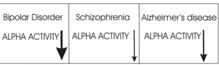
Decrease of spontaneous alpha activity is one of the common EEG parameters reported in Alzheimer's disease, bipolar disorder and schizophrenia (Itil et al., 1972, 1974; Iacono, 1982; Miyauchi et al., 1990; Sponheim et al., 1994, 2000; Alfimova and Uvarova, 2008, see
Fig. 1for a graphical summary). Since the cause and pathology behind these three diseases differ considerably, the generalization of this finding needs further exploration.
In this manuscript, we will therefore discuss the similarities and differences of reductions in spontaneous alpha activity in patients with Alzheimer's disease, bipolar disorder, and schizophrenia. As a first step, we will review some previously published results on sponta-neous alpha activity in patients with bipolar disorder with respect to an accompanying, new study that includes patients with schizophrenia.
Our previous study (Başar et al., 2012) was one of thefirst to show that spontaneous alpha activity (8–13 Hz) is highly reduced in drug-free, euthymic patients with bipolar disorder. In this study (Başar et al., 2012), we analyzed the spontaneous EEG activity (4 min eyes closed, 4 min eyes open) of eighteen drug-free DSM-IV euthymic bipolar patients and compared it to eighteen healthy controls. The re-sults showed that spontaneous EEG alpha power of healthy participants was significantly higher than the spontaneous EEG alpha power of euthymic patients during both the eyes open and eyes closed conditions.Fig. 2(modified fromBaşar et al., 2012) specifically depicts reduced alpha power in bipolar patients at the right and left occipital region for the eyes closed condition.
Fig. 1. Decrease of spontaneous alpha activity in bipolar disorder, schizophrenia and Alzheimer's disease.
2.1. Spontaneous alpha response in patients with schizophrenia 2.1.1. Participants
A total of nineteen inpatients (four female, mean age: 31.1 ± 2.0 years) and nineteen healthy controls (six female, mean age: 32.7 ± 2.2 years) gave informed consent and participated in the study. Seventeen patients andfifteen healthy controls were right hand-ed. Two patients and two healthy controls were left-handhand-ed. Handed-ness of two healthy controls is unknown. All patients met DSM-IV criteria for schizophrenia (APA, 1994) and displayed mainly negative symptoms. Mean illness duration was 6.4 ± 1.4 years. Fourteen patients were medicated on atypical antipsychotics, one patient with typical antipsychotics, and two patients on mixtures of atypical and typical antipsychotic drugs.
2.1.2. Experimental procedure and analysis
EEG was recorded during eyes open and eyes closed resting condi-tions from standard locacondi-tions (F3, F4, C3, C4, P3, P4, O1, O2) of the
interna-tional 10–20 system. Linked earlobes served as reference. The signal was amplified using a Nihon Kohden system (EEG-4421G), with band limits between 0.1 and 70 Hz (24 dB/octave) and an additional notch fil-ter at 50 Hz. Data was digitized at a 500 Hz sampling rate and analyzed
off-line. The EOG was registered from medial upper and lateral orbital rim of the right eye.
Data analysis was comparable to the study byBaşar et al. (2012), where a detailed description of all analytical steps is provided. In short, EEG signals were fragmented in artifact-free epochs of one second. The digital FFT-based (10% Hanning window) power spectrum analysis was performed for each epoch. The power values of each epoch were subsequently averaged for each participant for both conditions, eyes open and eyes closed. For the statistical analysis, the in-dividual maximum of the alpha power (as defined between 8 to 13 Hz) was determined. An analysis of variance (ANOVA) (mixed design) was performed separately for both conditions (eyes open and eyes closed), including two groups (patients and controls) as between-subject factors and eight electrodes as within-subject factors.
2.1.3. Results
For both conditions, alpha power was increased at posterior sites (eyes closed: F(7,252) = 26.2, pb .001; eyes open: F(7,252) = 5.6, pb .001). Although visual inspection ofFigs. 3 and 4indicated slight differences between groups, the statistical analysis did not reveal any significant differences (for all group and group × electrode effects: p≥ .27).
2.2. Comparison of spontaneous alpha activity in Alzheimer's disease, schizophrenia, and bipolar disorder
The reduction of alpha activity in patients with bipolar disorder is profound. As can be seen inFig. 2, the mean alpha power at occipital sites during the eyes closed condition in healthy participants was approximately 4μV2. In patients with bipolar disorder the mean
ampli-tude was reduced to approximately 1μV2. In contrast, no significant
differences were found between healthy participants and patients with schizophrenia. Although the studies had to be conducted in differ-ent EEG laboratories (Bipolar study: Istanbul, Turkey; schizophrenia study: Bremen, Germany), the healthy participants measured in Bre-men showed similar mean alpha power compared to the healthy partic-ipants measured in Istanbul. In patients with schizophrenia, the mean alpha power at occipital sites during the eyes closed condition was approximately 3.5μV2. This indicates comparability between the two
groups of healthy participants and, accordingly, that a decrease in alpha activity seems to be more severe in patients with bipolar disorder than those with schizophrenia.
The literature suggests that alpha activity in patients with schizo-phrenia is also reduced in comparison to healthy participants (Itil et al., 1972, 1974; Iacono, 1982; Miyauchi et al., 1990; Sponheim et al., 1994, 2000; Alfimova and Uvarova, 2008) and our results are similar, but there is not enough power to significantly demonstrate the differ-ences. It is also important to note that bipolar disorder patients were drug-free, whereas the schizophrenia patients were medicated, which might have had an influence on alpha power (Tislerova et al., 2008; Hyun et al., 2011). The main goal of this chapter was to evaluate similar-ities and differences of spontaneous alpha power between different psy-chiatric and neurological diseases. Taking the study byBaşar et al. (2012)and the above-presented data together, these results indicate that alpha power might be similar in bipolar disorder and schizophre-nia, even though its pathological decrease is more apparent in bipolar disorder. Future studies are needed that consequently compare different neuropsychiatric and neurological diseases to further explore the potential of spontaneous alpha power as a biomarker for differenti-ating between pathologies.
3. Discrepancy between schizophrenia and bipolar disorder: beta response oscillations
InSection 2, we presented the decrease of spontaneous alpha
activity as a similarity between patients with schizophrenia and those
Fig. 2. Grand averages of power spectra of drug-free euthymic bipolar patients and healthy controls for the eyes closed condition at O1, Oz,and O2electrode sites. Solid lines represent
healthy participants, dashed lines represent drug-free euthymic bipolar patients. (Modified fromBaşar et al., 2012).
with bipolar disorder. Although this is a similarity between these two diseases, it is important to note that the decrease of spontaneous alpha activity was more pronounced in bipolar disorder patients. On the other hand, beta oscillatory responses appear to be one of the dis-crepancies between these two diseases. Since it is very important to
focus on the discrepancies in search of biomarkers for neuropsychi-atric diseases, beta response oscillations could be a good candidate (Fig. 5).
Beta response reduction was reported in schizophrenia patients during different paradigms (Spencer et al., 2003; Krishnan et al., 2005; Fig. 3. Grand averages of power spectra of schizophrenia patients and healthy controls for the eyes open recording session over occipital electrodes. Black lines represent healthy subjects, red lines represent patients with schizophrenia.
Uhlhaas et al., 2006; Pachou et al., 2008; Barr et al., 2010; Riečanský et al., 2010; Arnfred et al., 2011). On the other hand, an increase of beta response oscillations was found in bipolar disorder patients during the visual oddball paradigm (Özerdem et al., 2008), auditory oddball paradigm (Ethridge et al., 2012), as well as during auditory paired stimuli (Hamm et al., 2012).
Spencer et al. (2003)andUhlhaas et al. (2006)showed a reduction of phase synchrony in beta oscillations during “Gestalt” stimuli.
Pachou et al. (2008)andBarr et al. (2010)showed reduced beta
responses during N-back tasks. During visual steady state responses, schizophrenia subjects showed reduced beta power compared to healthy controls (Krishnan et al., 2005; Riečanský et al., 2010).
Fig. 4. Grand averages of power spectra of schizophrenia patients and healthy controls for the eyes closed recording session over frontal, central, parietal and occipital electrodes. Black lines represent healthy subjects, red lines represent schizophrenia patients.
On the other hand in bipolar disorder patients' event-related beta re-sponses were increased compared to healthy subjects.Özerdem et al.
(2008)showed that manic bipolar disorder patients had increased
beta responses compared to healthy subjects upon application of the vi-sual oddball paradigm. Furthermore, in the same study, the authors re-ported normalized beta responses after valproate monotherapy. In contrast to the normalizing effect of valproate, lithium has a different ef-fect on beta response oscillations.Atagün et al. (2015)during auditory oddball paradigm andTan et al. (2015)during visual oddball paradigm showed that lithium treated bipolar disorder patients had increased beta responses compared to both drug-free bipolar disorder patients and healthy controls.
Hamm et al. (2012)reported increased beta power in bipolar disor-der patients compared to healthy controls and schizophrenia patients upon application of auditory paired stimuli. In a separate study, the same group of researchers also showed increased late (530–560 ms) beta-gamma power (around 37 Hz) in bipolar disorder patients relative to healthy controls and schizophrenia patients (Ethridge et al., 2012). Increased beta power for bipolar disorder patients compared to healthy controls upon applying facial expression stimuli has also been reported (Lee et al., 2010).Fig. 5schematically shows the increase of beta re-sponse oscillations in bipolar disorder patients compared to healthy controls and schizophrenia patients.
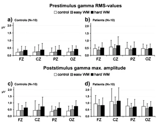
3.1. Increase of cognitive gamma in SCZ
The strategy referred to inFig. 6is important, since the alteration of gamma activity in schizophrenia is not manifested only with reduced gamma responses. However, during a working memory task paradigm, patients showed a sustained increase in gamma activity, whereas healthy subjects show a differentiation related to the varied degrees of difficulty in the working memory task.
4. Discrepancy between Alzheimer's disease and bipolar disorder: event-related gamma coherence
One of the other discrepancies that we will present in this manu-script is the event-related gamma coherence in Alzheimer's disease and bipolar disorder patients. Previously, our group showed lower values of event-related coherences in delta, theta, and alpha frequency ranges in AD subjects (Başar et al., 2010; Güntekin et al., 2008), and that event-related alpha coherence values recover with cholinergic treatment (Başar et al., 2010; Yener and Başar, 2010). In this manuscript we will present our new results on event-related gamma coherence in AD subjects and healthy controls upon application of the visual oddball paradigm. In contrast to a decrease in low frequency event-related coherences, event-related gamma coherence values displays higher values in AD subjects compared to healthy control subjects.
Fig. 5. The increase of beta response oscillations in bipolar disorder patients in comparison to healthy controls and schizophrenia patients.
Fig. 6. Upper panel represents prestimulus RMS gamma values in control subjects and in schizophrenia patients in the three tasks. Lower panel shows post-stimulus maximal gamma amplitudes.
(Modified fromBasar-Eroglu et al., 2007).
Fig. 7. Discrepancy between Alzheimer's disease and bipolar disorder: event-related gamma coherence.
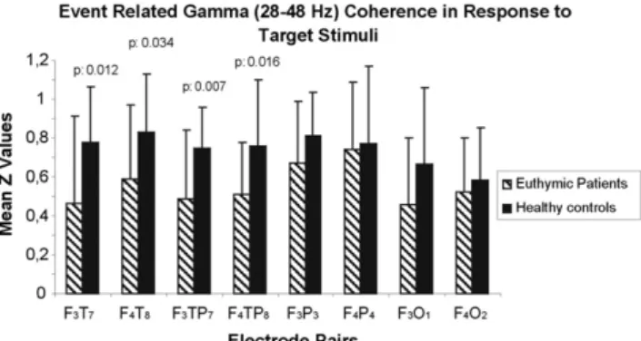
In contrast to AD subjects, manic (Özerdem et al., 2010) and euthymic bipolar disorder patients (Özerdem et al., 2011) displayed lower values of event-related gamma coherences upon application of the visual oddball paradigm. The next section briefly describes this reduction both in the manic and euthymic stage of bipolar disorder (Fig. 7).
4.1. Event-related gamma coherence in bipolar disorder patients In two separate experiments with different groups of subjects from our earlier reports, we analyzed the event-related gamma coherence in manic bipolar patients, drug-free euthymic bipolar patients, and healthy controls (Özerdem et al., 2010, 2011).Özerdem et al. (2010) an-alyzed evoked and event-related gamma coherence upon application of the simple light and visual oddball paradigms. The study subject group included 10 drug-free manic patients and 10 healthy controls. The EEG recordings of manic patients were performed a second time after six weeks valproate monotherapy; the healthy subjects also had a second set of EEG recordings performed six weeks after thefirst set. Event-related gamma coherence values for all subject groups and for all experimental recordings were analyzed in 28–48 Hz frequency band for intra-hemispheric pairs (F3–P3, F3–T5, F3–O1, C3–O1, F4–P4, F4–T6,
F4–O2,and C4–O2) and inter-hemispheric pairs (F3–F4, C3–C4, T3–T4,
T5–T6, P3–P4, and O1–O2). Event-related gamma coherence of the
drug-free manic bipolar patients were lower than the healthy controls in the first recording and analysis. After six weeks of valproate
monotherapy, the difference between event-related gamma coherence of bipolar patients and healthy controls became insignificant.
The study byÖzerdem et al. (2011)analyzed the event-related gamma coherence in drug-free euthymic patients. The study included 20 drug-free euthymic patients and 20 healthy controls. Event-related gamma coherence was analyzed for intra-hemispheric electrode pairs. (F3–T7, F4–T8, F3–TP7, F4–TP8, F3–P3, F4–P4, F3–O1, and F4–O2). The
results showed that patients had lower event-related gamma coherence compared to healthy controls, especially over fronto-temporal (F3–T7,
F4–T8) and fronto-temporoparietal (F3–TP7, F4–TP8) locations. In
con-trast to studies byÖzerdem et al. (2010, 2011), a study byVelasques
et al. (2013)showed increased gamma coherence in manic bipolar
disorder patients during prosaccadic attention task. The authors analyzed the event-related gamma coherence for inter-hemispheric short distance electrode pairs and found increased gamma coherence values for bipolar patients. On the other hand,Özerdem et al. (2010)
did notfind differences in inter-hemispheric electrode pairs. However, the increased event-related gamma coherence in bipolar disorder patients was seen in long distance intra-hemispheric electrode pairs (Özerdem et al., 2010, 2011).
4.2. Event-related gamma coherences in Alzheimer's disease 4.2.1. Subjects
Changes in coherence in Alzheimer's disease during a cognitive task (P300 Oddball) were analyzed by the visual oddball paradigm. Twelve healthy participants (7 M, 5 F, mean age: 67.5, SD: 4.77) and twelve un-treated patients with Alzheimer's disease according to DSM-IV criteria and the NINCDS-ADRDA criteria (5 M, 7 F, mean age: 75.15, SD: 8.81) participated in the study. The mean educational years was 9.5 (SD = 5.45) for control group and 5.58 (SD = 2.64) for AD subjects. The mean of MMSE scores was 28.91 (SD = 1.08) for control group and 23.27 (SD = 1.61) for AD patients out of a possible 30 points. 4.2.2. Statistical analysis
Statistical analyses were performed in SPSS Statistics 15.0. Repeated Measures Analysis of Variance (ANOVA) was used to investigate the changes in coherence in Alzheimer's disease and healthy subjects. Five locations (fronto-temporal, fronto-temporoparietal, fronto-parietal, fronto-occipital, and centro-occipital) and two lateralizations (left and right hemispheres) were included as within-subject factors. Sex and group were included as between-subjects factors. Greenhouse–Geisser corrected p-values are reported. Post-hoc comparison tests were
Fig. 8. Event-related gamma coherence in response to target stimulation for euthymic bipolar disorder patients and healthy controls.
(Modified fromÖzerdem et al., 2011).
Fig. 9. Mean z values for event-related target coherence in response to the visual oddball paradigm at all electrode pairs in 24.4–30 Hz frequency range. Untreated AD patients have increased right and left hemispheric z values at all electrode pairs for target detection. However, the difference is not statistically significant.
done by using Bonferroni correction. All results are reported with a sig-nificance level p = b.05. Oscillatory activity in the 0–800 ms time win-dow was compared in two groups in the 24.4–30 Hz, 30–35.2 Hz, and 40–48 Hz frequency bands at ten electrode pairs (F3–T7, F4–T8, F3–TP7,
F4–TP8, F3–P3, F4–P4, F3–O1, F4–O2, C3–O1and C4–O2).
4.2.3. Results
4.2.3.1. Event-related gamma coherence in 24.4–30 Hz frequency range. The importance of higher frequency responses was emphasized as early as byBaşar (1972)andBaşar and Ungan (1973).
Although the AD patients showed higher coherence values than healthy participants, the between group difference was not statistically significant. Repeated Measures ANOVA showed that location is statisti-cally significant in groups (F: 12.69; df: 2.37; p: 0.000). Coherence at both the fronto-parietal and centro-occipital locations is significantly higher than coherence at the temporoparietal and fronto-occipital locations, pb 0.05. Two-tailed t-test results show that AD patients have significantly higher coherence only at the C3O1electrode
pair, pb 0.05 compared to healthy subjects. After Bonferroni correction, none of the electrode pairs are significantly different between groups (Fig. 9).
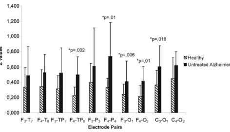
4.2.3.2. Event-related gamma coherence in 30–35.2 Hz frequency range. The between group difference for evoked-coherence values was signif-icant, showing that untreated AD patients have increased coherence compared to healthy participants, p = 0.004. Repeated Measures ANOVA showed that location is statistically significant in groups (F: 10.63; df: 2.16; p: 0.000). The fronto-parietal location has the highest coherence in both groups, whereas the lowest coherence is seen at the fronto-temporoparietal location. Coherence at fronto-parietal and centro-parietal locations is significantly higher than coherence at fronto-temporal, fronto-temporo parietal and fronto-occipital locations, pb 0.05. Two-tailed t-test results show that AD patients have signifi-cantly higher coherence only at F3–T7,F3–TP7,and F4–O2electrode
pairs, pb 0.05 compared to healthy subjects. After Bonferroni correction, F3–TP7,and F4–O2electrode pairs are significantly different between
groups (Fig. 10).
Fig. 10. Mean z values for event-related target coherence in response to the visual oddball paradigm at all electrode pairs in 30–35 Hz frequency range. Untreated AD patients have increased right and left hemispheric z values at all electrode pairs for target detection. Significant change was observed in F4–P4(decrease: 56%), F4–TP8(decrease: 58%), F3–O1(decrease:
48%), F4–O2(decrease: 50%), and C3–O1(decrease: 38%) electrode pairs.
Fig. 11. Mean z values for even-related target coherence in response to the visual oddball paradigm at all electrode pairs in 40–48 Hz frequency range. Untreated AD patients have increased right and left hemispheric z values at all electrode pairs for target detection. Significant change was observed in F4–P4(decrease: 48%), F3–O1(decrease: 47%), F4–O2(decrease:
4.2.3.3. Event-related gamma coherence in 40–48 Hz frequency RANGE. The between group difference for visual event-related target coherence values was significant, indicating that untreated AD patients have increased coherence compared to healthy participants, p = 0.018. ANOVA showed that location is statistically significant in groups (F: 14.69; df: 2.69; p: 0.000). Coherence at fronto-parietal and centro-occipital locations is significantly higher than coherence at fronto-temporal, fronto-temporoparietal, and fronto-occipital loca-tions, pb 0.05. ANOVA revealed the significant effect of location and group (F: 4.38; df: 2.69; p: 0.01). However, post-hoc analysis with Bonferroni correction showed that none of the values are significantly different from each other. Two-tailed t-test results show that AD pa-tients have significantly higher coherence at F4–P4, F3–O1, F4–O2,and
C3–O1electrode pairs, pb 0.05 compared to healthy subjects. After
Bonferroni correction, only C3–O1electrode pair is significantly different
between groups (Fig. 11).
4.2.3.3.1. What does the coherence change in pathology mean?. Accord-ing to the results inFig. 8, euthymic patients showed bilaterally-diminished long-distance gamma coherence between frontal and tem-poral regions, as well as between frontal and temporoparietal regions, compared to healthy controls. The decrease in event-related coherence differed topologically, and ranged between 28.85% and 44.44%. Oscilla-tory responses to target stimuli are manifestations of working memory processes. Therefore, the coherence decrease in response to target stimuli suggests inadequate connectivity between different parts of the brain while under cognitive load. The discovery of a large coherence decrease under cognitive load, but not in response to simple sensory stimuli (Özerdem et al., 2011), is a majorfinding with regard to the well-documented cognitive dysfunction across all states of bipolar disorder (Martínez-Arán et al., 2004).
Structural imaging studies showed abnormalities in the prefrontal cortex, medial temporal lobe, and subcortical structures in bipolar disor-der (Strakowski et al., 2005), which suggests a diminished prefrontal modulation of subcortical and medial temporal structures within the anterior limbic network (e.g., amygdala, anterior striatum, and thala-mus). The reduction is usually seen as a negative correlation between ventral prefrontal cortex (vPFC) and amygdala in patients with bipolar disorder compared to healthy controls.Chepenik et al. (2010)supports this. The previously reported decrease in gamma coherence between frontal and temporal regions also reveals a functional fronto-temporal connectivity disturbance in mania (Özerdem et al., 2010), which is in line with the above-mentioned imagingfindings. Taken together, struc-tural and functional data highlight the importance of fronto-temporal circuits in bipolar disorder.
4.2.3.3.2. The association between gamma oscillations and GABA/gluta-mate neurotransmission. Gamma oscillations are essential in information processing during sensory perception, motor behavior, and memory formation. They coordinate neuronal activity in hippocampal and neo-cortical networks (Kann et al., 2014). Cortico-cortical communication and the large-scale integration of distributed sets of neurons are needed for a well-functioning cognitive ability and require synchronous neural gamma oscillations (Rodriguez et al., 1999).
Gamma oscillations originate within networks of inhibitory GABAergic interneurons (Gray and McCormick, 1996). This causes a membrane-potential oscillation in long-axoned projection neurons (i.e., pyramidal cells in the neocortex, hippocampus, and thalamo-cortical neurons) to provide communication between spatially separate sites and control brain function. Gamma oscillations are known to have a network-inhibitory effect and were proposed to be driven by metab-otropic glutamate receptor activation (Whittington et al., 1995). GABAergic modulation is required for synchronization of glutamatergic firing (Whittington et al., 2000). Taken together, there is an interplay between the GABA/glutamate system and the gamma oscillations.
Over activation of the glutamatergic system and hypoactivity of the GABAergic neurotransmission are reported to be involved in the pathogenesis of mood disorders (Frisardi et al., 2011). In addition to
that, low GABA activity (Petty, 1995) and abnormalities affecting GABAergic inhibitory neurotransmission (Benes and Berretta, 2001; Levinson et al., 2007) were reported in bipolar disorder. In accordance with the multidimensional model of electrical signals (Başar and Güntekin, 2008), oscillatory activity obtained by various input modali-ties such as visual, auditory, somatosensory, cognitive, and emotional are capable of displaying the relationship between any given neuro-psychiatric disturbance and different neurotransmitter systems. Be-cause of the role the neurotransmitter systems play in neural synchroni-zation, a dysfunctional neurotransmitter system (i.e., GABA/glutamate system) may lead to cognitive and affective integration deficits, like those seen in bipolar disorder.Özerdem et al. (2010)have previously noted an increase in fronto-temporal coherence and decrease in error
Fig. 12. Visualized magnitude changes of post-stimulus gamma power in relation to the prestimulus baseline (−200 to 0 ms) in dB. Blue indicates lower and red indicates higher gamma power compared to the baseline level. A: Grand averages of the single trial-based gamma activity in controls (upper panel) and patients with schizophrenia (lower panel) elicited by passively listening to auditory stimuli (auditory evoked gamma response, AEGR task, 1st column) and by target stimuli during the auditory oddball task (auditory event-related gamma response, AERGR, 2nd column). (Modified fromBaşar-Eroğlu et al., 2011).
rates while reporting target stimuli during the oddball paradigm after six weeks of valproate treatment compared to pre-treatment values in patients during manic state of illness. Based on the fact that valproate is a GABA/glutamate-modulating mood stabilizer, thefindings support the suggested association between gamma oscillations and GABA/ glutamate neurotransmission.
The contradictoryfinding of increased gamma coherence in AD patients may result from a different GABAergic system dysfunction in AD where the GABAergic system is relatively unaffected compared to glutamatergic and cholinergic systems. Despite changes in GABAA receptor subunits, the overall GABAergic system remains intact in AD, which can induce compensatory increases in GABA(A) receptor
subunits within surrounding cells (Rissman et al., 2007). The existing hippocampal neurons may be increasing to synthesize GABAA receptor subunits to maintain inhibitory hippocampal circuitry (Frisardi et al., 2011).
5. Paradoxical changes in auditory processing in schizophrenia 5.1. Increase of posterior auditory gamma responses in schizophrenia
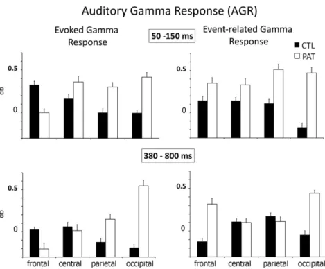
Başar-Eroğlu et al. (2011)investigated changes in gamma oscilla-tions during auditory sensory processing and target detection in pa-tients with schizophrenia using both single-trial averaged data. They found that single-trial gamma responses in patients were altered in magnitude and topographic pattern during both the AEGR and AERGR experimental conditions, whereas no differences were found for the av-eraged evoked gamma response. At the single-trial level, auditory stim-uli elicited higher gamma responses at both anterior and occipital sites in patients with schizophrenia compared to controls. Furthermore, in patients with schizophrenia, target detection compared to passive lis-tening to stimuli was related to increased single-trial gamma power at frontal sites. Enhancement of the gamma response in controls was only apparent for the averaged gamma response, with a distribution largely restricted to anterior sites. The differences in oscillatory activity between healthy controls and patients with schizophrenia were not reflected in the behavioral measure (i.e., counting targets). (SeeFigs. 12 and 13.)
5.2. Results SZ (alpha)
The study of alpha response to auditory stimuli in patients with schizophrenia was published byBasar-Eroglu et al. (2013). Their results show that patients have a larger alpha amplitude at the occipital location under auditory stimulus (Fig. 14). The authors interpreted this result as“…a combination of both a deficient mechanism related to sensory processing (i.e., reduced anterior response) and a deficient
Fig. 13. Means and standard deviations of post-stimulus total gamma-band enhancement within the analyzed time windows of 50–150 ms (top) and 380–800 ms (bottom) elicited by auditory stimuli during passive listening (AEGR, left column) and by target stimuli during the auditory oddball task (AERGR, right column) for controls (CTL) and patients (PAT). (Modified fromBaşar-Eroğlu et al., 2011).
Fig. 14. Maximum amplitudes of alpha oscillations within thefirst 250 ms after stimulus onset averaged over single subjects (Control: N = 10, Patients: N = 9). Error bars represent the standard error. Note that the occipital alpha is higher in patients than the controls under simple auditory stimulation. (PzN Oz, p b 0.05 for all post hoc). (Modified fromBasar-Eroglu et al. 2013).
mechanism engaging long-range inhibition of task-irrelevant cortical areas, (i.e., larger alpha amplitudes at occipital sites)” (Fig. 14). 6. Synopsis: we need profound comparisons and thoughts to approach brain function and dysfunctions
1. Our rationale in jointly analyzing controversial brain oscillation and connectivities in three common diseases is based on our desire to raise new questions about the description of function and dysfunc-tion. In this report, we did not aim to demonstrate all of the differences in the analysis of oscillations and connectivities in all different diseases. Instead, we focused our analysis on developing
strategies that may lead to future progress in understanding the web of oscillations and the possible influence of neurotransmitters. 2. A common property of all three diseases is a decrease in delta re-sponse. Regarding this point, the reader is referred to the volume by (Başar et al., 2013). A common feature of Mild Cognitive Impairment (MCI) and Alzheimer's disease is the gradual de-crease of delta response in MCI and Alzheimer's disease as they progress (see paper byYener et al., 2008, 2009, 2012, 2013,
2016-in this issue). Loss of memory (or dynamic memory) and
focused attention is a common deficit in AD, BD, and SZ. Accord-ingly, in the near future, common decreased delta response must undergo a profound analysis (see alsoEmek-Savaş et al., 2016-in Table 1
The possible biomarkers and related neurotransmitters in several neuropsychiatric disorders. (Modified fromYener and Başar, 2013b).
phrenia. In bipolar disorder, the spontaneous activity is tremendously reduced (approx. 80%), whereas in schizophrenia, the spontaneous alpha activity is only slightly reduced, as shown inFigs. 3 and 4. Thisfinding may serve later as a marker for dif-ferentiation between the two diseases.
4. Increased beta activity in bipolar disorder is also a marked change compared to schizophrenia. Is the increased beta activity in BD a compensation for the reduced alpha? Does beta activity take over the alpha functionality?
5. Hyper-connectivity in schizophrenia: Auditory stimulation evokes enhancement of alpha response in occipital areas com-pared to healthy subjects. Babiloni (comments of c. Babiloni in
Yener and Başar, 2013a,b) explains that this “hyper-connectivi-ty” controversy is one of the most interesting findings.Ba şar-Eroğlu et al. (2011)showed that schizophrenic patients were characterized by“paradoxical” occipital EEG oscillatory re-sponses to auditory oddball targets in two different experi-ments. This is further evidence that schizophrenia patients can display maladapted hyper-connectivity; it has been speculated that in these patients, abnormal auditory information is distrib-uted and triggers excitation in the occipital visual cortex, possi-bly producing abnormal visual imagery or visual processing. This intriguing working hypothesis needs to be tested with control experiments in schizophrenic patients to evaluate possible relationships between the“paradoxical” occipital EEG oscillatory responses to auditory oddball targets and structural neuroimaging indexes (i.e., tractography, diffusion tensor imag-ing) (comments of c. Babiloni inYener and Başar, 2013b). 6. Although the results are not yet significant, we have a similar
situation in BD patients. The auditory target stimulations evoke increased power spectra in posterior visual areas, thus indicat-ing a type of hyper-connectivity similar to SZ patients. In addition to spectral analysis, it is recommended to start a single trial analysis for more efficient analysis.
7. In patients with schizophrenia, upon auditory stimulation, the gamma response is increased in posterior areas compared to healthy subjects. This again demonstrates a type of hyper-connectivity in the gamma frequency range. This lastfinding is also an important controversy when comparing neural behavior in psychiatric diseases (seeFig. 12).
8. In the gamma frequency band, connectivity is low in bipolar disorder compared to healthy subjects, whereas in Alzheimer patients the connectivity in gamma band is superior compared to healthy subjects.
9. As it was shown in several reports, neurotransmitters and oscillations are interrelated in brain oscillation analysis. The differences detected in neurotransmitter release must be considered relative to different diseases (seeTable 1).
10. In diseases, oscillation amplitudes are not always decreased. There are cases with decreased oscillations and increased coherences, as shown in the present paper in AD.
7. Plasticity and compensation
In this report, we have shown that brain oscillations may show plasticity or compensation in some cases. In BD, the decrease of alpha activity does occur in parallel with the increase of beta activity com-pared to healthy subjects. In AD, event-related coherences in gamma frequency band are increased while we detect lower coherences in delta, theta, and alpha frequency ranges compared to healthy subjects.
patients.
8. What is new memory continuum in time space?
In the present report, we have discussed cognitive impairment in three diseases within the scope of oscillatory brain dynamics. Are they similar activation patterns in all three diseases? What correlates to memory impairment in pathology?
a) In bipolar disorder, alpha responses are highly decreased and beta responses are increased. In other words, there is a paradox upon comparison to healthy control subjects. Changes in the alpha responses or beta responses are recorded in time space between 50 and 200 ms.
b) Gamma coherence is decreased in bipolar disorder, whereas in Alzheimer's disease it is increased. These results show that gamma coherence is not necessarily a sign of attenuation or working memory attenuation.
c) With similar reasoning, it can be assumed that changes in spontane-ous alpha activity do not necessarily indicate memory loss. d) Is the“delta response” in all the three pathologies a common
response indicating a partial memory deficit?
In a parallel report byBaşar and Düzgün (2016-in this issue), we ten-tatively assumed that all types of memories (phyletic memory, working memory, psychological memory, semantic memory, and episodic mem-ory) are activated as a continuum in a hypertime space ranging from milliseconds to decades in the inhomogeneous time space of episodic memory. According to the present data, this time space is appreciably altered in the analyses of the diseases. However, it is highly recom-mended that in the future, changes (frequency reductions, amplitude reductions, response delays) should be analyzed in neuropsychiatric diseases.
References
Alfimova, M.V., Uvarova, L.G., 2008.Changes in EEG spectral power on perception of neu-tral and emotional words in patients with schizophrenia, their relatives, and healthy subjects from the general population. Neurosci. Behav. Physiol. 38 (5), 533–540.
Andreasen, N.C., 1997.Improvements of negative symptoms: concepts, definition and assessment. Int. Clin. Psychopharmacol. 12 (Suppl. 2), S7–S10.
APA, 1994.Diagnostic And Statistical Manual Of Mental Disorders IV From The American Psychiatric Association. American Psychiatric Press.
Arnfred, S.M., Mørup, M., Thalbitzer, J., Jansson, L., Parnas, J., 2011.Attenuation of beta and gamma oscillations in schizophrenia spectrum patients following hand posture perturbation. Psychiatry Res. 185 (1–2), 215–224.
Atagün, M.I., Güntekin, B., Tan, D., Tülay, E., Başar, E., 2015.Lithium excessivel yenhances event related beta oscillations in patients with bipolar disorder. J. Affect. Disord. 170, 59–65.
Babiloni, C., Roberta, L., Nicola, M., Paolo, C., Andrea, S., Ivanoi, T.A., Susanna, C., Claudio, D.P., 2016.Brain neural synchronization and functional coupling in Alzheimer's disease as revealed by resting state EEG rhythms. Int. J. Psychophysiol. 103, 88–102 (in this issue).
Barr, M.S., Farzan, F., Tran, L.C., Chen, R., Fitzgerald, P.B., Daskalakis, Z.J., 2010.Evidence for excessive frontal evoked gamma oscillatory activity in schizophrenia during working memory. Schizophr. Res. 121 (1–3), 146–152.
Başar, E., 1972.A study of the time and frequency characteristics of the potentials evoked in the acoustical Cortex. Kybernetik 10, 61–64.
Başar, E., 1980.EEG-Brain Dynamics: Relation Between EEG And Brain Evoked Potentials. Elseiver, Amsterdam (412 pp.).
Başar, E., Düzgün, A., 2015a. Prologue. Int. J. Psychophysiol.http://dx.doi.org/10.1016/j. ijpsycho.2015.02.008(in this volume).
Başar, E., Düzgün, A., 2016b.The brain as a working syncytium and memory as a contin-uum in a hyper timespace: Oscillations lead to a new model. Int. J. Psychophysiol. 103, 199–214 (in this issue).
Başar, E., Güntekin, B., 2008.A review of brain oscillations in cognitive disorders and the role of neurotransmitters. Brain Res. 1235, 172–193.
Başar, E., Ungan, P., 1973.A component analysis and principles derived for the under-standing of evoked potentials of the brain: studies in the hippocampus. Kybernetik 12, 133–140.
Başar, E., Rahn, E., Demiralp, T., Schürmann, M., 1998.Spontaneous EEG theta activity controls frontal visual evoked potential amplitudes. Electroencephalogr. Clin. Neurophysiol. 108 (2), 101–109.
Başar, E., Güntekin, B., Tülay, E., Yener, G.G., 2010.Evoked and event related coherence of Alzheimer patients manifest differentiation of sensory-cognitive networks. Brain Res. 1357, 79–90.
Başar, E., Güntekin, B., Atagün, İ., Turp-Gölbaşı, B., Tülay, E., Özerdem, A., 2012.Brain's alpha activity is highly reduced in euthymic bipolar disorder patients. Cogn. Neurodyn. 6 (1), 11–20.
Başar, E., Başar-Eroglu, C., Özerdem, A., Rossini, P.M., Yener, G.G., 2013.Application of brain oscillations in neuropsychiatric diseases. Suppl. Clin. Neurophysiol 62. Elseiver, Amsterdam.
Başar-Eroglu, C., Başar, E., Schmielau, F., 1991.P300 in freely moving cats with intracranial electrodes. Int. J. Neurosci. 60 (3–4), 215–226.
Başar-Eroglu, C., Strüber, D., Stadler, M., Kruse, P., Başar, E., 1993.Multistable visual perception induces a slow positive EEG wave. Int. J. Neurosci. 73 (1–2), 139–151.
Başar-Eroglu, C., Demiralp, T., Schürmann, M., Başar, E., 2001.Topological distribution of oddball‘P300’ responses. Int. J. Psychophysiol. 39 (2–3), 213–220.
Basar-Eroglu, C., Brand, A., Hildebrandt, H., Karolina Kedzior, K., Mathes, B., Schmiedt, C., 2007.Working memory related gamma oscillations in schizophrenia patients. Int. J. Psychophysiol. 64 (1), 39–45.
Başar-Eroğlu, C., Mathes, B., Brand, A., Schmiedt-Fehr, C., 2011.Occipital gamma response to auditory stimulation in patients with schizophrenia. Int. J. Psychophysiol. 79 (1), 3–8.
Basar-Eroglu, C., Schmiedt-Fehr, C., Mathes, B., 2013.Auditory evoked alpha oscillations imply reduced anterior and increased posterior amplitudes in schizophrenia. Suppl. Clin. Neurophysiol. 62, 121–129.
Benes, F.M., Berretta, S., 2001.GABAergic interneurons: implications for understanding schizophrenia and bipolar disorder. Neuropsychopharmacology 25 (1), 1–27.
Berger, H., 1929.Über das elektrenkephalogramm des menschen. I. Bericht. Arch. Psychiatr. Nervenkr. 87 (1), 527–570.
Bora, E., Yucel, M., Pantelis, C., 2009.Cognitive endophenotypes of bipolar disorder: a meta-analysis of neuropsychological deficits in euthymicpatients and their first-degree relatives. J. Affect. Disord. 113 (1–2), 1–20.
Bora, E., Lin, A., Wood, S.J., Yung, A.R., McGorry, P.D., Pantelis, C., 2014.Cognitive deficits in youth with familial and clinical high risk to psychosis: a systematic review and meta-analysis. Acta Psychiatr. Scand. 130 (1), 1–15.
Chepenik, L.G., Raffo, M., Hampson, M., Lacadie, C., Wang, F., Jones, M.M., Pittman, B., Skudlarski, P., Blumberg, H.P., 2010.Functional connectivity between ventral prefron-tal cortex and amygdala at low frequency in the resting state in bipolar disorder. Psychiatry Res. 182, 207–210.
Emek-Savaş, D.D., Güntekin, B., Yener, G.G., Başar, E., 2016.Decrease of delta oscillatory responses is associated with increased age in healthy elderly. Int. J. Psychophysiol. 103, 103–109 (in this issue).
Ethridge, L.E., Hamm, J.P., Shapiro, J.R., Summerfelt, A.T., Keedy, S.K., Stevens, M.C., Pearlson, G., Tamminga, C.A., Boutros, N.N., Sweeney, J.A., Keshavan, M.S., Thaker, G., Clementz, B.A., 2012.Neural activations during auditory oddball processing discriminating schizophrenia and psychotic bipolar disorder. Biol. Psychiatry 72, 766–774.
Ferri, C.P., Prince, M., Brayne, C., Brodaty, H., Fratiglioni, L., Ganguli, M., Hall, K., Hasegawa, K., Hendrie, H., Huang, Y., Jorm, A., Mathers, C., Menezes, P.R., Rimmer, E., Scazufca, M., Alzheimer's Disease International, 2005.Global prevalence of dementia: a Delphi consensus study. Lancet 366 (9503), 2112–2117.
Frisardi, V., Panza, F., Farooqui, A.A., 2011.Late-life depression and Alzheimer's disease: the glutamatergic system inside of this mirror relationship. Brain Res. Rev. 67 (1–2), 344–355.
Gold, J.M., 2004.Cognitive deficits as treatment targets in schizophrenia. Schizophr. Res. 72, 21–28.
Goldberg, J.F., Chengappa, K.N., 2009.Identifying and treating cognitive impairment in bipolar disorder. Bipolar Disord. 11 (Suppl. 2), 123–137.
Gray, C.M., McCormick, D.A., 1996.Chattering cells: superficial pyramidal neurons con-tributing to the generation of synchronous oscillations in the visual cortex. Science 274, 109–113.
Güntekin, B., Başar, E., 2016.Review of evoked and event-related delta responses in the human brain. Int. J. Psychophysiol. 103, 43–52 (in this issue).
Güntekin, B., Saatçi, E., Yener, G., 2008.Decrease of evoked delta, theta and alpha coherences in Alzheimer patients during a visual oddball paradigm. Brain Res. 1235, 109–116.
Hamm, J.P., Ethridge, L.E., Shapiro, J.R., Stevens, M.C., Boutros, N.N., Summerfelt, A.T., Keshavan, M.S., Sweeney, J.A., Pearlson, G., Tamminga, C.A., Thaker, G., Clementz, B.A., 2012.Spatiotemporal and frequency domain analysis of auditory paired stimuli processing in schizophrenia and bipolar disorder with psychosis. Psychophysiology 49, 522–530.
Hyun, J., Baik, M.J., Kang, U.G., 2011.Effects of psychotropic drugs on quantitative EEG among patients with schizophrenia-spectrum disorders. Clin. Psychopharmacol. Neurosci. 9 (2), 78–85.
Iacono, W.G., 1982.Bilateral electrodermal habituation–dishabituation and resting EEG in remitted schizophrenics. J. Nerv. Ment. Dis. 170 (2), 91–101.
Itil, T.M., Saletu, B., Davis, S., 1972.EEGfindings in chronic schizophrenics based on digital computer period analysis and analog power spectra. Biol. Psychiatry 5 (1), 1–13.
Itil, T.M., Hsu, W., Saletu, B., Mednick, S., 1974.Computer EEG and auditory evoked potential investigations in children at high risk for schizophrenia. Am. J. Psychiatry 131 (8), 892–900.
Kann, O., Papageorgiou, I.E., Draguhn, A., 2014.Highly energized inhibitory interneurons are a central element for information processing in cortical networks. J. Cereb. Blood Flow Metab. 34 (8), 1270–1282.
Koch, M., Schmiedt-Fehr, C., Mathes, B., 2016.Neuropharmacology of altered brain oscillations in schizophrenia. Int. J. Psychophysiol. 103, 62–68 (in this issue).
Krishnan, G.P., Vohs, J.L., Hetrick, W.P., Carroll, C.A., Shekhar, A., Bockbrader, M.A., O'Donnell, B.F., 2005.Steady state visual evoked potential abnormalities in schizo-phrenia. Clin. Neurophysiol. 116 (3), 614–624.
Leboyer, M., Kupfer, D.J., 2010.Bipolar disorder: new perspectives in health care and pre-vention. J. Clin. Psychiatry 71 (12), 1689–1695.
Lee, P.S., Chen, Y.S., Hsieh, J.C., Su, T.P., Chen, L.F., 2010.Distinct neuronal oscillatory responses between patients with bipolar and unipolar disorders: a magnetoencephalographic study. J. Affect. Disord. 123, 270–275.
Lehmann, D., 1989.From mapping to the analysis andınterpretation of EEG/EP maps. In: Maurer, K. (Ed.), Topographic Brain Mapping Of EEG And Evoked Potentials. Springer, Berlin (53-75 pp.).
Levinson, A.J., Young, L.T., Fitzgerald, P.B., Daskalakis, Z.J., 2007.Cortical inhibitory dys-function in bipolar disorder: a study using transcranial magnetic stimulation. J. Clin. Psychopharmacol. 27 (5), 493–497.
Lewis, D.A., Lieberman, J.A., 2000.Catching up on schizophrenia: natural history and neurobiology. Neuron 28 (2), 325–334.
Martínez-Arán, A., Vieta, E., Colom, F., Torrent, C., Sánchez-Moreno, J., Reinares, M., Benabarre, A., Goikolea, J.M., Brugué, E., Daban, C., Salamero, M., 2004.Cognitive impairment in euthymic bipolar patients: implications for clinical and functional outcome. Bipolar Disord. 6 (3), 224–232.
Mathes, B., Wood, S.J., Proffitt, T.M., Stuart, G.W., Buchanan, J.A., Velakoulis, D., Brewer, W.J., McGorry, P.D., Pantelis, C., 2005.Early processing deficits in object working memory infirst-episode schizophreniform psychosis and established schizophrenia. Psychol. Med. 35 (7), 1053–1062.
Merikangas, K.R., Akiskal, H.S., Angst, J., Greenberg, P.E., Hirschfeld, R.M., Petukhova, M., Kessler, R.C., 2007.Lifetime and 12-month prevalence of bipolar spectrum disorder in the National Comorbidity Survey replication. Arch. Gen. Psychiatry 64 (5), 543–552.
Miyauchi, T., Tanaka, K., Hagimoto, H., Miura, T., Kishimoto, H., Matsushita, M., 1990.
Computerized EEG in schizophrenic patients. Biol. Psychiatry 28 (6), 488–494.
Murray, C.J.L., Lopez, A.D., 1996.The Blobal Burden Of Disease: A Comprehensive Assess-ment Of Mortality And Disease. Harvard School of Public Health, Massachusetts.
Nunez, P.L., Wingeier, B.M., Silberstein, R.B., 2001.Spatial–temporal structures of human alpha rhythms: theory, microcurrent sources, multiscale measurements, and global binding of local networks. Hum. Brain Mapp. 13 (3), 125–164.
Özerdem, A., Güntekin, B., Tunca, Z., Başar, E., 2008.Brain oscillatory responses in patients with bipolar disorder manic episode before and after valproate treatment. Brain Res. 1235, 98–108.
Özerdem, A., Güntekin, B., Saatçi, E., Tunca, Z., Başar, E., 2010.Disturbance in long distance gamma coherence in bipolar disorder. Prog. Neuropsychopharmacol. Biol. Psychiatry 34 (6), 861–865.
Özerdem, A., Güntekin, B., Atagün, I., Turp, B., Başar, E., 2011.Reduced long distance gamma (28–48 Hz) coherence in euthymic patients with bipolar disorder. J. Affect. Disord. 132 (3), 325–332.
Pachou, E., Vourkas, M., Simos, P., Smit, D., Stam, C.J., Vasso, T., Micheloyannis, S., 2008.
Working memory in schizophrenia: an EEG study using power spectrum and coher-ence analysis to estimate cortical activation and network behavior. Brain Topogr. 21 (2), 128–137.
Pantelis, C., Velakoulis, D., Wood, S.J., Yücel, M., Yung, A.R., Phillips, L.J., Sun, D.-Q., McGorry, P.D., 2007. Neuroimaging and emerging psychotic disorders: the Melbourne ultra-high risk studies. Int. Rev. Psychiatry 19 (4), 371–381.
Petty, F., 1995.GABA and mood disorders: a brief review and hypothesis. J. Affect. Disord. 34 (4), 275–281.
Rehn, A.E., Rees, S.M., 2005.Investigating the neurodevelopmental hypothesis of schizophrenia. Clin. Exp. Pharmacol. Physiol. 32, 687–696.
Riečanský, I., Kašpárek, T., Rehulová, J., Katina, S., Přikryl, R., 2010.Aberrant EEG responses to gamma-frequency visual stimulation in schizophrenia. Schizophr. Res. 124 (1–3), 101–109.
Rissman, R.A., De Blas, A.L., Armstrong, D.M., 2007.GABA(A) receptors in aging and Alzheimer's disease. J. Neurochem. 103 (4), 1285–1292.
Rodriguez, E., George, N., Lachaux, J.P., Martinerie, J., Renault, B., Varela, F.J., 1999.
Perception's shadow: long-distance synchronization of human brain activity. Nature 397 (6718), 430–433.
Saha, S., Chant, D., Welham, J., McGrath, J., 2005.A systematic review of the prevalence of schizophrenia. PLoS Med. 2 (5), e141.
Sakowitz, O.W., Quiroga, R.Q., Schürmann, M., Başar, E., 2001.Bisensory stimulation increases gamma-responses over multiple cortical regions. Brain Res. Cogn. Brain Res. 11 (2), 267–279.
Sanchez-Alaveza, M., Ehlers, C.L., 2016.Event-related oscillations (ERO) during an active discrimination task: effects of lesions of the nucleus basalis magnocellularis. Int. J. Psychophysiol. 103, 53–61 (in this issue).
Schürmann, M., Başar-Eroglu, C., Başar, E., 1997.Gamma responses in the EEG: elementa-ry signals with multiple functional correlates. Neuroreport 8 (7), 1793–1796.
Spencer, K.M., Nestor, P.G., Niznikiewicz, M.A., Salisbury, D.F., Shenton, M.E., McCarley, R.W., 2003.Abnormal neural synchrony in schizophrenia. J. Neurosci. 23 (19), 7407–7411.
Sponheim, S.R., Clementz, B.A., Iacono, W.G., Beiser, M., 1994.Resting EEG infirst-episode and chronic schizophrenia. Psychophysiology 31 (1), 37–43.
Sponheim, S.R., Clementz, B.A., Iacono, W.G., Beiser, M., 2000.Clinical and biological concomitants of resting state EEG power abnormalities in schizophrenia. Biol. Psychi-atry 48 (11), 1088–1097.
2006.Dysfunctional long-range coordination of neural activity during Gestalt percep-tion in schizophrenia. J. Neurosci. 26 (31), 8168–8175.
Vawter, M.P., Freed, W.J., Kleinman, J.E., 2000.Neuropathology of bipolar disorder. Biol. Psychiatry 48 (6), 486–504.
Velasques, B., Bittencourt, J., Diniz, C., Teixeira, S., Basile, L.F., Salles, J.I., Novis, F., Angélica Silveira, L., de Assis da Silva, R., de Lima Teixeira, A., Nardi, A.E., Akiskal, H.S., Cagy, M., Piedade, R., Cheniaux, E., Kapczinki, F., Ribeiro, P., 2013.Changes in saccadic eye movement (SEM) and quantitative EEG parameter in bipolar patients. J. Affect. Disord. 145 (3), 378–385.
Whittington, M.A., Traub, R.D., Jefferys, J.G., 1995.Synchronized oscillations in interneu-ron networks driven by metabotropic glutamate receptor activation. Nature 373 (6515), 612–615.
Whittington, M.A., Traub, R.D., Kopell, N., Ermentrout, B., Buhl, E.H., 2000. Inhibition-based rhythms: experimental and mathematical observations on network dynamics. Int. J. Psychophysiol. 38, 315–336.
Wood, S.J., Proffitt, T., Mahony, K., Smith, D.J., Buchanan, J.A., Brewer, W., Stuart, G.W., Velakoulis, D., McGorry, P.D., Pantelis, C., 2002.Visuospatial memory
ders following an interactive panel discussion and synopsis. Suppl. Clin. Neurophysiol. 62, 343–363.
Yener, G.G., Emek-Savaş, D.D., Lizio, R., Çavuşoğlu, B., Carducci, F., Ada, E., Güntekin, B., Babiloni, C.C., Başar, E., 2016.Frontal delta event-related oscillations relate to frontal volume in mild cognitive impairment and healthy controls. Int. J. Psychophysiol. 103, 110–117 (in this issue).
Yener, G.G., Güntekin, B., Öniz, A., Başar, E., 2008.Event related delta oscillatory responses of Alzheimer patients. Eur. J. Neurol. 15, 540–547.
Yener, G.G., Güntekin, B., Tülay, E., Başar, E., 2009.A comparative analysis of sensory visual evoked oscillations with visual cognitive event related oscillations in Alzheimer's dis-ease. Neurosci Lett. 462 (3), 193–197.
Yener, G.G., Güntekin, B., Örken, D.N., Tülay, E., Forta, H., Başar, E., 2012.Auditory delta event-related oscillatory responses are decreased in Alzheimer’s disease. Behav. Neurol. 25 (1), 3–11.
Yener, G.G., Kurt, P., Emek-Savas, D.D., Güntekin, B., Başar, E., 2013.Reduced visual event-related delta oscillatory responses in amnestic mild cognitive impairment. J. Alzheimers Dis. 37 (4), 759–767.