PRETREATMENT WITH CYPRESS CONES’ WATER
ExTRACT ENHANCES SURVIVAL OF ISCHEMICALLY
CHALLENGED SKIN FLAPS A PRELIMINARY STUDY
*Betül Gözel ULUSAL, **Hale TUFAN, ***Ali Engin ULUSAL, ****Cevahir HABERAL, *Tamer SEYHAN, *Hüseyin BORMAN, *****Mehmet HABERAL*Plastic and Reconstructive Surgery **Pharmacology
***Orthopedics and Traumatology ****Cardiovascular Surgery
*****General Surgery, Baskent University Faculty of Medicine, Ankara, Turkey abstract
The pathophysiology of ischemic skin flap necrosis is complex, due primarily to vascular thrombosis and insufficient angiogenesis. Nitric oxide can significantly increase angiogenic response and protect the endothelium from ischemia-reperfusion injury. Also, anticoagulants can prevent or reverse skin flap ischemia. In vitro assessment of endothelial cell function in isolated aortic rings of rats pretreated with cypress cones’ water extract showed increased production of endothelium-derived nitric oxide. Additionally, we have shown its anticoagulant properties. Based on these observations, we hypothesized that pretreatment with cypress cones’ water extract would enhance survival of random extensions of ischemic axial flaps via its increased endothelial nitric oxide release and its anticoagulant effect.
Twenty four Sprague-Dawley rats were randomly assigned as pretreatment (n=12) and control (n=12) groups. The pretreated group received 30% of cypress cones’ water extract treatment orally 7 days before flap elevation and for 3 days afterward. The control group received tap water. The ischemic target was a 6 × 7 cm islanded epigastric artery flap based on the right inferior epigastric pedicle. After the observation period, hemodynamic variables including mean arterial pressure and heart rate were assessed. Flap survival and perfusion rates were determined by microangiography and laser Doppler flowmetry. In twelve rats, in vitro isometric tension of the aortic segments isolated from the control and pretreated groups was monitored to reflect vascular responsiveness. Using isolated tissue baths, the dose-response relations to acetylcholine was determined and compared between the two groups.
SErvİ KoZALAğI SU EKSTrESİYLE
ÖNKoşULLANDIrMA İSKEMİK CİLT FLEPLErİNDE SAğKALIMI ArTTIrIr
özet
İskemik cilt flebi nekrozunun patofizyolojisi karmaşıktır ve öncelikle vasküler tromboz ve yetersiz anjiogenez sonucunda gerçekleşir. Nitrik oksit anjiogenik cevabı belirgin biçimde arttırarak endoteli iskemi-reperfüzyon hasarından korur. Aynı zamanda antikoagülanlar da cilt flep iskemisini önler ya da düzeltebilir. Servi kozalağı su ekstresiyle önkoşullandırma yapılmış sıçanlardan izole edilen aort halkalarının In vitro değerlendirilmesi endotel kökenli nitrik oksit üretiminde artış olduğunu ortaya koymuştur. Buna ilaveten önceki çalışmalarımızda ekstrenin antikogülan özelliği olduğunu da göstermiştik. Bu gözlemlere dayanarak servi kozalağının su ekstresiyle önkoşullandırmanın endotelyal nitrik oksit salınımında artış ve antikogülan özellikleriyle iskemik aksiyel fleplerin random uzantılarının sağkalımını arttırabileceği hipotezinde bulunduk.
Yirmi dört adet Sprague-Dawley türü sıçan gelişigüzel şekilde önkoşullandırma grubu (n=12) ve kontrol grubu (n=12) olarak ayrıldı. Önkoşullandırma yapılmış grup %30’luk servi kozalağı ekstresini oral yoldan almaya flebin kaldırılmasından 7 gün evvel başladı ve operasyonu takiben 3 gün de almaya devam etti. Kontrol grubuna çeşme suyu verildi.Gözlem süresinin sonunda ortalama arter basıncı ve kalp hızını içeren hemodinamik değişkenler değerlendirildi. Flep sağkalım ve perfüzyon oranları mikroanjiografi ve lazer Doppler akım ölçerle çalışıldı. Vasküler cevabı saptamak için kontrol grubu ve önkoşullandırma yapılmış gruplardan izole edilen aort segmentlerin izometrik gerimleri
There were no significant differences between the hemodynamic variables. In the pretreated group, microangiograms revealed increased angiogenesis and capillary density and enhanced flap perfusion (as blood perfusion units) in the right distal and proximal parts (P < .05). Endothelium-derived nitric oxide – dependent maximal relaxation (Emax) and the EC50
value to Acetylcholine were significantly greater in the pretreated group compared to that of the controls.
These data suggest that pretreatment with cypress water extract enhances the viability of ischemically challenged flaps.
Keywords: cupressus sempervirens cones, nitric oxide, anticoagulant, water extract, ischemic flap, angiogenesis, isolated tissue bath.
in vitro koşullarda kaydedilerek değerlendirildi İzole doku banyosu kullanarak aortic segmentlerin asetilkoline karşı oluşturdukları doz bağımlı cevapları saptandı ve iki grup arasında karşılaştırma yapıldı.
Hemodinamik değişkenler arasında anlamlı fark bulunamadı. Tedavi grubunda flebin sağ distal ve proksimal kısımlarında artmış anjiogenez ve kapiller yoğunluğunun yanında artmış flep perfüzyonu saptandı (P < .05). Endotel kökenli nitrik oksit bağımlı maksimum gevşeme cevabı (Emax) ve EC50 değerleri kontrol grubunda anlamlı derecede yüksekti.
Bu bulgular servi kozalağı su ekstresinin iskemik fleplerde sağkalımı arttırdığını desteklemektedir.
Anahtar Kelimeler : Servi kozalağı, nitrik oksit, antikoagüln, su ekstresi, iskemik flep, anjiogenez, izole doku banyosu.
IntroductIon
Cupressus sempervirens (cypress) is a common tree in the Mediterranean basin. Its cone is an organ commonly used in phytotherapy owing to its high content of bioactive components. Skin flap necrosis can be a devastating complication, and the potential of various pharmacological agents to induce development of angiogenesis in ischemic skin flaps has stimulated considerable research.1,2 The principle of therapeutic
angiogenesis is to achieve adequate blood flow by enhancing capillary formation so that nutrients and oxygen are provided to ischemia tissues, thus preventing ischemic tissue necrosis.3From this perspective,
angiogenesis treatment can be regarded as a promising approach to enhance survival of ischemic tissues.3
Nitric oxide (NO) is an endothelium-derived relaxing factor released from endothelial cells known to mediate a wide range of biologic functions in various cells. Nitric oxide has a key role in promoting angiogenesis by increasing vasodilation, vascular permeability, endothelial cell proliferation and migration, and by modifying the activities of angiogenic mediators.4,5
We showed anticoagulant properties of this extract.6
Using the rat epigastric extended skin island flap, we sought to explore ischemic skin tissue survival after pretreatment with cypress cones’ water extract.
MaterIals and Methods
This study was approved by the Research Council and Ethical Committee of Research on Animals of Baskent University. The guidelines outlined in the Guide for the Care and Use of Laboratory Animals were followed. Twenty four female Sprague Dawley rats weighing 200 to 300 grams were used; each was randomized to either the control or the experimental (pretreatment) group. Animals were housed individually in polycarbon cages at 20°C ± 2°C in a humidity controlled environment (50% ± 10%) with a 12 hour dark/light cycle. Water and commercial rat chow were supplied ad libitum.
The cones obtained from the Mediterranean coast of Turkey in March 2007 were cleaned with water to remove any contamination. Partially crushed fresh cones were boiled in tap water for 10 minutes. The yellow-brown colored extract (yield of 30% w/v) was filtered off and cooled down for 30 minutes. One mL of the extract
(which was prepared daily) was administrated by an orogastric tube, once a day at 10:00 AM.
Animals in the pretreated group (n=12) received cypress cones’ water extract for 7 days before the flap elevation and continuing for 3 days afterward. Animals in the control group (n=12) received tap water. At the end of pretreatment period, rats were anesthetized with ketamine (IM, 50 mg/kg) and xylazine (IM, 7 mg/kg ), and the abdominal region was shaved and prepared. After marking with Indian blue dye, an extended epigastric skin flap measuring 6 × 7 cm was elevated based on the right epigastric vessels; the left counterpart was ligated to induce ischemic necrosis in the contralateral flap side. The underlying panniculus carnosus was included. During the observation period, animals’ general health and wound status, including activity, weight loss, infection, and wound dehiscence were recorded. Survival of the flaps was determined by gross examination by the color, texture, and vascular supply of the flap.
On postoperative day 3, animals were anesthetized and data were collected including mean arterial pressure, heart rate, and laser doppler flowmetry. Mean arterial pressure was recorded by a data acquisition system (BIOPAC, MP100A model, CA) via a 24-guage catheter placed in the abdominal aorta. Laser doppler flowmetry (BIOPAC, MP100A) was used to measure flap perfusion. Four fixed reference points were tattooed using a needle puncture before elevation of the flap. A laser probe was placed, and readings were recorded at four distinct points: the right proximal, right distal, left proximal, and left distal parts of the skin flap. During these measurements, the animals’ body temperatures were kept steady to avoid a possible interference in the flap circulation. The results are expressed as blood perfusion units.
Following all data collection, the animals were killed with an overdose of ketamine (IM, 150 mg/kg). The abdominal cavity was opened, and the inferior vena cava and abdominal aorta exposed immediately. In twelve animals, (6 animals from each group), the descending thoracic aorta was removed and placed in Kreb’s Henseleit solution (in mM): (NaCl 118.4; KCl 4.7; CaCl2.2H2O 1.9; KH2PO4 0.9; MgSO4.7H2O 1.2; NaHCO3 2.1; Glucose 11.1 at +4°C). The aorta was cleaned of adhering fatty tissues and cut into rings
3-4 mm in length. Each ring was suspended between two stainless steel wire hooks in a jacketed isolated tissue bath contained 10 ml of Kreb’s Henseleit solution at 37°C and aerated with 95% O2 and 5% CO2. The changes in isometric tension of the rings were recorded by a computerized data acquisition system (BIOPAC, MP 100A model, Ca, USA). Aorta rings were allowed to equilibrate under an initial tension of 1 g for 60 min before any experimentation. The segments were washed out with fresh Kreb’s Henseleit solution every 15 min during the equilibration period. All experiments were performed in the presence of indomethacin (10-5 M) in the solution. After the equilibration period, KCl (60 mM) was added into the solution to test the viability of the preparations. The contractile response of the aorta rings to KCl was recorded. Each ring was tested twice and KCl response was allowed to reach plateau before being washed out. After returning the base-line tensions, the segments were permitted to stabilize for 30 min. Then, the segments precontracted induced with PE (10-6 M) and Acetylcholine (ACh) (10-9 – 10-4 M) was added into the baths in a concentration-dependent manner after the rings reached to the plateau. This protocol was repeated in the presence of NOS inhibitor L-NAME (10-4 M) for 15 min. At the end of the experimental protocol, the segments were exposed to PE (10-6 M) and Sodium nitroprusside to test the viability and the ability of contraction and relaxation. The reference chemicals were obtained from the source specified: acetylcholine chloride, Nω-nitro-L-arginine methyl ester hydrochloride (L-NAME), phenylephrine hydrochloride and potassium chloride (Sigma Chemical Company, St Louis, MO). All chemicals used were of the highest purity grade. Stock solutions of all chemicals were prepared in distilled water and the dilutions were made freshly on the day of experiment.
In the remaining twelve animals (6 from each group), the aorta was perfused with 200 mL heparinized (100 U/ mL) warm saline, which was followed by an injection of a mixture of lead oxide and gelatin powder. Thereafter, total skin flap was harvested and preserved in a saline-soaked sponge at 4°C for 5 days. Microangiographic radiographs were obtained using a radiograph machine
(General Electric, DMR) and a mammography film. The radiographs were scanned for images and compared.
statIstIcal analYses
All data were expressed as mean ± standard error of mean (SEM). Data were analyzed using student’s t and one way ANOVA, post hoc Bonferroni’s test. “n” was the number of the animals used in each group. P < 0.05 was considered as statistically significant. The maximal response (Emax) to ACh was expressed as percentage of relaxation relative to the PE (10-6 M) induced maximum contraction. The negative logarithmic concentration of ACh that elicited 50% of the maximal response (EC50) was expressed as the sensitivity of the aorta rings to ACh, which was calculated separately for each concentration-response curve. EC50 values were given as negative Log M and “n” was the number of the segments used in each group.
results Flap Survival
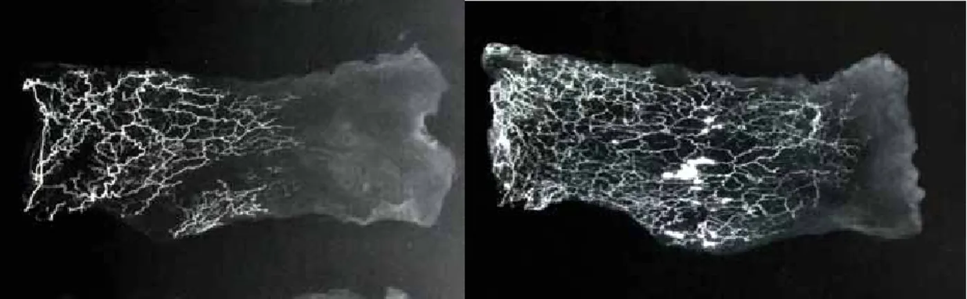
The surviving parts of the flaps were pink, pliable, and appeared well-perfused; the necrosed parts were black. Although exact quantification could not be done, the average percentage of flap necrosis was markedly higher in the ischemic, nonpretreated control group (Figures 1 and 2). Interestingly, animals in the pretreated group tolerated the surgical trauma better than controls did. During the observation period, subjects seemed more active and healthy, and total body weight loss was less. Mean weight loss was 6.50 grams and 15.17 grams in the pretreated and control groups, respectively. In the pretreated group, the lead oxide microangiogram showed a rich vascular network and newly formed connections crossing the midline of the flap. The sprouting vessels reached very close to the distal end (Figure 3). In the control group, the capillaries were relatively sparse with simple branching (Figure 4). In the pretreated group, laser doppler flowmetry measurements showed statistically significantly higher perfusion rates compared with controls. Mean flap perfusions (as blood perfusion units) in the right distal and proximal parts of the flaps in the pretreated and Figure 1: Macroscopic appearance of the skin flap from the
control groups were 131.50 ± 49.71 blood perfusion units (right distal), 274.0 ± 14.36 blood perfusion units (right proximal), 35.20 ± 10.24 blood perfusion units (right distal), and 121.0 ± 38.03 blood perfusion units (right proximal) (P < .05) (Table 1). There were no significant differences between the hemodynamic variables (Table 2).
Endothelium-derived nitric oxide – dependent relaxation (expressed as percentage contraction relative to PE (10-6M) - induced maximum contraction) to ACh was significantly greater in the pretreated group than the control group (Figure 5 and Table 3). Treatment with L-NAME, NOS inhibitor, inhibited of ACh-induced relaxation in all groups. In the case of the sensitivity of muscarinic receptors, the values of EC50 (-Log M) for the segments from pretreated rats was significantly higher than those for the control group (Table 3).
dIscussIon
Ischemia can be defined as inadequate perfusion to meet tissue needs. Skin flap necrosis due to microvascular perfusion failure is a major concern in reconstructive surgery. Various pharmacological agents have been investigated for their effectiveness in preventing or reversing skin flap ischemia including anticoagulants, vasodilators, sympatholytics, glucocorticoids, prostaglandin inhibitors, and calcium channel blockers.7-10 This article introduces cupressus
sempervirens (cypress) cones as a new herbal alternative for preventing the tissue from ischemic damage. The increased viability of random extensions of ischemic axial skin flaps was clearly demonstrated in subjects pretreated with cypress cones’ extract. The results obtained from laser doppler flowmetry and microangiographic radiographs were consistent with the macroscopic findings and verified by increased angiogenesis and flap perfusion.
Cupressus sempervirens cones are used in traditional Figure 3: Microangiographic radiograph of the skin flap depicting
enhanced angiogenesis from the pretreated group. Note the rich vascular network and newly formed connections crossing the midline of the flap.
Figure 4: Microangiographic radiograph of the skin flap from the control group depicting capillaries with simple branching.
Figure 5: Concentration-response curves induced by ACh (10-9-10-4M) in aortic rings obtained from the control (♦, n=6), and pretreated (▲, n=6) groups. Values are expressed as mean ±SEM.
control (n=5) pretreatment (n=4)
grouPs
Tablo 1: Flap perfusion in the right distal (RD), right proximal (RP), and left distal (LD) parts of the skin flaps from the control and pretreatment groups.
35.20 ± 10.24 p < .05 131.50 ± 49.71 p < .05 rd 274.00 ± 14.36 84.80 ± 6.87 22.25 ± 3.57 p < .05 121.00 ± 38.03 rP ld regIons
Tablo 2: Hemodynamic parameters of subjects from the control and pretreatment groups control (n=5) pretreatment (n=4) grouPs 35.20 ± 10.24 p < .05 131.50 ± 49.71 p < .05 MAp 274.00 ± 14.36 121.00 ± 38.03 heArt rAte
Tablo 3: Maximal relaxations (Emax) and EC50 values of ACh in aortic rings obtained from the rats (control and pretreated). Values are expressed as mean ± SEM. and pretreatment groups.
control (n=6) pretreatment (n=6) p < 0.01 grouPs 56.53 ± 13.23 98.18 ± 2.23 Emax 7.36 ± 0.09 6.69 ± 0.13 ec50 Ach
medicine for many purposes.11Nitric oxide mediates
endothelium-dependent relaxation and plays a major role in the physiology of microcirculation by improving blood flow to the flap by vasodilatation of preexisting vessels.12,13 The result of our experiments in aortic
rings suggests that oral administration of cypress cone extract augments ACh-induced dilator response in-vitro condition. The mechanisms of these responses are needed to be clarified. However, in this preliminary study, the improvement in the survival of ischemic flaps may be due at least in part to an increase in endothelial nitric oxide. The lack of a statistically significant difference between the groups suggests that our results were independent of the hemodynamic variables.
Microangiography and laser Doppler flowmetry studies have shown enhanced angiogenesis and flap perfusion as early as on the third day after the ischemic challenge. The timing for the detection of angiogenesis was similar to that of previous reports, which shows formation of new capillary blood vessels within 48 hours of an ischemic challenge of a flap tissue.14The
concentration of the extract and the pretreatment period choosen was based on our previous experiments and trials. In pilot studies, 30% concentration of the extract was established to be more effective than its dilute concentrations. The most effective and non-toxic concentration has yet to be determined. Further, the longer the pretreatment period is the more effective results are likely obtained regarding the flap perfusion. Given the multiple chemical constituents in cypress cones, the actual active ingredient for angiogenesis and ischemic tolerance is unknown. Although the primary focus of the responsible mechanism is on the NO-related mechanisms on which the hypothesis was based, the anticoagulant properties of this herb extract or other mechanisms also might have triggered an early protective response by unknown pathways. This issue remains to be explored in future studies.
Because the water extract of cypress can be safely consumed by humans, it has the potential for clinical use to prevent or salvage flap failures, once the most-effective therapeutic dosage and duration of therapy has been established. Further in vivo and in vitro studies about the effects of cypress cones’ water extract are ongoing in our laboratory.
Conclusion
Oral pretreatment with cupressus sempervirens cones’ water extract induces regional angiogenesis and improves survival of random extensions in ischemic axial skin flaps.
Acknowledgements
We thank Hatice Lakadamyali, MD, for microangiographic radiographs and Safiye Karagozlu for the technical assistance.
DR.BETÜL GÖZEL ULUSAL
HASAN BASRİ ÇANTAY MH. TANKÇİFTLİĞİ CD. ALTINKENT SİTESİ, A BLOK, KAT :3, DAİ:7
10050, BALIKESİR Tel: 90.505.7484390 e-mail: [email protected]
REFERENCES
1. Khouri RK, Brown DM, Leal-Khouri SM, Tark KC, Shaw WW. The effect of basic fibroblast growth factor on the neovascularisation process: skin flap survival and staged flap transfers.
Br J Plast Surg,44:585-588;1991
2. Hom DB, Assefa G. Effects of endothelial cell growth factor on vascular compromised skin flaps.
Arch Otolaryngol Head Neck Surg; 118:624 628;1992.
3. Radomski MW, Palmer RM, Moncada S. An L-arginine/nitric oxide pathway present in human platelets regulates aggregation. Proc Natl Acad Sci U S A,87:5193-5197;1990 4. Furchgott RF, Zawadzki JV. The obligatory role of
endothelial cells in the relaxation of arterial smooth muscle by acetylcholine.
Nature,288:373-376;1980 5. Singh JP. Dimethylarginine
dimethylaminohydrolase: a new therapeutic target for the modulation of nitricoxide and angiogenesis.
Curr Opin Investig Drugs,8:736-741;2007 6. Ulusal BG, Arikan S, Durusoy C. Anticoagulant
effect of Cupressus sempervirens. Phytother Res,21:1116;2007
7. Shalom A, Friedman T, Westreich M. Effect of aspirin and heparin on random skin flap survival in rats.
Dermatol Surg,34:785-790;discussion790; 2008 8. Nichter LS, Sobieski MW. Efficacy of verapamil in
the salvage of failing random skin flaps. Ann Plast Surg,21:242-245;1988 9. Nichter LS, Sobieski MW, Edgerton MT.
Augmentation of critical skin flap survival following ibuprofen therapy.
Ann Plast Surg16:305-312;1986
10. Kerrigan CL, Daniel RK. Pharmacologic treatment of the failing skin flap.
Plast Reconstr Surg,70:541-549;1982 11. Said O, Khalil K, Fulder S, Azaizeh H.
Ethnopharmacological survey of medicinal herbs in Israel, the Golan Heights and the West Bank region. J Ethnopharmacol,83:251-65;2002 12. Palmer RM, Ferrige AG, Moncada S. Nitric
oxide release accounts for the biological activity of endothelium-derived relaxing factor.
Nature,327:524-526;1987
13. Moncada S, Palmer RM, Higgs EA. Nitric oxide: physiology, pathophysiology, and
pharmacology.
Pharmacol Rev,43:109-142;1991 14. Ghali S, Butler PE, Teper OM, Gurtner
GC. Vascular delay revisited.