Accepted: 2017.07.18 Published: 2017.08.08
2361
3
—
25
High Prolactin Level as a Predictor of Vasospasm
in Aneurysmal Subarachnoidal Hemorrhage
AB 1
Ilker Çöven
DE 2Atilla Kırcelli
BF 3
Enes Duman
BFG 4Huseyin Ulas Pınar
BCF 5Betul Basaran
Corresponding Author: Ilker Çöven, e-mail: [email protected]
Source of support: Departmental sources
Background: Aneurysmal subarachnoid hemorrhage (aSAH) is a destructive syndrome with a mortality rate of 50%. Recent studies have also suggested a high pervasiveness of hypothalamic-pituitary insufficiency in up to 45% of pa-tients after aSAH. Prolactin has been associated with the pathogenesis of hypertensive irregularities that are linked to pregnancy.
Material/Methods: We identified a group of 141 patients with spontaneous SAH due to a ruptured cerebral aneurysm; these pa-tients were operated on at our institution’s Neurosurgery and Interventional Radiology Department between 2011 and June 2015. All of the data were obtained retrospectively from medical records.
Results: The hormonal abnormalities observed in the initial 24 h after ictus in subjects with subarachnoid SAH were caused by stressful stimulation aggravated by intracranial bleeding.
Conclusions: The elevated prolactin levels that occur in patients with aSAH can be used in conjunction with other auxiliary factors that we believe may be beneficial to vasospasm.
MeSH Keywords: Prolactin • Subarachnoid Hemorrhage • Vasospasm, Intracranial Full-text PDF: https://www.medscimonit.com/abstract/index/idArt/906010 Authors’ Contribution: Study Design A Data Collection B Statistical Analysis C Data Interpretation D Manuscript Preparation E Literature Search F Funds Collection G
1 Department of Neurosurgery, Konya Training and Research Hospital, Konya, Turkey
2 Department of Neurosurgery, Baskent University, Ankara, Turkey 3 Department of Interventional Radiology, Baskent University, Ankara, Turkey 4 Department of Anesthesiology, Baskent University, Ankara, Turkey 5 Department of Anesthesiology, Konya Training and Research Hospital, Konya,
Background
Aneurysmal subarachnoid hemorrhage (aSAH) is a destructive syndrome with a mortality rate of 50%. In spite of all medical care efforts, 30% of survivors experience perpetual neurologi-cal impairment [1]. A considerable factor influencing outcome following treatment of a fractured aneurysm is the neurolog-ical condition of the accepted patients, which largely reflects the severity of the hemorrhage. Other risks include recurrent bleeding, initial and secondary vasospasm, and the complex-ity of the therapeutic intervention [1,2].
Recent studies have also suggested that hypothalamic-pi-tuitary insufficiency is very common following aSAH [3–5]. Hyperprolactinemia (HPRL) is physiological condition that oc-curs after intercourse and stressful activity, and during lacta-tion and pregnancy. The expected rate of HPRL is 0.4–3% in a healthy population. Prolactin has a crucial role in modulating the immune response [2].
Previous studies have linked dysfunction and subarachnoid hemorrhage (SAH). A number of studies have suggested that endocrine disorders are induced by compression of the hypo-thalamus-pituitary complex by the aneurysm. Other reported causes are SAH due to perfusion changes, toxins from erupt-ed blood, vasospasm-relaterupt-ed ischemia, hydrocephalus, and high intracranial pressure. One additional cause is injury dur-ing the surgical procedure [3,4,6,7].
A relationship between prolactin and the pathogenesis of preg-nancy-related hypertensive disorders has been reported [8,9]. Reports on the hormone levels in these disorders show they are increased, within normal limits, or decreased [10,11]. There is scant evidence regarding the dominant blood flow and resis-tance effects of prolactin in intact blood vessels, but reports have associated prolactin with vascular effects. Prolactin as an i.v. infusion has been reported to boost arterial pressure in de-cerebrate rabbits [12] and to decrease renal blood flow, elevate body fluid level, and increase the pressor response to norepi-nephrine in rats. The N-terminal 16-kDa particle of prolactin was proved to prevent coronary vessel relaxation of isolated perfused animal hearts. There are also reports of deterioration of endothelial vasodilatory function and insulin susceptibility in subjects with pituitary adenoma and hyperprolactinemia. Prolactin i.v. infusion primarily leads to a decline in blood flow and elevation in coronary vascular resistance, mesenteric, re-nal, and iliac vascular beds. The structure behind these ef-fects was proven to include beta 2-adrenergic receptor-me-diated effects linked to the intracellular NO pathway [13,14]. Pituitary prolactin is a 200-amino acid peptide hormone synthesized in and secreted from the anterior pituitary [9]. Several lines of evidence suggest that prolactin is secreted
in the periphery by lymphohematopoietic cells. A number of endogenous and exogenous factors modulate prolac-tin secretion but, for the purposes of this review, estrogen and dopaminergic agonism are pivotal. Circulating prolac-tin binds to the prolacprolac-tin receptor and its various isoforms with specific downstream effects, as would be expected for a receptor that belongs to the growth hormone, erythropoi-etin, and cytokine receptor superfamily. Signal transduction of the prolactin receptor leads to gene transcriptional reg-ulation. Interestingly, prolactin and the immunosuppressive drug cyclosporine appear to be antagonistic through a re-ceptor-based mechanism.
Material and Methods
We identified a cohort of 141 patients with spontaneous SAH due to a ruptured cerebral aneurysm; these patients were oper-ated on in Baskent University Neurosurgery and Interventional Radiology Department between September 2011 and June 2015. This study was approved by Baskent University Institutional Review Board (Project no: KA 17/165) and supported by Baskent University Research Fund. All of the data were obtained retro-spectively from medical records.
Cerebral angiography was conducted in all patients to deter-mine localization of the aneurysm. All patients that had an SAH underwent brain computed tomography (CT) and results of the CT were noted.
According to the findings of cranial digital subtraction angi-ography, the localization of the aneurysm, the aneurysm neck diameter, length, and the ratio of the dome and neck length were all recorded. Basal hormone levels of the patients were taken within 12 h of their admission.
Patients that used confounding drugs earlier, such as substi-tuting hormonal therapy of all types, were excluded from this study. Patients using drugs that might affect pituitary func-tion, such as glucocorticoids or dopamine, were additionally ex-cluded. We measured fundamental hormone status along with free (f) T3, fT4, TSH, PRL, cortisol, FSH, LH, and macroprolactin. We made a diagnosis of spontaneous SAH according to an-amnesis, physical findings, and brain CT data. Then, dilatation was detected using four-vessel conventional x-ray angiogra-phy (all carotids and all vertebrals) via femoral artery cathe-terism. When the localization of the aneurysm was visualized, the image of the neck was recorded. The Fisher CT score cor-responds to: (1) No blood detected; (2) A diffuse disposition or thin layer with all vertical layers of blood (interhemispheric fissure, insular cistern, ambient cistern) less than 1 mm thick; (3) Localized clots and/or vertical layers of blood 1 mm or more
in thickness; and (4) Diffuse or no subarachnoid blood, but in-tracerebral or intraventricular blood [10].
Following hospital admissions, the patients were clinically as-sessed and categorized in accordance with the World Federation of Neurosurgical Societies (WFNS) SAH grading scale, which is used exclusively in patients with SAH for clinical and predictive assessment. All of the patients were tracked until they were released from the hospital. Prior to release, the patients were re-evaluated using the prognostic WFNS scale [15].
We collected blood samples at 8 AM and 9 AM, 24 h upon ictus, centrifuged at 3000 rpm, for 10 min. We stored the serum at –20° C until we conducted the assays. We measured hormones using commercial kits: T4 by RIA (INEP, Zemun, Serbia); TSH by IRMA (INEP); PRL, LH, and FSH by IRMA (Cis BioInternational, France); and cortisol by RIA (Cis BioInternational).
Statistical analysis
Statistical analysis was performed using SPSS 20.0.
Normality of distributions of continuous variables was deter-mined by Kolmogorov-Smirnov test. Descriptive statistics for continuous variables are expressed as mean ±SD or medi-an (25th–75th) percentiles, where applicable. Numbers of
cas-es and percentagcas-es were used for categorical data. The mean differences between groups were compared by the t test, and Mann-Whitney U test was used for comparisons of non-nor-mally distributed data.
Results
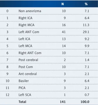
We identified 141 eligible patients, but 17 patients who were lost to follow-up were excluded. We included 124 patients in the analysis. There were 49 females and 75 males, with an average age of 56.33±11.17 years, and there were no statis-tically significant differences between the average age of fe-males (52.5±14.8 years) and fe-males (51±3.3 years). Aneurysm location sites are listed in Table 1. Fifty-three subjects were categorized as grade I in the WFNS grading scale (asymptom-atic or with minimal to discrete headache), 37 patients were classified as grade II (headache levels moderate to severe, stiff neck, and absence of focal neurological signs, excluding crani-al nerves parcrani-alysis), and 19 patients were classified as grade III (sleepiness, mental confusion, and discreet focal neurolog-ical deficits), 28 patients were classified as grade IV, and 4 pa-tients were classified as grade V.
We assessed endocrine levels at 7.4±6.6 h after SAH. Prolactin levels were significantly correlated with Fischer grade on CT (Pearson correlation coefficient: 0.59, p<0.0001). The mean
prolactin levels were 8.23±5.15 (15 patients) in Fischer grade I, 12.93±12.60 (48 patients) in Fischer grade II, 36.08 ± 19.39 (50 patients) in Fischer grade III, and 43.29±24.33 (28 patients) in Fischer grade IV. There were significant differences between prolactin levels with Fischer grades and WFNS grades of the patients (Pearson correlation coefficient 0.496, p<0.0001). The mean prolactin levels were lower for patients with lower Fischer grades (p<0.0001), and there were no differences be-tween prolactin levels and sex (p=0.328). Radiologically, va-sospasm was observed in patients with high prolactin levels in diagnostic cerebral angiography, and the aneurysm was closed within 24 h.
The TSH, T3, and T4 levels in men and women were compara-ble. TSH levels were higher than normal in 5 patients (14.2%). T3 levels characterized by a mean value of 33.8±9 ng/dL, were found lower than normal in 5 patients (14.2%). The T4 levels were low in 2 patients (5.6%). Antithyroglobulin and antimi-crosomal antibodies were not identified in any of the subjects. We divided the patients according to their aneurysmal site, in terms of anterior circulation and posterior circulation. There were 83 anterior circulation aneurysm patients and 41 poste-rior circulation aneurysm patients. The mean prolactin levels were 29.98±23.43 and 16.31±15.42 in anterior and posterior circulation aneurysms, respectively. We found significant differ-ences between aneurysmal site and prolactin levels (p=0.001). There were no differences in aneurysm site between aneurysm neck diameter, dome length/neck size ratio, and Fischer and WFNS grades. Radiologic vasospasm detected in diagnostically
N %
0 Non anevrizma 10 7.1
1 Right ICA 9 6.4
2 Right MCA 16 11.3
3 Left ANT Com 41 29.1
4 Left ICA 13 9.2
5 Left MCA 14 9.9
6 Right ANT Com 10 7.1
7 Post cerebral 2 1.4 8 Post Com 10 7.1 9 Ant cerebral 3 2.1 10 Basiler 9 6.4 11 PICA 3 2.1 12 Left SCA 1 0.7 Total 141 100.0
constructed cerebral angiographies was observed in Fischer grade and WFNS grades with high prolactin levels. There was a statistically significant positive correlation between pro-lactin and cortisol (p<0.05) (Table 2). There was a statistical-ly significant positive correlation between prolactin, cortisol, and Fischer grade, D/N ratio, and size (mm) (p<0.05). Fischer grade, D/N, and cortisol increased as size increased (Table 3).
Discussion
The pathophysiology of neuroendocrine dysfunction upon SAH is obscure. It has been suggested that the genesis of these adjustments may be the ischemic or hemorrhagic lesions in the hypothalamus or pituitary gland. Increased intracrani-al pressure, locintracrani-al tissue pressure changes upon hemorrhage, vasospasm-based ischemia, microinfarctions of the pituitary gland, venous stasis, and surgical procedures may damage the pituitary gland [3]. The pathogenesis of aSAH [10,15–18] can also include traumatic brain injuries, genetic predisposi-tion, and autoimmunity and neuroinflammatory responses. Insufficiencies in all pituitary hormones following SAH have been reported to differ from none to 68% in previous stud-ies [5,11,17,19]. Our results revealed increased prolactin lev-els beginning 12 h after SAH.
The increase in cortisol during stress is a necessary mecha-nism for the defense of the human body in the perpetuation of its homeostasis. Alterations of the excitability of the neu-ronal membranes [20] can be immediately activated by glu-cocorticoids. Cerebrovascular permeability and the choroidal transport of water and electrolytes may also be affected, and they are crucial for the regulation of liquor synthesis and vol-ume homeostasis in the brain [19]. Complicated intercom-munication between the immune system and the hypothala-mus-pituitary-adrenal axis can also be of crucial physiological significance in the stress response [20]. Glucocorticoids duce a multistep restriction of the immune system that pro-tects against the aftereffects of extreme inflammatory reac-tions [20,21]. Our study also revealed that higher levels of cortisol were correlated with higher Fisher grades.
It has been shown that prolactin IV infusion mainly causes a decline in blood flow and elevates vascular protection in the coronary, mesenteric, renal, and iliac vascular beds. The struc-tures of these reactions have been proven to include beta 2-ad-renergic receptor-mediated reactions that are connected with the intracellular NO pathway [13].
The decline in the tracked blood flow against prolactin IV infu-sion might be related to a main reaction of the hormone and not to concomitant alterations in hemodynamic variables. The heart rate and arterial blood pressure of the patients were kept consistent, and there were no important alterations in heart fill-ing pressures and left ventricular dP/dtmax. This case prohib-ited any secondary intervention from reflexes, local metabolic, or natural effects on the reaction of tracked blood flows to pro-lactin immersion. Moreover, IV immersion of the vehicle only at the identical rate with prolactin did not result in the same reactions of the immersed hormone. Also, the candid connec-tion between the hormone and its coronary, mesenteric, renal, and iliac reactions was validated in the dose-response study, which proved that the increase in regional blood flow could be developed by enhancing the dosage amount of the immersed hormone. Several lines of evidence suggest that prolactin is secreted peripherally by lymphohematopoietic cells [12]. The role of prolactin in lymphoproliferation and its effects on cy-tokine production are also important. Prolactin acts through potentiation of T and B cell function [22].
Henderson et al. showed that the coronary, mesenteric, renal, and iliac vasoconstriction caused by prolactin immersion con-tained both the endothelial discharge of NO and its intracellu-lar direction. Although we did not measure the real discharge of NO, the regional vasoconstriction drawn out by hormone infusion was counteracted by the local intra-arterial adminis-tration of L-NAME, which has been proven to prevent the for-mation of NO [21].
Cortisol
r P n
Prolactine .784** .000* 141
Table 2. Prolactine and cortisol level.
* p<0.05. There were statistically significant positive relationship between prolactin and cortisol (p<0.05). As prolactin increases, cortisol increases. Prolactine r P n Fischer grade .502** .000* 141 Age .106 .210 141 D/N .189* .025* 141 Size (mm) .126 .135 141
Table 3. Relationship between prolactine levels and other variables.
* p<0.05. The correlation between prolactin levels, Fisher grade and D/N ratio were statistically significant (p<0.05). As Fisher grade increased, prolactin levels also rised. There was no statistically significant relationship between age and size of the aneursym (p>0.05).
In our analysis, patients who presented a more severe clinical status on upon WFNS evaluations and greater bleeding on CT (Fisher grade) exhibited a higher propensity for endocrinolog-ical disorders. Klose et al. [23] observed a correlation between lower GCS and the presence of hydrocephalus with hormonal changes in the first days after SAH.
In patients with SAH, delayed cerebral ischemia with arterial va-sospasm can be an important cause of disability and death. In our treatment protocol, we used the previous version of these guidelines, which focused on prevention with oral nimodip-ine and maintenance of euvolemia, as well as treatment with triple-H and endovascular therapy with selective intra-arteri-al vasodilators and/or bintra-arteri-alloon angioplasty [24]. We intra-arteri-also treat-ed the patients with acute hydrocephalus by external ventric-ular drainage (EVD) and/or lumbar drainage.
Mangieri et al. [21] studied the hormonal profiles of patients in the hyperacute phase of SAH (the first 24 h). These au-thors observed an increase in cortisol in all patients studied (n=35), a finding that was most likely due to the physiologi-cal response to stress. Mangieri et al. also observed a higher incidence of high prolactin (14.2%). In terms of thyroid hor-mones, these authors observed high levels of TSH in 14.2% of patients, low levels T3 in 14.2% of patients, and low levels of T4 in 5.6% of patients; FSH and LH were unaffected [21]. In
our study, we found high prolactin and cortisol levels in early-stage SAH. Nakagava et al. [25] described the clinical features of cerebral blood flow (CBF) data obtained from single-photon emission computed tomography (SPECT) during the risk peri-od for DCI after SAH. We performed cerebral angioplasty and/ or selective intra-arterial vasodilator therapy in the region of hypoperfusion of arterial territories after SPECT in the patients with high GCS and neurological regression.
Conclusions
Early detection of vasospasm may prevent morbidity and mor-tality after aneurysm rupture, and it improve outcomes. On the contrary, better selection of patients prevents complica-tions of aggressive treatment such as induced hypertension and hypervolemia, as well as interventional therapies. Elevated prolactin levels have a potentially valuable role in identifying SAH patients at higher risk of developing vasospasm beyond Fischer and WFNS scales. Future studies of related mechanism of prolactin secretion in SAH patients may also identify new targets of treatment.
Conflicts of interest None.
Referances:
1. Bederson JB, Connolly ES Jr., Batjer HH et al: Guidelines for the manage-ment of aneurysmal subarachnoid hemorrhage: A statemanage-ment for healthcare professionals from a special writing group of the Stroke Council, American Heart Association. Stroke, 2009; 40: 994–1025
2. Molyneux AJ, Kerr RS, Yu LM et al: International subarachnoid aneurysm tri-al (ISAT) of neurosurgictri-al clipping versus endovascular coiling in 2143 pa-tients with ruptured intracranial aneurysms: a randomised comparison of effects on survival, dependency, seizures, rebleeding, subgroups, and an-eurysm occlusion. Lancet, 2005; 366(9488): 809–17
3. Tölli A, Borg J, Bellander BM et al: Pituitary function within the first year after traumatic brain injury or subarachnoid haemorrhage. J Endocrinol Invest, 2017; 40(2): 193–205
4 Hannon MJ, Behan LA, O’Brien MM et al: Chronic hypopituitarism is uncom-mon in survivors of aneurysmal subarachnoid haemorrhage Clin Endocrinol (Oxf), 2015; 82(1): 115–21
5. Vieira G Jr., de Albuquerque LA, de Avellar AB et al: Long-term follow-up of anterior pituitary deficiency after aneurysmal subarachnoid hemorrhage: Prospective cohort. J Stroke Cerebrovasc Dis, 2016; 25(10): 2405–14 6. Kronvall E, Sonesson B, Valdemarsson S et al: Reduced quality of life in
pa-tients with pituitary dysfunction after aneurysmal subarachnoid hemor-rhage: A prospective longitudinal study. World Neurosurg, 2016; 88: 83–91 7. Sharshar T, Bastuji-Garin S, Polito A et al: Hormonal status in pro-tracted critical illness and in-hospital mortality. Crit Care, 2011; 15: R47 8. Oney T, Bellmann O, Kaulhausen H: Relationship between serum prolactin
concentration, vascular angiotensin sensitivity and arterial blood pressure during third trimester pregnancy. Arch Gynecol Obstet, 1988; 243: 83–90 9. Cabrera-Reyes EA, Limón-Morales O, Rivero-Segura NA et al: Prolactin
func-tion and putative expression in the brain. Endocrine, 2017; 57(2): 199–213 10. Fisher CM, Kistler JP, Davis JM: Relation of cerebral vasospasm to
subarachnoid hemorrhage visualized by computerized tomographic scan-ning. Neurosurgery, 1980; 6: 1–9
11. Weant KA, Sasaki-Adams D, Dziedzic K et al: Acute relative adrenal insuf-ficiency after aneurysmal subarachnoid hemorrhage. Neurosurgery, 2008; 63: 645–49; discussion 649–50
12. Horrobin DF, Manku MS, Burstyn PG: Effect of intravenous prolactin infu-sion on arterial blood pressure in rabbits. Cardiovasc Res, 1973; 7: 585–87 13. Molinari C, Grossini E, Mary DA et al: Prolactin induces regional vaso-constriction through the beta2-adrenergic and nitric oxide mechanisms. Endocrinology, 2007; 148: 4080–90
14. Chrousos GP: The hypothalamic-pituitary-adrenal axis and immune-medi-ated inflammation. N Engl J Med, 1995; 332: 1351–62
15. Teasdale GM, Drake CG, Hunt W et al: A universal subarachnoid hemor-rhage scale: Report of a committee of the World Federation of Neurosurgical Societies. J Neurol Neurosurg Psychiatry, 1988; 51: 1457
16. Tanriverdi F, Unluhizarci K, Kelestrimur F: Persistent neuroinflammation may be involved in the pathogenesis of traumatic brain injury (TBI)-induced hy-popituitarism: potential genetic and autoimmune factors. J Neurotrauma, 2010; 27: 301–2
17. Parenti G, Cecchi PC, Ragghianti B et al: Evaluation of the anterior pituitary function in the acute phase after spontaneous subarachnoid hemorrhage. J Endocrinol Invest, 2011; 34: 361–65
18. Pereira JL, Albuquerque LA, Dellaretti M et al: Pituitary deficiency after aneu-rysmal subarachnoid hemorrhage. Clinics (Sao Paulo), 2013; 68(6): 745–49 19. Can A, Gross BA, Smith TR et al: Pituitary dysfunction after aneurysmal sub-arachnoid hemorrhage: A systematic review and meta-analysis. Neurosurgery, 2016; 79(2): 253–64
20. Chen J, Wang ZZ, Zuo W et al: Effects of chronic mild stress on behavior-al and neurobiologicbehavior-al parameters – role of glucocorticoid. Horm Behav, 2016; 78: 150–59.
21. Mangieri P, Suzuki K, Ferreira M et al: Evaluation of pituitary and thyroid hormones in patients with subarachnoid hemorrhage due to ruptured in-tracranial aneurysm. Arq Neuropsiquiatr, 2003; 61: 14–19
22. Correale J, Farez MF, Ysrraelit MC: Role of prolactin in B cell regulation in multiple sclerosis. J Neuroimmunol, 2014; 269: 76–86
23. Klose M, Brennum J, Poulsgaard L et al: Hypopituitarism is uncommon af-ter aneurysmal subarachnoid haemorrhage. Clin Endocrinol (Oxf), 2010; 73: 95–101
24. Connolly ES Jr., Rabinstein AA, Carhuapoma JR et al: Guidelines for the man-agement of aneurysmal subarachnoid hemorrhage: A guideline for health-care professionals from the American Heart Association/american Stroke Association. Stroke, 2012; 43(6): 1711–37
25. Nakagawa M, Mutoh T, Takenaka S et al: Asymptomatic mild hyperperfu-sion for the prediction of clinical outcome in postoperative patients after subarachnoid hemorrhage. Med Sci Monit, 2017; 23: 285–91