Is the presence of linear fracture a predictor of
delayed posterior fossa epidural hematoma?
Atilla Kırcelli, M.D.,1 Ömer Özel, M.D.,2 Halil Can, M.D.,3 Ramazan Sarı, M.D.,4 Tufan Cansever, M.D.,1 İlhan Elmacı, M.D.51Department of Neurosurgery, Başkent University Faculty of Medicine, İstanbul-Turkey 2Department of Orthopaedic Surgery, Başkent University Faculty of Medicine, İstanbul-Turkey 3Department of Neurosurgery, Private Medicine Hospital, İstanbul-Turkey
4Department of Neurosurgery, Memorial Hizmet Hospital, İstanbul-Turkey 5Department of Neurosurgery, Memorial Şişli Hospital, İstanbul-Turkey
ABSTRACT
BACKGROUND: Though traumatic posterior fossa epidural hematoma (PFEDH) is rare, the associated rates of morbidity and mortality are higher than those of supratentorial epidural hematoma (SEDH). Signs and symptoms may be silent and slow, but rapid de-terioration may set in, resulting in death. With the more frequent use of computed tomography (CT), early diagnosis can be achieved in patients with cranial fractures who have suffered traumatic injury to the posterior fossa. However, some hematomas appear in-significant or are absent on initial tomography scans, and can only be detected by serial CT scans. These are called delayed epidural hematomas (EDHs). The association of EDHs in the supratentorial-infratentorial compartments with linear fracture and delayed EDH (DEDH) was presently investigated.
METHODS: A total of 212 patients with SEDH and 22 with PFEDH diagnosed and treated in Göztepe Training and Research Hos-pital Neurosurgery Clinic between 1995 and 2005 were included. Of the PFEDH patients, 21 underwent surgery, and 1 was followed with conservative treatment. In this group, 4 patients underwent surgery for delayed posterior fossa epidural hematoma (DPFEDH). RESULTS: Mean age of patients with PFEDH was 12 years, and that of the patients with SEDH was 18 years. Classification made ac-cording to localization on cranial CT, in order of increasing frequency, revealed of EDHs that were parietal (27%), temporal (16%), and located in the posterior fossa regions (approximately 8%). Fracture line was detected on direct radiographs in 48% of SEDHs and 68% of PFEDHs. Incidence of DPFEDH in the infratentorial compartment was statistically significantly higher than incidence in the supra-tentorial compartment (p=0.007). Review of the entire EDH series revealed that the likelihood of DEDH development in the infraten-torial compartment was 10.27 times higher in patients with linear fractures than in patients with suprateninfraten-torial fractures (p<0.05). CONCLUSION: DPFEDH, combined with clinical deterioration, can be fatal. Accurate diagnosis and selection of surgery modality can be lifesaving. The high risk of EDH development in patients with a fracture line in the posterior fossa on direct radiographs should be kept in mind. These patients should be kept under close observation, and serial CT scans should be conducted when necessary. Key words: Delayed epidural hematoma; head trauma; posterior cranial fossa.
toma (SEDH). PFDEH accounts for 0.3% of all intracranial hematomas and 4–12% of all epidural hematomas (EDHs). [1–6] PFDEH usually develops secondary to trauma to the oc-cipital, subococ-cipital, and retromastoid regions. Though clinical progression is slow, neurological progression is rapid and fatal in the absence of timely intervention. Diagnosis of PFEDH can be established by cranial computed tomography (CT) or
magnetic resonance imaging.[7] Hematoma that induces mass
effect should be surgically treated.
Delayed posterior fossa epidural hematoma (DPFEDH) is ex-tremely rare, but can occur following head injury. DPFEDH is defined as the absence of EDH with or without linear frac-ture on initial CT, followed by development or deterioration of clinical symptoms, or detection of EDH on serial CT scans.
Address for correspondence: Atilla Kırcelli, M.D.
Oymacı Sok, No: 7, Başkent Üniversitesi İstanbul Hastanesi, 34662 Altunizade, İstanbul, Turkey
Tel: +90 216 - 554 15 00 E-mail: [email protected]
Qucik Response Code Ulus Travma Acil Cerrahi Derg 2016;22(4):355–360
doi: 10.5505/tjtes.2015.52563 Copyright 2016
TJTES
INTRODUCTION
Traumatic posterior fossa epidural hematoma (PFEDH) is less common, compared to supratentorial epidural
hema-Presence of linear fracture is a demonstrated risk factor for delayed supratentorial epidural hematomas (DSEDHs). In the literature, DSEDH accounts for 9–10% of all EDHs. However, its potential as a risk factor for DPFEDH has yet to be ad-dressed.[8–13]
The present patient series included 234 patients with trau-matic cranial EDH treated in Göztepe Training and Research Hospital Neurosurgery Clinic between 1995 and 2005, 22 of whom were diagnosed with PFEDH, and 212 of whom were diagnosed with SEDH. Patients were retrospectively com-pared and evaluated based on clinical and radiological find-ings. The present objective was to investigate whether linear fracture was a risk for DPFEDH development, and whether there was a difference in risk severity between linear fracture in the infratentorial and supratentorial compartments.
MATERIALS AND METHODS
A total of 234 patients treated for traumatic cranial EDH be-tween 1995 and 2005 were retrospectively analyzed based on clinical and radiological findings. From this series, findings of 22 patients with PFEDH (9.4%) and 212 (90.6%) with SEDH were retrospectively evaluated.
Delayed EDH was detected in 4 of the 22 PFEDH patients and 2 of the 212 SEDH patients. Underlying causes were in-vestigated. Direct radiographs were obtained in the lateral and anteroposterior planes in the 234 patients with head injury. Those with trauma to the occipital, suboccipital, or retromastoid regions were also examined with Towne’s radi-ography. In patients with fractures, fracture type and whether the fracture had crossed venous sinuses were noted. CT scan was performed in all patients, and recorded findings included enlargement of the trigone, temporal horn, and 3rd ventricle, 4th ventricle compression and shift, basal-ambient and quad-rigeminal cistern compressions, and whether hematomas were bilateral. In addition, EDH volume was calculated using the equation: 0.5 height x depth x length.
Upon admission, severity of head injury was classified accord-ing to the Glasgow Coma Scale (GCS) by Narayan,[14] as mild (GCS: 13–15), moderate (GCS: 9–12), or severe (GCS: 3–8).
SEDH patients with a hematoma of less than 30 cm3 in
vol-ume, less than 15 mm in thickness, and with a midline shift of less than 5 mm were clinically observed and followed up, as were those with GCS greater than 8 but with no focal neuro-logical deficit. In patients with PFEDH of greater than 5 mm in thickness on CT, and greater than 15 cm3 in volume, with mass effect resulting in fourth ventricle shift, ventricle com-pression and perimesencephalic cistern comcom-pression were surgically evacuated.
Level of consciousness and severity of head injury on admis-sion were considered during postoperative follow-up. Initial Glasgow Outcome Score (GOS) and examination results of
all patients were recorded. Of the patients diagnosed with PFEDH, 21 received surgical treatment, and 1 received con-servative treatment. While type of surgery varied, depend-ing on hematoma localization, paramedian or median suboc-cipital craniotomy was performed, as was evacuation of the EDH, followed by detection of the bleeding site and bleed-ing control, after which surgery was terminated. Of the 22 PFEDH patients, only 4 had DPFEDH, all of whom had linear fractures. Two SEDH patients developed DSEDH, none of whom had linear fractures.
Statistical Analysis
Statistical analysis was performed using the GraphPad Prism program (version 3; GraphPad Software, Inc., San Diego, CA, USA). Mann-Whitney U-test was used to compare data with descriptive statistical methods (mean, SD), as well as with paired data. Fisher’s exact test and relative risk were used to compare qualitative data. A p value of p<0.05 was considered statistically significant.
RESULTS
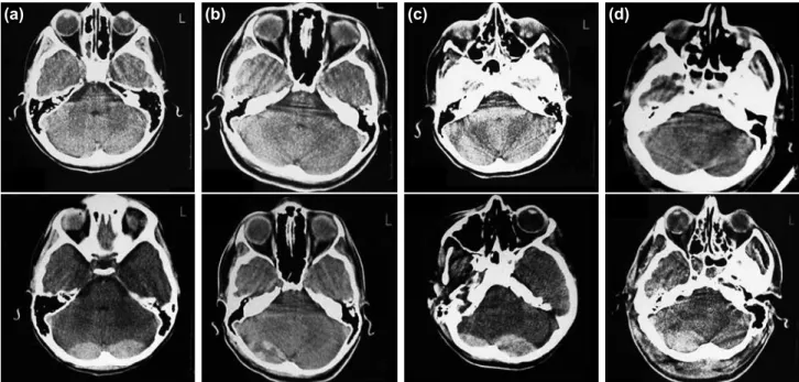
A total of 234 patients were analyzed retrospectively based on clinical and radiological features. Eight patients diagnosed with PFEDH (36%) were female, and 14 (64%) were male, with a ratio of 2:3. Fifty-one patients with SEDH were fe-male (24%), and 161 (76%) were fe-male, with a ratio of 1:3. Of the patients with EDH, 22 were diagnosed with PFEDH, and 212 with supratentorial EDH. Of the patients with PFEDH, 4 were diagnosed with DPFEDH (Fig. 1a–d). Of the patients with SEDH, 2 were diagnosed with DSEDH.
Mean age of patients with PFEDH was 12 years, while that of patients with SEDH was 18 years. Incidence of supraten-torial and infratensupraten-torial EDH in the 1st decade of life was significantly high (p<0.05), while incidence in the 2nd decade of life was quite considerably high: 56% and 82% in SEDH and PFEDH patients, respectively. Demographic characteristics are presented in Table 1.
The most common causes of traumatic posterior fossa EDH include fall from a height, traffic accident, assault, and colli-sion of the head with a solid object. Signs and symptoms of the 22 PFEDH patients were headache (17 patients), nausea and vomiting (15), impaired consciousness (14), and retro-mastoid, occipital, and suboccipital swelling (12). Though less common, symptoms including loss of consciousness, cerebel-lar dysfunction, diplopia, abducens paralysis, otorrhagia, neck stiffness, and anisocoria were encountered in otherwise as-ymptomatic patients (Table 2). Two of the 4 patients with DPFEDH presented with headache and nausea/vomiting, the other 2 with neurological disorientation. All were discharged in GOS 5 condition.
of injury. The other most common symptoms included head-ache, nausea/vomiting, impaired consciousness, loss of
con-sciousness, lateralizing signs (hemiparesis, Babinski reflex), and herniation. Two patients had delayed SEDH in this group. There was no need to evacuate the hematoma, and a conser-vative approach was adopted. Both patients were discharged in GOS 5 condition.
Classification according to localization on cranial CT, in order of increasing frequency, revealed EDH in the temporal (16%), temporoparietal (22%), and parietal (27%) regions, while in-cidence of EDH in the posterior fossa was approximately 8% in the present series (Fig. 2). Overall incidence of DPFEDH was 2%. Regarding localization of PFEDH in the infratentorial compartment on CT scan, 5 EDHs showed bilateral and 17 unilateral localization. Two DPFEDHs showed bilateral and 2 showed unilateral localization. Mean PFEDH volume was 13.5 cm3 (range: 2.3–45 cm3).
Fracture line was detected on direct radiography of 102 (48%) of the 212 SEDH patients, while direct radiographs of 110 (52%) patients showed normal findings. DSEDH was detected during follow-up of 2 patients with no fracture line on direct radiography (0.09%). Linear fracture was identified in 15 (68%) of the 22 PFEDH patients. Twelve patients had paramedian occipital fracture and 3 had occipital fracture crossing the transverse sinus at the midline. DPFEDH was noted in 4 patients (18%) whose direct radiographs revealed the presence of a fracture line.
No statistically significant difference was found in fracture distribution between patients with PFEDH and those with SDEH (p=0.11). However, occurrence of EDH was 2.14 times more likely in the infratentorial compartment than in the supratentorial compartment in patients with linear fracture (p>0.05). Incidence of DPFEDH was statistically significantly higher in the infratentorial compartment than in the supratentorial compartment (p=0.007). Probability of DPFEDH development in the presence or absence of fracture was 4.66 times higher in the infratentorial com-partment than in the supratentorial comcom-partment (p<0.05). Incidence of DPFEDH in the infratentorial compartment, in the presence of fracture, was statistically significantly higher than in the supratentorial compartment (p=0.0002). A re-view of the entire EDH series revealed that the probability of delayed EDH (DEDH) development in the infratento-rial compartment in patients with linear fracture was 10.27 times higher than in patients with supratentorial fracture (p<0.05).
PFEDHs and DPFEDHs were compared in terms of CT find-ings including ventricle compression and shift, and enlarge-ment of the trigone, temporal horn, and third ventricle, indi-cating development of hydrocephalus. In cases of DPFEDH, enlargement of the trigone and compression of the ambient cistern, among basal cisterns, were significantly increased (p=0.01 and p=0.029, respectively) (Table 2).
Table 1. Demographic factors
Supratentorial Infratentorial EDH EDH Gender Male 161 14 Female 51 8 Age (median) 18 12 Trauma type
Fall from height 63 10 Traffic accident 90 4 Assaults & collisions 59 8
ED: Epidural hematomas.
Table 2. Comparison of cranial tomographic findings of posterior fossa epidural hematomas (PFEDHs) vs delayed posterior fossa epidural hematomas
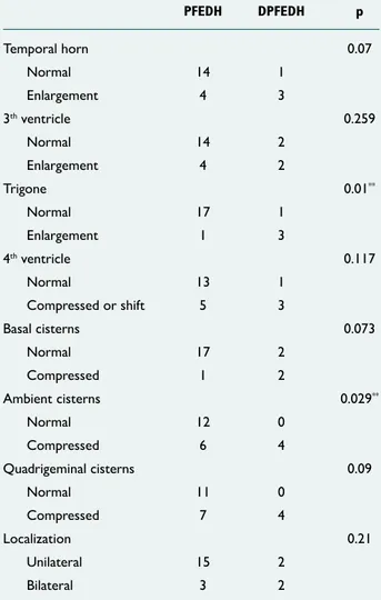
(DPFEDHs) PFEDH DPFEDH p Temporal horn 0.07 Normal 14 1 Enlargement 4 3 3th ventricle 0.259 Normal 14 2 Enlargement 4 2 Trigone 0.01** Normal 17 1 Enlargement 1 3 4th ventricle 0.117 Normal 13 1 Compressed or shift 5 3 Basal cisterns 0.073 Normal 17 2 Compressed 1 2 Ambient cisterns 0.029** Normal 12 0 Compressed 6 4 Quadrigeminal cisterns 0.09 Normal 11 0 Compressed 7 4 Localization 0.21 Unilateral 15 2 Bilateral 3 2
DISCUSSION
EDHs are defined as lesions that typically develop immediate-ly after trauma and expand in volume in minutes. Following the alleviation of the tamponade effect on intracranial vol-ume, EDHs constitute a threat to life.[15,16] These lesions may develop slowly and can be manageable, though prediction of the course of EDH is challenging.
The posterior fossa is a rare location for EDH. In PFEDH, the deterioration of symptoms may be slow and silent. However, neurological progression is rapid and can be fatal if left un-treated. Early diagnosis is crucial for survival. Diagnosis and cure of PFEDH was possible following the first case of suc-cessful surgery, reported by Coleman and Thompson in 1941. [17] However, until recently, diagnosis was challenging. As in the
first published case series, documented by Campbell, Fisher, Hooper, and Petit-Dutaillis et al., the difficulty in establish-ing diagnosis resulted in the deaths of almost half the patient population.[18–21] These authors emphasized that half of the population was in the pediatric age group and had occipital fractures crossing the sinus. Recently, the frequent use of CT has facilitated PFEDH diagnosis in patients with head injury, and the number of patients diagnosed with DEDH has increas-ingly grown. Some authors suggest that a CT scan should be performed in each patient with swelling in the occipital region or fracture of the occipital bone.[22,23] In the present series, CT scan was performed, after neurological progression, in 4 patients with linear fracture, and DPFEDH was noted in all. EDH is an accumulation of blood resulting from bleeding of extracerebral vessels, leading to an extra axial mass. Irrevers-ible damage is difficult to prevent, due to epidural hemor-rhage resulting in brain herniation and increased intracranial pressure. Today, the increasingly frequent use of CT in the differential diagnosis of patients with head injury has facili-tated the diagnosis of EDH, resulting in a decreased mortality rate.[24] A control CT scan performed in the first 24 hours in patients kept under observation following head trauma can detect DPFEDH. Radiological changes precede clinical pro-gression.[25] Though the mortality rate of DEDH has been re-ported as 5%, it was approximately 4.5% in the present series. The majority of the present PFEDH patients were in the sec-ond decade of life. Harwood and Nash reported that 30% of patients with linear fracture developed EDH, and that EDH can develop following trauma to the occipital region due to abundant diploic and dural vascularization in infants and
chil-(a) (b) (c) (d)
Figure 1. (a) Case 1: S.K., 9 years old, initial computed tomography (CT) view (above) and view after 24 hours (below). (b) Case 2: Ş.S.,
13 years old, initial CT view (above) and view after 10 hours (below). (c) Case 3: S.Y.; 10 years old, initial CT view (above) and view after 12 hours (below). (d) Case 4: T.Ş., 33 years old, initial CT view (above) and view after 12 hours (below).
60 70 50 40 30 20 10 Temporal Temporo-parietal Frontal Parietal
Posterior parietal Frontoparietal
0
Figure 2. Distribution of epidural hematomas (EDHs) according to
dren. Most of the present PFEDH patients (68%) were in the pediatric age group. Surgical intervention was not performed in 1 patient with PFEDH based on cranial CT findings, which
showed a hematoma less than 5-mm thick, 10 cm3, and
with-out mass effect. Of the patients who underwent surgery, those with bilateral EDH underwent median craniectomy, while those with unilateral EDH underwent suboccipital cra-niectomy on the side where the lesion was located.
According to reports, incidence of bilateral PFEDH is approx-imately 30%, while it was approxapprox-imately 23% in the present series.[8] In most cases, bleeding arises from the venous sinus, from posterior branches of the meningeal artery or diploic veins, and from the dural sinuses. The transverse or sigmoid sinus is usually responsible for acute bleeding.[26] A review of the literature revealed that prognosis of EDH is better in chronic and subacute cases. Prognosis is poor in acute cases, and perimesencephalic cistern-basal cistern compressions due to rapid mass effect caused by hematoma can be associ-ated with mortality. In the present study, basal cistern com-pression was significantly higher in cases of DPFEDH, among all cases of PFEDH. Likewise, in spite of a significant increase in the width of the trigone, it is likely that mortality is as-sociated with acute brain stem-pons compression and acute hydrocephalus due to ventricular occlusion.
The presence of a linear fracture is a risk factor for EDH. Results of a case series comparing 77 patients with primary EDH and DEDH by Poon et al. showed that primary brain damage was associated with linear fracture, and that hemor-rhage arising from torn dura mater or venous injury was more common, compared to meningeal artery injuries. The authors also made clear that DEDH-related symptoms including hy-perventilation, osmotic diuretics, otorrhea, surgical decom-pression, hypovolemia, or hypotension were not included in their study. Several studies have reported incidence of DEDH in patients with minor head trauma and linear fracture to be
approximately 1%.[27] In most series, incidence of DEDH in
patients with or without fractures was approximately 9–10%. This number is likely to increase as CT becomes is more widely used. Poon et al. reported that incidence of DEDH
in traumatic EDH was about 30%.[24] Adeloye and Onabanjo
suggested that DEDH could be of venous origin, and that administration of hyperosmolar agents such as mannitol may result in the formation of DEDH through loss of the intracra-nial pressure-related tamponade effect on small dural venules. [9] A case report by Koulouris and Rizzoli demonstrated that contralateral EDH developed after alleviation of the tampon-ade effect of EDH following surgical decompression.[22] Goodkin and Zahniser,[16] in 1978, reported a case in which DEDH was documented by serial angiography. They suggest-ed that DEDH could have been caussuggest-ed by increassuggest-ed blood pressure in a previously hypotensive and hypovolemic patient. A review of relevant studies showed that linear fracture line was present in 70% of PFEDH patients, and that most
pa-tients were in the pediatric age group. This finding supports the hypothesis that DEDH can be caused by a linear fracture, and is likely of diploic venous origin in this compartment.[4] In the present series, a linear fracture was present in 50% of EDH patients. In the present study, risk of PFEDH develop-ment in the presence of a linear fracture was 10.27 times higher than in patients with supratentorial fracture.
Though the posterior cranial fossa is the largest of the 3 cra-nial fossae, the comparison of supratentorial and infratento-rial compartments reveals differences in the development of EDH in the presence of a linear fracture. In the present study, probability of EDH development in patients with fracture was 2.14 times higher in the infratentorial compartment than in the supratentorial compartment, and 64% of patients with infratentorial EDH were in the pediatric age group. In addi-tion, risk of DEDH development was 4.66 times higher in the infratentorial compartment, compared to the supratentorial compartment. In cases of head trauma, severity is correlated with the presence of linear fracture and primary brain dam-age. In a study by Roda et al.,[28] infratentorial and supratento-rial lesions were found in 39.7% of patients with head-injury-related EDH. In the present series, PFEDH accompanied 45% of lesions. Outcome of traumatic EDH, an extracerebral le-sion, is much better than those of other traumatic intracranial bleedings, when treated properly. Early diagnosis and surgery, if necessary, is the key point of treatment. Patients should be closely observed and followed up with CT, particularly those with head injury and linear fracture in the posterior fossa.
Conclusion
DPFEDH, combined with clinical deterioration, can be fatal. Accurate diagnosis and choice of surgical modality can be life-saving. However, the high risk of EDH in patients with a frac-ture line in the posterior fossa on direct radiography should be kept in mind. These patients should be kept under close observation and serial CT scans should be performed when necessary.
Conflict of interest: None declared.
REFERENCES
1. Erşahin Y, Mutluer S, Güzelbag E. Extradural hematoma: analysis of 146 cases. Childs Nerv Syst 1993;9:96–9. Crossref
2. Ammirati M, Tomita T. Posterior fossa epidural hematoma during child-hood. Neurosurgery 1984;14:541–4. Crossref
3. Duthie G, Reaper J, Tyagi A, Crimmins D, Chumas P. Extradural haema-tomas in children: a 10-year review. Br J Neurosurg 2009;23:596–600. 4. Bozbuğa M, Izgi N, Polat G, Gürel I. Posterior fossa epidural
hemato-mas: observations on a series of 73 cases. Neurosurg Rev 1999;22:34–40. 5. Baykaner K, Alp H, Ceviker N, Keskil S, Seçkin Z. Observation of 95
patients with extradural hematoma and review of the literature. Surg Neurol 1988;30:339–41. Crossref
6. Jamieson KG, Yelland JD. Extradural hematoma. Report of 167 cases. J Neurosurg 1968;29:13–23. Crossref
mag-OLGU SUNUMU
Lineer kırık varlığı, geç gelişen posterior fossa epidural hematomu açısından
bir risk faktörümüdür?
Dr. Atilla Kırcelli,1 Dr. Ömer Özel,2 Dr. Halil Can,3 Dr. Ramazan Sarı,4 Dr. Tufan Cansever,1 Dr. İlhan Elmacı5 1Başkent Üniversitesi Tıp Fakültesi, Beyin ve Sinir Cerrahisi Anabilim Dalı, İstanbul
2Başkent Üniversitesi Tıp Fakültesi, Ortopedi ve Travmatoloji Anabilim Dalı, İstanbul 3Özel Medicine Hastanesi, Beyin ve Sinir Cerrahisi Kliniği, İstanbul
4Memorial Hizmet Hastanesi, Beyin ve Sinir Cerrahisi Kliniği, İstanbul 5Memorial Şişli Hastanesi, Beyin ve Sinir Cerrahisi Kiniği, İstanbul
AMAÇ: Travmatik posterior fossa epidural hematomları ender görülmelerine karşın mortalite ve morbiditesi suratentorial yerleşimli epidural hema-tomlardan daha yüksektir. Belirti ve bulguları silik ve belirsiz olmalarının yanında hızlı bir kötüleşme göstererek bilinç bozukluğundan komaya ve so-nuçta ölüme yol açabilir. Bilgisayarlı tomografinin (BT) kullanımının posterior fossa travması geçirmiş kranium kırığı bulunan olgularda yaygınlaşması ile erken tanı konulabilmektedir. Ancak ilk çekilen tomografide görülmeyip seri çekimlerde yakalanan hematomlara geç gelişen epidural hematom’lar denmektedir. Çalışmamızda supratentorial-infratentorial kompartmanlarda gelişmiş epidural hematomların (EDH) lineer kırık varlığı ile ilişkisi ve geç dönemde gelişen epidural hematomlarla ilişkisi incelendi.
GEREÇ VE YÖNTEM: Bu çalışmada 1995–2005 yıllarında kliniğimizde 212 supratentorial epidural hematom (SEDH) ve 22 posterior fossa epidural hematomu (PFEDH) tanısı ile tedavi edilen olgular sunuldu. PFEDH olgularından 21’i ameliyat edildi, 1 olgu konservatif takip edildi, bu grubun için-den 4 olgu ise gecikmiş posterior fossa epidural hematomu (GPFEDH) neiçin-deniyle ameliyat edildi.
BULGULAR: Posterior fossa epidural hematomu nedeniyle tedavi ettiğimiz hastaların yaş ortalamaları 12, SEDH nedeniyle tedavi ettiğimiz hastaların yaş ortalamaları 18 idi. Hematomlar, parietal bölgede %27, temporal bölgede %16 ve posterior fossada %8 oranlarında görülmekteydi. Supratento-rial epidural hematom olan 212 hastanın %48’inde, PFEDH olgularının %68’inde lineer kırık mevcuttu. Posterior fossada geç gelişen eipdural hema-tom görülme sıklığı %2 idi. İnfratentorial kompartmanda geç gelişen epidural hemahema-tom görülme olasılığı ileri derecede supratentorial kompartmana nazaran anlamlıydı (p=0.007). Çalışmamızda lineer kırığı olupta posterior fossada EDH gelişme oranı 10.27 kat daha bulundu (p<0.05).
TARTIŞMA: Geç gelişen posterior fossa epidural hematomları klinik detoryantasyonla beraber hastayı ölümcül sonuçlara götürebilmektedir. Ge-cikmiş posterior fossa epidural hematomlarının tanısı ve cerrahi modalite hayat kurtarıcı olmakla beraber posterior fossa direkt grafilerinde kırık hattı tespit edilen hastalarda yüksek oranda EDH gelişebileceği göz önünde bulundurulmalı, bu tarzda olan hastaları yakın gözlem altında tutmalı gerektiğinde seri BT çekimleri yapılmalıdır.
Anahtar sözcükler: Geç gelişen epidural hematom; kafa travması; posterior kranial fossa.
Ulus Travma Acil Cerrahi Derg 2016;22(4):355–360 doi: 10.5505/tjtes.2015.52563
ORİJİNAL ÇALIŞMA - ÖZET
netic resonance imaging in the conservative management of posterior fossa epidural haematomas: case illustration. Acta Neurochir (Wien) 2000;142:717–8. Crossref
8. Abdul-Azeim SA, Binitie OP. Delayed traumatic epidural hematoma of the posterior fossa. Neurosciences (Riyadh) 2002;7:198–200.
9. Adeloye A, Onabanjo SO. Delayed post-traumatic extradural haemor-rhage: a case report. East Afr Med J 1980;57:289–92.
10. Chandrasekaran S, Zainal J. Delayed traumatic extradural haematomas. Aust N Z J Surg 1993;63:780–3. Crossref
11. Domenicucci M, Signorini P, Strzelecki J, Delfini R. Delayed post-trau-matic epidural hematoma. A review. Neurosurg Rev 1995;18:109–22. 12. Fankhauser H, Kiener M. Delayed development of extradural
haemato-mas. Acta Neurochir (Wien) 1982;60:29,35.
13. Fankhauser H, Uske A, de Tribolet N. Delayed epidural hematoma. Apropos of a series of 8 cases. [Article in French] Neurochirurgie 1983;29:255–60. [Abstract]
14. Narayan RK, Greenberg RP, Miller JD, Enas GG, Choi SC, Kishore PR, et al. Improved confidence of outcome prediction in severe head in-jury. A comparative analysis of the clinical examination, multimodality evoked potentials, CT scanning, and intracranial pressure. J Neurosurg 1981;54:751–62. Crossref
15. Gudeman SK, Kishore PR, Miller JD, Girevendulis AK, Lipper MH, Becker DP. The genesis and significance of delayed traumatic intracere-bral hematoma. Neurosurgery 1979;5:309–13. Crossref
16. Goodkin R, Zahniser J. Sequential angiographic studies demonstrating delayed development of an acute epidural hematoma. Case report. J Neu-rosurg 1978;48:479–82. Crossref
17. Coleman CC, Thomson JL. Extradural hemorrhage in the posterior fossa. Surgery 1941;10:985–90.
18. Campbell E, Whıtfıeld RD, Greenwood R. Extradural hematomas of the posterior fossa. Ann Surg 1953;138:509–20. Crossref
19. Fisher RG, Kim JK, Sachs E Jr. Complication in posterior fossa due to occipital trauma; their operability. J Am Med Assoc 1958;167:176–82. 20. Hooper R. Observations on extradural haemorrhage. Br J Surg
1959;47:71–87. Crossref
21. Petit-Dutaillis D, Guiot G, Pertuıset B, Le Besneraıs Y. Extradural he-matoma of posterior fossa; after a series of 6 cases. [Article in French] Neurochirurgie 1956;2:221–2. [Abstract]
22. Koulouris S, Rizzoli HV. Acute bilateral extradural hematoma: case re-port. Neurosurgery 1980;7:608–10. Crossref
23. Karasu A, Civelek E, Aras Y, Sabanci PA, Cansever T, Yanar H, et al. Analyses of clinical prognostic factors in operated traumatic acute subdu-ral hematomas. Ulus Travma Acil Cerrahi Derg 2010;16:233–6. 24. Poon WS, Rehman SU, Poon CY, Li AK. Traumatic extradural
hema-toma of delayed onset is not a rarity. Neurosurgery 1992;30:681–6. 25. Roberson FC, Kishore PR, Miller JD, Lipper MH, Becker DP. The value
of serial computerized tomography in the management of severe head in-jury. Surg Neurol 1979;12:161–7.
26. Kabataş S, Civelek E, Sencer A, Sencer S, Barlas O. A case of superior sagittal sinus thrombosis after closed head injury. Ulus Travma Acil Cer-rahi Derg 2004;10:208–11.
27. Onal MB, Civelek E, Kırcelli A, Yakupoğlu H, Albayrak T. Re-formation of acute parietal epidural hematoma following rapid spontaneous resolu-tion in a multitraumatic child: a case report. Ulus Travma Acil Cerrahi Derg 2012;18:524–6. Crossref
28. Roda JM, Giménez D, Pérez-Higueras A, Blázquez MG, Pérez-Alvarez M. Posterior fossa epidural hematomas: a review and synthesis. Surg Neurol 1983;19:419–24. Crossref