Erkan Topkan, Ozan Cem Guler, Yurday Ozdemir Department of Radiation Oncology, Baskent University Adana Treatment and Research Center, Adana, Turkey For correspondence: Prof. Erkan Topkan, Department of Radiation Oncology, Baskent University, Adana Treatment and Research Center, Adana 01120, Turkey. E‑mail: docdretopkan@ gmail.com
Definitive concurrent chemoradiotherapy
outcomes in Stage IIIB nonsmall cell lung
cancer patients younger than 45 years:
A retrospective analysis of 145 patients
ABSTRACTPurpose: To assess the survival outcomes and prognostic factors of young (≤45 years) Stage IIIB nonsmall cell lung cancer (NSCLC) patients treated with definitive concurrent chemoradiotherapy (C‑CRT).
Materials and Methods: Medical records of 145 Stage IIIB NSCLC patients (≤45 years) who received 60–66 Gy thoracic radiotherapy and concurrent 1–3 cycles of cisplatin‑based doublet chemotherapy were retrospectively evaluated. The primary endpoint was overall survival (OS), while locoregional progression‑free survival (LRPFS), progression‑free survival (PFS), and evaluation of potential prognostic factors constituted the secondary endpoints.
Results: At median 21.6 months (range: 7.3–62.5) of follow‑up, the median and 4‑year survival estimates were 24.8 months and 24.2% for OS, 15.7 months and 18.9%, for LRPFS and 12.0 months and 11.2% for PFS, respectively. On univariate analyses, among all factors, the smaller tumor size (≤7.0 cm; P = 0.03), lower T‑stage (T1–T2; P = 0.02), lower N‑stage (N2; P = 0.01), absence of anemia before C‑CRT (hemoglobin [Hb] ≥12 g/dL; P < 0.001), and lower/no pretreatment weight loss (WL ≤5%; P < 0.001) were found to be associated significantly with longer median OS durations, which also retained their independent significance on multivariate analyses, except for tumor size category.
Conclusions: The encouraging median 24.8 months OS duration observed here in young NSCLC patients accords well with the results of recent landmark locally advanced NSCLC series without age stratification. Other than the well‑established T and N stages, extra exhibit of superior OS in patients with initial Hb ≥12 g/dL and ≤5% WL levels suggests a noteworthy prognostic role for these two latter variables in the stratification of such patients.
KEY WORDS: Concurrent chemoradiotherapy, nonsmall cell lung cancer, young patients
INTRODUCTION
Median age of nonsmall cell lung cancer (NSCLC) patients is 69 years, and the incidence tends to further increase in adults over 70 years according to surveillance, epidemiology, and end results database.[1] Starkly contrasting with elderly, NSCLC is documented to be rare in younger individuals with only <6% of all NSCLC patients being ≤45 years old.[1‑7] The information on treatment outcomes and prognostic factors of young NSCLC patients was chiefly got from the review single‑institutional experiences in small cohorts.[2‑15] In such series, study cohorts were usually not homogeneous in terms of ethnical differences, tumor histology, disease stage, and treatment modalities including the
surgery, radiotherapy (RT), and/or chemotherapy; moreover, the investigators of such studies usually concentrated on protocol tolerance and survival outcomes with limited impact on prognostic factors, which might potentially point out and stratify younger from older NSCLC population. Results of large phase III randomized trials firmly established the concurrent chemoradiotherapy (C‑CRT) as the standard of care for medically fit locally advanced NSCLC (LA‑NSCLC) patients.[16,17] In
Access this article online Website: www.cancerjournal.net DOI: 10.4103/jcrt.JCRT_1063_16 Quick Response Code: This is an open access journal, and articles are distributed under the terms of the
Creative Commons Attribution‑NonCommercial‑ShareAlike 4.0 License, which allows others to remix, tweak, and build upon the work non‑commercially, as long as appropriate credit is given and the new creations are licensed under the identical terms.
For reprints contact: [email protected]
Cite this article as: Topkan E, Guler OC, Ozdemir Y. Definitive concurrent chemoradiotherapy outcomes in Stage IIIB nonsmall cell lung cancer patients younger than 45 years: A retrospective analysis of 145 patients. J Can Res Ther 2020;16:757‑63.
Submitted: 26‑Sep‑2016 Accepted in Revised Form: 24‑Feb‑2018 Published: 24‑Oct‑2018
addition, regardless of the patients’ age, emerging data suggest the C‑CRT as the current treatment choice for all fit LA‑NSCLC patients in the absence of comorbid conditions.[18,19] The current expanding enthusiasm on C‑CRT studies in elderly LA‑NSCLC patients is presumably based on a higher NSCLC incidence in elderly compared to younger counterparts though unfortunate is the lack of solid data in younger cohorts.
Although the LA‑NSCLC represents the most common form of nonmetastatic disease stage, no study in young patients with Stage IIIB NSCLC has been reported to specifically investigate the outcomes and prognostic factors during the definitive C‑CRT era in this age group, to the best of our information. Hence, to be constructive in decision‑making in this emerging literature without such studies, we planned to retrospectively analyze the survival outcomes and prognostic factors in our cohort of 145 patients ≤45 years old with Stage IIIB NSCLC who received definitive C‑CRT at our institution.
MATERIALS AND METHODS
Patients
LA‑NSCLC patients treated with definitive C‑CRT between May 2007 and October 2014 were retrospectively evaluated by a database search. One hundred forty‑five patients with the diagnosis of Stage IIIB adenocarcinoma (AC) or squamous cell carcinoma (SCC) who met the following inclusion criteria were included: age ≤45 years, available baseline staging 18F‑fluorodeoxyglucose positron emission tomography (FDG‑PET/CT) and brain magnetic resonance imaging (MRI), Eastern Cooperative Oncology Group (ECOG) performance status of 0–1, body mass index (BMI) ≥20 kg/m2, and treated with at least one cycle of platinum‑based chemotherapy during the thoracic RT (TRT). All patients received their first‑line treatment at our institution, and none had previous history of other cancers, oncological surgery for presenting NSCLC, chemotherapy, and/or RT. Patients with superior sulcus tumors, bronchoalveolar histology, malignant pleural and/or pericardial effusion, contralateral supraclavicular lymphatic involvement, synchronous/metachronous tumors, or inadequate respiratory function test (forced expiratory volume 1s <1200 ml and partial oxygen pressure <60%) were excluded at the initial database search. Patients with serious comorbid conditions and those judged to be not able to tolerate C‑CRT upon clinical evaluations and dosimetric calculations were also excluded. The study protocol was approved by the institutional review board before data collection.
Concurrent chemoradiotherapy
According to our institutional standards, both the FDG‑PET/CT and diagnostic thoracic CT images were utilized for initial staging evaluations and RT planning (RTP) procedure. All patients received a total dose of 60–66 Gy TRT (2 Gy/fx/day) with high‑energy linear accelerators. The RTP was based on gross tumor volume (GTV), which was restricted to all primary tumors and abnormally enlarged hilar or
mediastinal lymph nodes >1 cm in shortest diameter seen on CT images or metabolically active areas of any size on PET‑CT. Clinical target volumes (CTVs) were defined by adding respective 0.6 and −0.8 cm margins to GTV for SCC and AC histologies. Planning target volume (PTV) was created by adding an additional 1‑cm margin to CTVs at all directions. Three‑dimensional conformal RT technique was utilized providing coverage of PTVs by at least 95% isodose surfaces; the “100% isodose” was defined at each isocenter. Minimizing the risk of radiation pneumonitis, mean lung and V20 dose (lung volume that received >20 Gy) were kept under 14 Gy and 30%, respectively. TRT was administered through anteroposterior and posteroanterior (AP/PA) or multiple‑field design portals utilizing individualized multi‑leaf collimator blocks. If AP/PA fields were utilized as the initial field design and the spinal cord was involved partially or totally than, in an effort to meet the critical tolerance limits, an off‑spinal cord oblique boost dose plan was created at 46 Gy and dose was carried up to 60–66 Gy with this final plan with no elective nodal irradiation being permitted.
TRT was given concurrently with at least one of the following prescribed chemotherapy cycles: cisplatin (80 mg/m2) and vinorelbine (30 mg/m2) on days 1 and 8 (CV treatment, n = 95) and/or cisplatin (80 mg/m2) and docetaxel (80 mg/m2) on days 1, 22, and 43 (CD treatment, n = 50). All patients received standard chemotherapy premedication and supplementary nutritional arrangements throughout the C‑CRT duration, as indicated.
Response and toxicity assessment
Common Toxicity Criteria for Adverse Events version 3.0 (CTCAE v3) was used to score both acute and late toxicities. During C‑CRT, the patients were evaluated once weekly or more frequently for acute toxicities. Reported toxicity scores reflected the highest toxicity at acute or late phase of follow‑up. We performed the first FDG‑PET‑CT and brain MRI evaluation at the 3rd month of C‑CRT completion to assess the response of treatment by utilizing Response Evaluation Criteria in Solid Tumors before 2009 and PET Response Criteria in Solid Tumors thereafter. For the first 2 years, eligible patients were followed up every 3 months, every 6 months between 3 and 5 years, and annually thereafter or more frequently if necessitated.
Statistical analysis
The primary endpoint was overall survival (OS) outcomes, while locoregional progression‑free‑(LRPFS), progression‑free survival (PFS), and evaluation of potential prognostic factors constituted the secondary endpoints. OS, LRPFS, and PFS were defined as the intervals between the first C‑CRT day and the dates of death/last visit, local or regional relapse, and any type of local/regional or distant progression of the disease, respectively. Categorical and quantitative variables were described as frequency distributions and mean, median, and ranges, respectively. Chi‑square test was performed for
subgroup comparisons regarding the demographic features and toxic‑event incidences. Survival was analyzed using the Kaplan–Meier method, and the curves of subgroups were compared by two‑sided log‑rank test. To evaluate the relationship between different variables and survival, a Cox proportional hazard model was used. All tests were two‑tailed. A P ≤ 0.05 was considered statistically significant.
RESULTS
The institutional review database search identified 1739 Stage IIIB NSCLC patients referred for definitive C‑CRT and 155 (8.9%) were ≤45 years old. Of these, 10 were judged to be not suitable for C‑CRT because of inadequate respiratory capacity (n = 4), diabetic nephropathy (n = 1), cardiac problems (n = 2), inability to keep lung V20 <30% or mean lung dose ≤14 Gy (n = 1), and self‑refusal (n = 2). Remaining 145 patients formed the study cohort for this analysis. The patient demographics and treatment characteristics of eligible 145 patients are depicted in Table 1. C‑CRT was relatively well tolerated with only 3 (2.1%) Grade‑4 leukopenia during C‑CRT. Although toxicity or other health‑related treatment breaks of 3–22 days was mandated in 27 patients, all patients were able to receive prescribed TRT dose and 127 (87.6%) patients could receive 2 cycles (n = 89; 61.4%) or 3 cycles (n = 38; 26.2%) of prescribed chemotherapy regimen concurrently. Reasons for inability to receive more than 1 cycle chemotherapy were Grade‑4 leukopenia (n = 3), Grade‑3 esophagitis (n = 7), pneumonia (n = 4), nausea and vomiting resistant to available antiemetics (n = 2), and patients’ self‑choice (n = 2). Overall, 76 (52.4%) patients experienced various types of Grade‑3 hematologic or nonhematologic acute toxicities with no significant difference between CD and CV cohorts (56% vs. 50.5%; P = 0.63). The most common Grade‑3 hematological and nonhematological toxicities were leukopenia (35.9%) and nausea (26.2%). Grade‑3 late toxicities were reported in only 7 patients (4.8%): esophagitis (n = 3), pericarditis (n = 2), and peripheral neuropathy (n = 2).
Median follow‑up was 21.6 (7.3–62.5) and 27.4 months (14.4–62.5) for the whole study cohort and surviving 58 (40%) patients, respectively. Eleven of surviving 58 (19%) were progression‑free at the time of this analysis which befits 7.6% of all study population. Distant metastasis was the most common relapse type (51.0%), followed by locoregional (26.2%) relapses. Of the latter group, there were only 2 cases (1.4%) with isolated nodal failures. Locoregional plus distant metastasis was detected in 22 patients (15.2%).
The median OS, LRPFS, and PFS durations were 24.8 (95% confidence interval [CI]: 22.3–27.3), 15.7 (95% CI: 12.2–19.2), and 12.0 months (95% CI: 9.9–14.1), respectively. The corresponding 4‑year survival estimates were 24.2%, 18.9%, and 11.2% for OS, LRPFS, and PFS, respectively [Figure 1].
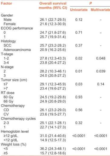
On univariate analyses [Table 2], among all factors, the smaller tumor size (≤ vs. >7.0 cm), lower tumor stage (T1–T2 vs. T3‑4), lower nodal status (N2 vs. N3), absence of anemia before C‑CRT (hemoglobin [Hb] ≥ vs. <12 g/dL), and lesser degree or no weight loss[7] during the 6‑month pretreatment period (≤5% vs. >5%) were found to be associated with significantly longer median OS [Table 2]. On multivariate analysis, although the tumor size category lost its significance, other four variables retained their significant association with longer OS times as depicted in Table 2 and Figure 2, respectively.
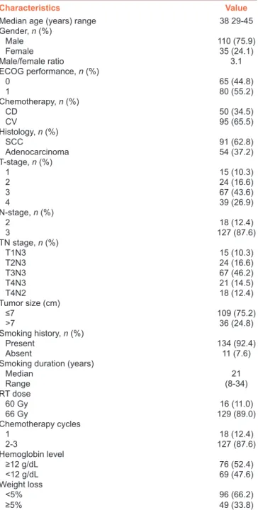
Table 1: Pretreatment and treatment patient characteristics
Characteristics Value
Median age (years) range 38 29‑45
Gender, n (%) Male 110 (75.9) Female 35 (24.1) Male/female ratio 3.1 ECOG performance, n (%) 0 65 (44.8) 1 80 (55.2) Chemotherapy, n (%) CD 50 (34.5) CV 95 (65.5) Histology, n (%) SCC 91 (62.8) Adenocarcinoma 54 (37.2) T-stage, n (%) 1 15 (10.3) 2 24 (16.6) 3 67 (43.6) 4 39 (26.9) N-stage, n (%) 2 18 (12.4) 3 127 (87.6) TN stage, n (%) T1N3 15 (10.3) T2N3 24 (16.6) T3N3 67 (46.2) T4N3 21 (14.5) T4N2 18 (12.4) Tumor size (cm) ≤7 109 (75.2) >7 36 (24.8) Smoking history, n (%) Present 134 (92.4) Absent 11 (7.6)
Smoking duration (years)
Median 21 Range (8-34) RT dose 60 Gy 16 (11.0) 66 Gy 129 (89.0) Chemotherapy cycles 1 18 (12.4) 2-3 127 (87.6) Hemoglobin level ≥12 g/dL 76 (52.4) ﹤12 g/dL 69 (47.6) Weight loss <5% 96 (66.2) ≥5% 49 (33.8)
C-CRT=Concurrent chemoradiotherapy, CD=Cisplatin + docetaxel, CV=Cisplatin + vinorelbine, CI=Confidence interval, ECOG=Eastern Cooperative Oncology Group, N=Node, SCC=Squamous cell carcinoma, T=Tumor, RT=Radiotherapy
DISCUSSION
We retrospectively analyzed the survival outcomes and prognostic factors following definitive C‑CRT in 145 Stage IIIB NSCLC patients under 45 years old. In the absence of early or late toxicity‑related deaths, the documented median OS of 24.8 months accords well with the results of recent benchmark C‑CRT series in LA‑NSCLC patients analyzed without age stratification.[20,21] Notwithstanding the customary lower T and N stages, the additional demonstration of favorable association between the pretreatment normal Hb levels and absence of significant weight loss (WL) (≤5%) with longer OS may prove further valuable in prognostic stratification of such patients group.
NSCLC is the leading cause of all cancer‑related morbidities and mortalities worldwide. The incidence of NSCLC is reported to be in the range of 3.8%–6.0% among patients ≤45‑years,[2‑4] but this rate tends to increase in young NSCLC population.[8,9] In our series, ≤45‑years patients constituted 8.9% of all LA‑NSCLC patients, which is in general a bit higher than the previously reported range.[2‑4] Although it is difficult to explain this difference with a single cause, based on the evidence demonstrating that >40% of all Turkish smokers begin smoking before 16 years of age,[22] we rationally anticipate that this higher incidence may be a result of early onset and longer exposure time to tobacco smoke and its carcinogenic ingredients in our country. Supporting this anticipation, 92.4% of our patients were active smokers for a median of 21 years at the time of diagnosis, which satisfactorily meets the reported range of 20–30 years of tobacco smoke exposure time needed for lung cancer development.[10]
Young NSCLC patients in the previous studies have been suggested as a distinct clinicopathological entity with more advanced stage at presentation, higher female proportion, and higher rate of AC histology.[4,6,7,11,12] We are not able to comment
on stage distribution due to our limited analysis of only Stage IIIB patients. Although male to female ratios differ between various series due to the ethnic and environmental effects, yet a ratio of 3.1 observed here falls in the 1.5–4.5 range reported for such patients.[2,3,11,15] In our study, histologically the SCC (62.8%) was more common than AC (37.2%). Although this finding appears to contrast with studies reporting the AC as the dominant histology in the United States and Japan, yet it is in accordance with European studies reporting SCC as the leading histology.[2,5,23‑27] Although SCC dominance in our cohort may probably be associated with the presence of early‑onset and long‑term smoking status, which is more important for SCC development than AC, the potential impact of unidentified causes and the ethnicity cannot yet be eliminated.
The median 24.8 months OS duration presented here is comparable with the reported 21 and 26 months in two recent benchmark C‑CRT trials without age stratification.[20,21] The outcomes of young, middle‑aged, and senior NSCLC patients had been repeatedly compared with inconsistent results.[3,6,7,28] Some researchers noted that young patients had longer OS than the older ones,[6,7,12,25] while others noted superior outcomes
Figure 1: Survival outcomes for whole study population: (solid line: overall survival; dotted line: locoregional progression-free survival; dashed line: progression-free survival)
Table 2: Results of univariate and multivariate analyses
Factor Overall survival
months (95% CI) Univariate MultivariateP Gender Male 26.1 (22.7‑29.5) 0.12 -Female 21.6 (12.3-30.9) ECOG performance 0 24.7 (21.8-27.6) 0.71 -1 25.7 (19.9‑31.4) Histology SCC 25.7 (23.2‑28.2) 0.37 -Adenocarcinoma 20.9 (16.2‑25.6) T-stage 1-2 27.8 (12.3-43.3) 0.02 0.048 3-4 23.8 (20.4-27.2) N-stage 2 28.2 (10.3-46.1) 0.01 0.039 3 24.0 (20.8-27.2) Tumor size (cm) ≤7 29.1 (12.3‑45.9) 0.03 0.14 >7 23.4 (19.6-27.2) RT dose 60 Gy 24.5 (19.2‑29.8) 0.93 -66 Gy 24.9 (20.8-29.0) Chemotherapy CD 26.1 (23.2-29.0) 0.56 -CV 23.6 (19.5‑27.7) Chemotherapy cycles 2-3 25.1 (22.1‑28.1) 0.32 -1 22.7 (14.1-27.3) Hemoglobin level ≥12 g/dL 31.0 (21.4-40.6) ﹤0.0001 ﹤0.0001 ﹤12 g/dL 14.9 (12.5‑17.3) Weight loss (%) <5 36.2 (24.3-48.1) ﹤0.0001 ﹤0.0001 ≥5 15.7 (12.8‑18.6)
C-CRT=Concurrent chemoradiotherapy, CD=Cisplatin + docetaxel, CV=Cisplatin + vinorelbine, CI=Confidence interval, ECOG=Eastern Cooperative Oncology Group, N=Node, SCC=Squamous cell carcinoma, T=Tumor, RT=Radiotherapy
in favor of older patients.[2,15] Interestingly, in the Bourke’s analysis, NSCLC patients under 45 years of age had shorter OS in Chicago but longer OS in Israel in comparison to older counterparts.[25] In stark contrast with aforementioned results, similar survival durations for young and old patients were also reported in other series.[28‑30] Despite the difficulties in assigning these contradictory outcomes to a few causes, the significant variations between different studies may probably be related with varying environmental, ethnic, and genetic factors. In a previous study in similarly treated 425 Stage IIIB NSCLC patients with a median age of 62 years (range; 33–70), we reported a median OS duration of 22.8 months which is similar with present 24.8 months.[31] In addition, we could not show any survival advantage favoring one age group over other after dichotomizing patients into ≤55 and >55 years groups (22.8 vs. 22.6 months;
P = 0.72). Considering these two studies together, our current
outcomes appear to land support for previous studies reporting similar survival times for young and old patients.[28‑30]
Besides landing support to the universally accepted prognostic values of the lower T and N stages, two other essential findings
of the present study were the emergence of the absence of anemia (Hb ≥12 g/dL; P < 0.001) before C‑CRT and WL (<5%; P < 0.001) as the additional factors being associated with significantly longer OS times. According to the Knight’s systematic literature review, anemia has a prevalence rate of 30%–90% among cancer patients.[32] Although the anemia has been intensely investigated and has been proved to be a negative prognosticator in various tumor sites,[33] surprisingly, its impact on young NSCLC patients undergoing C‑CRT has rarely been addressed. Anemia, and therefore tumor hypoxia, is an established condition that is directly associated with radioresistance and contributes to decreased efficacy of many chemotherapeutics via direct/indirect mechanisms.[34,35] In their quantitative review, Caro et al. reported that lung cancer patients presenting with anemia had 19% increased risk of death.[33] Similarly, in a recent study by Hsu et al.[2] in young NSCLC patients, anemia was reported to significantly shorten survival (hazards ratio: 2.08; 95% CI: 1.15–3.77). Supporting them, patients with anemia had significantly shorter median OS than those without (14.9 vs. 31 months; P < 0.001) in our series.
d c
b a
Weight loss, a key component of the irreversible and fatal cancer‑related anorexia‑cachexia syndrome, is present to some degree up to 80% of all NSCLC patients at presentation which has been unmistakably shown to negatively alter patients’ performance status, quality of life, response to treatment, and prognosis.[36,37] In our series, we observed 49 (33.8%) cases presenting with >5% WL over the past 6 months preceding initiation of C‑CRT [Table 1]. Present >5% WL cutoff was the one that was defined as the clinically significant WL cutoff value for patients presenting with a BMI ≥20 kg/m2 in a recent cancer‑specific consensus statement.[38] Our stratification patients into two groups according to their WL status demonstrated that the cohort undergoing C‑CRT with >5% WL had significantly shorter OS times than the cohort without (36.2 vs. 15.7 months; P < 0.001). This finding is not novel but valuable regarding its particular description in young patients in the absence of such specific evidence. Moreover, exhibition of a poor prognostic worth for WL in young NSCLC patients lands further support for the results of age‑unstratified and elderly studies revealing WL as a poor prognosticator[39,40] and suggests that WL’s prognostic value is somehow independent of age at presentation, although its extent may conceivably vary to some degree among different age groups.
The current article is powered by two factors compared to available young NSCLC literature: First, this is the first study including only Stage IIIB patients exclusively treated by definitive C‑CRT. And second, exclusion of lung pathologies other than SCC and AC prevents potential histology‑related biases. However, the present study has also some drawbacks: First, as common for any retrospective study, unpredictable biases might have influenced our results. Second, current results could not be generalized to all young patients with Stage IIIB NSCLC due to initial selection criteria of good ECOG and exclusive SCC/AC histologies. And third, the lack of mutation status determination due to limited availability in our country might have altered our outcomes in some patients, if not all, particularly at the salvage chemotherapy setting. These issues warrant to be addressed in appropriately designed prospective randomized trials with larger cohorts.
CONCLUSIONS
In the absence of any toxicity‑related death, the present median 24.8 months OS in young patients accords well with the results of recent benchmark C‑CRT series in LA‑NSCLC patients without age stratification.[20,21] In addition, superior OS in patients with normal Hb levels and <5% WL suggest that these factors could easily be used among major prognostic markers in further stratification of such patients in conjunction with the well‑recognized T and N stages and performance status.
Financial support and sponsorship Nil.
Conflicts of interest
There are no conflicts of interest.
REFERENCES
1. Wingo PA, Cardinez CJ, Landis SH, Greenlee RT, Ries LA, Anderson RN, et al. Long‑term trends in cancer mortality in the United States, 1930‑1998. Cancer 2003;97:3133‑275.
2. Hsu CL, Chen KY, Shih JY, Ho CC, Yang CH, Yu CJ, et al. Advanced non‑small cell lung cancer in patients aged 45 years or younger: Outcomes and prognostic factors. BMC Cancer 2012;12:241. 3. Subramanian J, Morgensztern D, Goodgame B, Baggstrom MQ, Gao F,
Piccirillo J, et al. Distinctive characteristics of non‑small cell lung cancer (NSCLC) in the young: A surveillance, epidemiology, and end results (SEER) analysis. J Thorac Oncol 2010;5:23‑8.
4. Mauri D, Pentheroudakis G, Bafaloukos D, Pectasides D, Samantas E, Efstathiou E, et al. Non‑small cell lung cancer in the young: A retrospective analysis of diagnosis, management and outcome data. Anticancer Res 2006;26:3175‑81.
5. McDuffie HH, Klaassen DJ, Dosman JA. Characteristics of patients with primary lung cancer diagnosed at age of 50 years or younger. Chest 1989;96:1298‑301.
6. Radzikowska E, Roszkowski K, Głaz P. Lung cancer in patients under 50 years old. Lung Cancer 2001;33:203‑11.
7. Ramalingam S, Pawlish K, Gadgeel S, Demers R, Kalemkerian GP. Lung cancer in young patients: Analysis of a surveillance, epidemiology, and end results database. J Clin Oncol 1998;16:651‑7.
8. Strand TE, Malayeri C, Eskonsipo PK, Grimsrud TK, Norstein J, Grotmol T, et al. Adolescent smoking and trends in lung cancer incidence among young adults in Norway 1954‑1998. Cancer Causes Control 2004;15:27‑33.
9. Marugame T, Yoshimi I, Kamo K, Imamura Y, Kaneko S, Mizuno S, et al. Trends in lung cancer mortality among young adults in Japan. Jpn J Clin Oncol 2005;35:177‑80.
10. Hanbury WJ. Bronchogenic carcinoma in young persons. Br J Cancer 1958;12:202‑6.
11. Sekine I, Nishiwaki Y, Yokose T, Nagai K, Suzuki K, Kodama T, et al. Young lung cancer patients in Japan: Different characteristics between the sexes. Ann Thorac Surg 1999;67:1451‑5.
12. Awadh‑Behbehani N, Al‑Humood K, Ayed A, Memon A, Ali A. Comparison between young and old patients with bronchogenic carcinoma. Acta Oncol 2000;39:995‑9.
13. Ganz PA, Vernon SE, Preston D, Coulson WF. Lung cancer in younger patients. West J Med 1980;133:373‑8.
14. Liam CK, Lim KH, Wong CM. Lung cancer in patients younger than 40 years in a multiracial Asian country. Respirology 2000;5:355‑61.
15. Zhang J, Chen SF, Zhen Y, Xiang J, Wu C, Bao P, et al. Multicenter analysis of lung cancer patients younger than 45 years in Shanghai. Cancer 2010;116:3656‑62.
16. Furuse K, Fukuoka M, Kawahara M, Nishikawa H, Takada Y, Kudoh S, et al. Phase III study of concurrent versus sequential thoracic radiotherapy in combination with mitomycin, vindesine, and cisplatin in unresectable stage III non‑small‑cell lung cancer. J Clin Oncol 1999;17:2692‑9.
17. Curran WJ Jr., Paulus R, Langer CJ, Komaki R, Lee JS, Hauser S, et al. Sequential vs. concurrent chemoradiation for stage III non‑small cell lung cancer: Randomized phase III trial RTOG 9410. J Natl Cancer Inst 2011;103:1452‑60.
18. Topkan E, Parlak C, Topuk S, Guler OC, Selek U. Outcomes of aggressive concurrent radiochemotherapy in highly selected septuagenarians with stage IIIB non‑small cell lung carcinoma: Retrospective analysis of 89 patients. Lung Cancer 2013;81:226‑30.
19. Jeremic B. Radiochemotherapy as the standard treatment for both elderly and non‑elderly fit patients with locally advanced (stage III)
nonsmall cell lung cancer. Lung Cancer 2013;82:176.
20. Hanna N, Neubauer M, Yiannoutsos C, McGarry R, Arseneau J, Ansari R, et al. Phase III study of cisplatin, etoposide, and concurrent chest radiation with or without consolidation docetaxel in patients with inoperable stage III non‑small‑cell lung cancer: The hoosier oncology group and U.S. oncology. J Clin Oncol 2008;26:5755‑60. 21. Govindan R, Bogart J, Stinchcombe T, Wang X, Hodgson L, Kratzke R,
et al. Randomized phase II study of pemetrexed, carboplatin, and thoracic radiation with or without cetuximab in patients with locally advanced unresectable non‑small‑cell lung cancer: Cancer and leukemia group B trial 30407. J Clin Oncol 2011;29:3120‑5. 22. Oztürk AB, Kiliçaslan Z, Işsever H. Effect of smoking and indoor air
pollution on the risk of tuberculosis: Smoking, indoor air pollution and tuberculosis. Tuberk Toraks 2014;62:1‑6.
23. Greenberg ER, Korson R, Baker J, Barrett J, Baron JA, Yates J, et al. Incidence of lung cancer by cell type: A population‑based study in new hampshire and vermont. J Natl Cancer Inst 1984;72:599‑603. 24. Kabat GC, Wynder EL. Lung cancer in nonsmokers. Cancer
1984;53:1214‑21.
25. Bourke W, Milstein D, Giura R, Donghi M, Luisetti M, Rubin AH, et al. Lung cancer in young adults. Chest 1992;102:1723‑9.
26. Roviaro GC, Varoli F, Zannini P, Fascianella A, Pezzuoli G. Lung cancer in the young. Chest 1985;87:456‑9.
27. Seddon DJ, Partridge MR. Carcinoma of the bronchus in young adults. Br J Clin Pract 1990;44:24‑5.
28. Zou B, Xu Y, Li T, Li W, Tang B, Zhou L, et al. A multicenter retrospective analysis of survival outcome following postoperative chemoradiotherapy in non‑small‑cell lung cancer patients with N2 nodal disease. Int J Radiat Oncol Biol Phys 2010;77:321‑8.
29. Ak G, Metintas M, Metintas S, Yildirim H, Erginel S, Alatas F, et al. Lung cancer in individuals less than 50 years of age. Lung 2007;185:279‑86. 30. Skarin AT, Herbst RS, Leong TL, Bailey A, Sugarbaker D. Lung cancer
in patients under age 40. Lung Cancer 2001;32:255‑64.
31. Topkan E, Parlak C, Selek U. Impact of weight change during the course of concurrent chemoradiation therapy on outcomes in stage IIIB non‑small cell lung cancer patients: Retrospective analysis of 425 patients. Int J Radiat Oncol Biol Phys 2013;87:697‑704. 32. Knight K, Wade S, Balducci L. Prevalence and outcomes of anemia
in cancer: A systematic review of the literature. Am J Med 2004;116 Suppl 7A:11S‑26S.
33. Caro JJ, Salas M, Ward A, Goss G. Anemia as an independent prognostic factor for survival in patients with cancer: A systemic, quantitative review. Cancer 2001;91:2214‑21.
34. Peitzsch C, Perrin R, Hill RP, Dubrovska A, Kurth I. Hypoxia as a biomarker for radioresistant cancer stem cells. Int J Radiat Biol 2014;90:636‑52.
35. Shannon AM, Bouchier‑Hayes DJ, Condron CM, Toomey D. Tumour hypoxia, chemotherapeutic resistance and hypoxia‑related therapies. Cancer Treat Rev 2003;29:297‑307.
36. Inui A. Cancer anorexia‑cachexia syndrome: Current issues in research and management. CA Cancer J Clin 2002;52:72‑91.
37. Werner‑Wasik M, Scott C, Cox JD, Sause WT, Byhardt RW, Asbell S, et al. Recursive partitioning analysis of 1999 radiation therapy oncology group (RTOG) patients with locally‑advanced non‑small‑cell lung cancer (LA‑NSCLC): Identification of five groups with different survival. Int J Radiat Oncol Biol Phys 2000;48:1475‑82.
38. Fearon K, Strasser F, Anker SD, Bosaeus I, Bruera E, Fainsinger RL, et al. Definition and classification of cancer cachexia: An international consensus. Lancet Oncol 2011;12:489‑95.
39. Ross PJ, Ashley S, Norton A, Priest K, Waters JS, Eisen T, et al. Do patients with weight loss have a worse outcome when undergoing chemotherapy for lung cancers? Br J Cancer 2004;90:1905‑11. 40. Fiorelli A, Vicidomini G, Mazzella A, Messina G, Milione R,
Di Crescenzo VG, et al. The influence of body mass index and weight loss on outcome of elderly patients undergoing lung cancer resection. Thorac Cardiovasc Surg 2014;62:578‑87.