Address for Correspondence / Yazışma Adresi: Merih Tepeoğlu, MD Baskent University, Faculty of Medicine Department of Pathology 79.sokak 7/4 Bahcelievler, 06490, Ankara, Turkey E-mail: [email protected]
©Telif Hakkı 2017 Gazi Üniversitesi Tıp Fakültesi - Makale metnine http://medicaljournal.gazi.edu.tr/ web adresinden ulaşılabilir. ©Copyright 2017 by Gazi University Medical Faculty - Available on-line at web site http://medicaljournal.gazi.edu.tr/
doi:http://dx.doi.org/10.12996/gmj.2017.35
A Case of Two Synchronous Cutaneous Collision Tumors
Eş Zamanlı Saptanan İki Kutanöz Kollizyon Tümör Olgusu
Sebnem Kupana Ayva
1, Merih Tepeoglu
1, Ozgur Gunduz
2, Ilker Yazici
3, Onder Bozdogan
41 Baskent University, School of Medicine, Department of Pathology, Ankara, Turkey 2 Kırıkkale University, School of Medicine, Department of Dermatology, Kırıkkale, Turkey 3 Kırıkkale University, School of Medicine, Plastic and Reconstructive Surgery, Kırıkkale, Turkey 4 Numune Training and Research Hospital, Department of Pathology, Ankara, Turkey
ABSTRACT
Cutaneous collision tumors are known as two independent tumors which are close anatomically and separated from one another by well boundaries. We, herein report a 83-year-old female patient with two cutaneous collision tumors in two different localizations at the same time. First cutaneous collision tumor located on left ala nasi was squamous cell carcinoma and basal cell carcinoma and second one located on the right commisure was composed of malignant melanoma (Clark Level IV) and basal cell carcinoma. However, the presence of collision tumors is not uncommon and is often reported in the literature, to the best of our knowledge, it is the first case which shows the association of two synchronous cutaneous collision tumor in the same individual.
Key words: Basal cell carcinoma, cutaneous collision tumor, malignant melanoma, squamous cell carcinoma
Received: 02.04.2016 Accepted: 01.14.2017
ÖZET
Kutanöz kollizyon tümörler, anatomik olarak yakın ancak birbirinden net sınırlarla ayrılan iki bağımsız tümördür. Biz burada aynı zamanda, farklı lokalizasyonlarda iki kutanöz kollizyon tümörü olan 83 yaşında bir kadın hastayı sunduk. Sol burun kanadında izlenen ilk kollizyon tümör skuamöz hücreli karsinom ve bazal hücreli karsinom’dan oluşmakta iken, ikinci kollizyon tümör sağ dudak kenarında izlenen Malign melanom (Clark Level IV) ve bazal hücreli karsinom idi. Kollizyon tümörler literatürde sıklıkla bildirilen, çok da nadir olmayan tümörler olmakla birlikte, bildiğimiz kadarıyla bu vaka aynı hastada eş zamanlı olarak saptanan iki kollizyon tümör birlikteliğini gösteren ilk vakadır.
Anahtar Sözcükler: Bazal hücreli karsinom, kutanöz kollizyon tümör, malign melanom, skuamöz hücreli karsinom
Geliş Tarihi: 04.02.2016 Kabul Tarihi: 14.01.2017
INTRODUCTION
Cutaneous cancers are the most common human cancer, mainly including basal cell carcinoma (BCC). It has been reported that there is an increasingly development risk of the second cutaneous cancer in the individuals, especially in sun-exposuring areas (1,2).The co-existence of two distinctly different neoplasms occuring in the same anatomic location is called as collision tumor. Various combinations of collision tumors have been described, mostly common BCC and melanocytic naevus (3). We, herein report a new case with two synchronous collision tumor in the face and discuss its clinical and histopathological features.
CASE REPORT
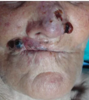
A 83-year-old-woman presented with an adjacent plaque (1.0x1.0 cm) and nodule (3.0x1.0 cm) with ulcerated surfaces overlying on the left ala nasi. Another accompanying feature was the solitary dark nodular lesion (4.0x1.3x0.3cm) with focal ulcerations on the right commissure of the patient’s lips (Fig.1).
Figure 1: Clinical appearance of the lesions that were seen on the left ala nasi and the right commisure of the lips.
The excisional biopsies were performed for the lesions. In histopathological examination, two different carcinoma adjacent to one another were observed on the left ala nasi. The first tumor was characterized by nests of atypical squamous epithelial cells arising from epidermis and extending into the reticular dermis. The second tumor was composed of islands and large nests of basaloid cells with hyperchromatic nuclei and poorly defined cytoplasm (Fig.2A). Actinic keratosis and solar elastosis were observed in the biopsy. In the histopathological sections of the dark nodule on the right labial commisure, there were again two adjacent malignant foci. One of these foci was composed of highly cellular discohesive nested groups of atypical melanocytic cells, extending from epidermis to reticular dermis (Clark Level IV). The second malignant foci adjacent to the first tumor was composed of basaloid cell proliferation with palisading in the periphery (Fig.2B). Actinic changes and marked solar elastosis were also seen in the biopsy. Finally, while the diagnosis of first collision tumor was squamous cell carcinoma (SCC) and BCC, second was malignant melanoma (MM) and BCC.
In immunohistochemical analysis, while Melan-A positivity was only seen in the sparsed melanocytes in BCC component of the first collision tumor (Fig. 2C), it is positive both in the MM component, and in some melanocyte groups with atypical findings in BCC component of the second collision tumor (Fig 2D).
Figure 2. (A) : The collision tumor seen on the left ala nasi composed of basaloid cells and atypical squamous cells nests (Hematoxylin and eosin, x 200), (B): The lesion located on the right commisure of the lips includes basaloid proliferation and atypical melanocyte nests (Hematoxylin and eosin, x 100), (C) The scattered melanocytes exhibiting Melan-A positivity in BCC component of the first collision tumor (Diaminobenzidine, x200), (D) Melanocytes which form the small groups revealing Melan-A positivity in BCC component of the second collision tumor (Diaminobenzidine, x200)
DISCUSSION
The collision tumor is refered to as two independent neoplasms occuring in close proximately to one another and maintaining sharp distinct boundaries (4).The combination of two benign neoplasms may have limited clinical significance, but the occurence of two cutaneous malignancies, each having the potential to metastasize, may pose a considerable health risk.
The presence of MM and BCC is rare, with only case studies reported (5,6). The first case described the collision of BCC and MM was in 1983 by Kao in the Annual meeting of the American Society of Dermatopathology and the greatest information about the incidence of these tumors had been reported by Pieard and et al (7). When Pieard et al., examined 78.000 excision materials, they found 11 collision tumors, including BCC and MM (7). Although, different combinations of both SCC and BCC with other tumors were mentioned in the literature, there were no co-existence of SCC and BCC in the literature. The presence of non-neoplastic melanocytes in the BCC have been mentioned in the literature (3,5). On the other hand, malignant melanocytes of melanoma in-situ (MIS) have populated within BCC (8,9). BCC colonised by MIS showed a much higher density of atypical melanocytes, with two or more melanocyte clusters, distributed throughout the lesion (8,9).
There are various views about the pathogenesis of collision tumors. Some authors consider to be merely coinsidental, whereas others except that the paracrine effects of one tumor may induce formation of a second tumor (5,9,10).Frequent damage to the skin by ultraviolet radiation may also induce different neoplasms adjacent to each other according to ‘field cancerization’ theory (10).In our case, the presence of actinic damage in the biopsies, let us think that, ultraviolet exposuring is the most important pathogenetic factor which triggers the mechanism of malignant transformation.
CONCLUSION
This case demonstrates the association of two synchronous cutaneous collision tumor in the same individual and to our knowledge, this is reported for the first time. The detailed dermatological examination may prevent from the skipping of these tumors especially in the elderly individuals having prominent actinic damage.
Conflict of interest
No conflict of interest was declared by the authors.
REFERENCES
1. Bhatia S, Estrada-Batres L, Maryon T, Bogue M, Chu D. Second primary tumors patient with cutaneous malignant melanoma. Cancer 1999;86:2014-20. 2. Hallaji Z, Rahimi H, Mirshams-Shahshahani M. Comparison of risk factors of
single basal cell carcinoma with multiple basal cell carcinomas. Indian J Dermatol 2011;56:398-402.
3. Giorgi V, Sestini S, Alfaioli B, Carelli G, Carli P. Cutaneous collision tumour (melanocytic naevus, basal cell carcinoma, seborrhoeic keratosis): a clinical, dermoscopic and pathological case report. Br J Dermatol 2005;152:787-90. 4. Satter EK, Metcalf J, Lountzis N, Elston DM. Tumors composed of malignant
epithelial and melanocytic populations: a case series and review of the literatüre. J Cutan Pathol 2009;36:211-9.
5. Papa G, Grandi G, Pascone M. Collision tumor of malignant skin cancers: A case of melanoma in basal cell carcinoma. Pathol Res Pract 2006;202:691-4. 6. Hirakawa E, Miki H, Kobayashi S, Nomura Y, Ohmori M. Collision tumor of
cutaneaous malignant melanoma and basal cell carcinoma. Pathol Res Pract 1998;194:649-53.
7. Piérard GE, Fazaa B, Henry F, Kamoun MR, Piérard- Franchimont C. Collision of
primary malignant neoplasms on the skin: the connection between malignant melanoma and basal cell carcinoma. Dermatology 1997;194:378-9.
8.Burkhalter A, White WL. Malignant melanoma in situ colonizing basal cell carcinoma. A simulator of invasive melanoma. Am J Dermatopathol 1997;19:303-7.
9. Belisle A, Gautier MS, Ghozali F, Plantier F, Wechsler J. A Collision Tumor Involving Basal Cell Carcinoma and Lentigo Maligna Melanoma. Am J Dermatopathol 2005;27:319-21.
10. Slaughter DP, Southwıck HW, Smejkal W. Field cancerization in oral
stratified squamous epithelium; clinical implications of multicentric origin. Cancer 1953;6:963-8.