Research Paper
The effect of bleomycin embolization on symptomatic improvement and
hemangioma size among patients with giant liver hemangiomas
Mahir Kirnap
a,*, Fatih Boyvat
b, Sedat Boyacioglu
c, Fatih Hilmioglu
c, Gokhan Moray
a,
Mehmet Haberal
aaGeneral Surgery Department, Baskent University Faculty of Medicine, Turkey bInterventional Radiology Department, Baskent University Faculty of Medicine, Turkey cGastroenterology Department, Baskent University Faculty of Medicine, Turkey
a r t i c l e i n f o
Article history:Received 19 March 2018 Accepted 24 May 2018 Available online 26 May 2018
1. Introduction
Hemangiomas are benign mesenchymal liver tumors with a prevalence of 3e20% in autopsy series [1]. Hemangiomas most commonly affect women in their fourth andfifth decades of life. Liver hemangiomas are usually asymptomatic, and liver function tests are typically within normal limits. Hence, they mostly detec-ted incidentally. Hemangiomas greater than 5 cm are referred to as giant hemangiomas, which can be symptomatic and require treatment[2]. Although surgery is the usual treatment method for symptomatic giant hemangiomas, surgical resection continues to be the classical paradigm[3]. Minimally invasive techniques have recently been introduced as alternatives to surgery. In this pro-spectively study, we aimed to determine the effects of the mini-mally invasive embolization with bleomycin mixed with lipiodol on mass size and symptoms of patients with giant liver hemangiomas. 2. Methods
In the present study 17 patients [10 women, 7 men; Age range 35e67 years (mean 46,41 ± 2,6 years)] with giant liver hemangi-omas who presented to our clinic between August 2014 and October 2016 were treated with minimally invasive approach using hemangioma embolization with bleomycin mixed with lipiodol. Two patients underwent a second session of embolization one
month later while three patients underwent simultaneous treat-ment of hemangiomas in two different lobes of the liver. Tumor characterization (length, lesion number) was performed with ul-trasonography, computerized tomography (CT) or magnetic reso-nance imaging. The diagnosis was confirmed by noninvasive examination and the typical angiographic characteristics noted during the Transarterial chemoembolization (TACE) procedure.
Following the placement of a 4F introducer into the right femoral artery under standard sterile conditions, the celiac root was selectively cannulized using a 4F shepherd hook catheter, and im-ages were taken after contrast material injection. The acquired cine films were used to assess large caliber hypervascular mass lesions supplied by the hepatic artery branch at the side of a liver hem-angioma. Following the acquisition of diagnostic images, super-selective catheterization of the above-described mass lesions was carried out using a microcatheter and a 0.016-inch guidewire. Bleomycin and lipiodol mixture was injected through a micro-catheter for the embolization procedure (Fig. 1). Fifteen milligrams of bleomycin sulphate [Bleosin-S (in 15 mg vials); Onko/Koçsel, Turkey] was dissolved in 5 ml saline solution and admixed with 10 ml lipiodol (Laboratoire Guerbet, Fransa) at 1:2 ratio. Lipiodol and bleomycin (10e20 mg/ml) dosages were determined by tumor diameter (cm), and a maximum of 30 mg bleomycin and 20 ml lipiodol were used for tumors greater than 10 cm.
During the follow-up, all patients were considered symptomatic due to an increase in hemangioma size. Additionally, eleven pa-tients were considered symptomatic owing to a sense of fullness in the right upper quadrant;five owing to pain; and one owing to liver failure. All patients were informed about therapeutic and surgical treatment methods currently available for giant liver hemangi-omas. All patients gave written informed consent, and the study was approved by the local ethics committee of Bas¸kent University. The main portal vein was patent in all patients. A scintigraphic examination proved the absence of an arterioportal or arteriove-nous shunt. All patients had an INR of less than 1.5 and a throm-bocyte count above 50.000/ml. Complete blood count a liver function tests were evaluated before and 1 day after the procedure. All patients were administered analgesics and sedatives prior to the * Corresponding author. FCIS Baskent University, Taskent Caddesi No: 77,
Bah-celievler, Ankara, 06490, Turkey.
E-mail address:[email protected](M. Kirnap).
Contents lists available atScienceDirect
International Journal of Surgery Open
j o u r n a l h o m e p a g e :w w w . e l s e v i e r. c o m / l o c a t e / i j s o
https://doi.org/10.1016/j.ijso.2018.05.003
2405-8572/© 2018 Published by Elsevier Ltd on behalf of Surgical Associates Ltd. This is an open access article under the CC BY-NC-ND license (http://creativecommons.org/ licenses/by-nc-nd/4.0/).
procedure. All patients were administered a single dose (cepha-zolin 1 g I.V) prophylactic antibiotic immediately before the pro-cedure. Analgesics and antiemetics were repeated as needed after the procedure. All patients were hospitalized for the embolization procedure. The range of hospital stay was 24e72 h (24 h in 15 patients; 48 h in 1 patient; and 72 h in 1 patient) and the mean duration of hospital stay was 28 h.
Bleomycin-lipiodol mixture was prepared as below:
Tumor size after embolization with bleomycin was assessed by computerized tomography at 1, 6, and 12 months. Hemangioma volume was calculated by multiplying its height, length, and width. The range of the duration of post-procedural clinical and radio-logical follow-up was 12e16 months (mean 14.47 ± 2.21 months). Treatment response was analyzed using SPSS 15.0 (Chicago, USA) software package. Wilcoxon test was used to test whether the distributions of two variables were equal, and the Firedman ANOVA test to compare the median values. Patients age and tumor size was expressed as mean± SD; statistical significance was set at p ¼ 0.05. 3. Results
Seven patients had hemangiomas in the right lobe; 2 in the left lobe, and 8 in both lobes. The diameter of the largest hemangioma was 28 cm. The volumes of the hemagiomas ranged between 480 and 9925.1 cm3. Eleven (64.8%) patients were diagnosed with
ul-trasonography (USG), and 6 (35.2%) with CT. A total of 22 emboli-zation sessions using the bleomycin lipiodol mixture were carried out for 19 hemangiomas of 17 patients. Radiological examination revealed a marked reduction in the mean hemangiomasize after embolization with bleomycin. The largest diameter of hemangi-omas was reduced from 14.72± 12.8 cm3to 7.63± 4.76 cm3. The
mean volume of the hemangiomas was 3716.276 cm3 (480.0e9925.1 cm3) prior to the procedure and 746.012 cm3 (38.7e3856 cm3) at 12 months. Thepre-procedural hemagioma volumes and diameters were significantly different from one-, six-, and twelve-month values. The mean tumor volume was 746± 405.7 cm3at 14 months of follow-up.
The mean volume of giant liver hemangiomas was reduced by 54.9% at 1 month after the procedure compared to the pre-procedural volume (p < 0.001). The volume reduction reached 79.6% at 6 and 12 months. The non-intervened hemangiomas, on the other hand, were reduced in volume by 50.8% at 1 month and 75.7% at 12 months, which was radiologically significant but not statistically significant owing to the low number of hemangiomas (Figs. 2 and 3).
All patients enjoyed a symptomatic improvement following embolization. Hemangioma-associated pain and sense of fullness
mostly started to regress from the first month and completely eliminated by 2 months. The regression of the sense of fullness was proportional to a reduction in the size of the mass. No symptom recurrence was observed during the follow-up period.
Seven patients developed post-procedural postembolization syndrome characterized by pain, fever, loss of appetite or nausea. The most prominent symptom of this syndrome was abdominal pain. Pain in the right upper quadrant started immediately following the procedure in all patients, and subsided by 24 h in 15 patients and 48 h in the remainders. Ten patients developed mild nausea and loss of appetite accompanied by pain. Four patients developed a subfebrile body temperature not exceeding 37.5 C, which also subsided by 24 h. No statistically significant increase occurred in liver enzymes (aspartate amino transferase, alanine amino transferase) at 24 h after the procedure. No clinically meaningful increase occurred in white blood cell count, either. No serious procedural complications developed during follow-up period, such as liver function disorders, liver abscess, or necrosis of normal liver tissue. No patient developed pulmonary symptoms after the procedure. A patient developed a drop in the hemoglobin level that necessitated the infusion of 2 packs of red blood cell suspension. No pathology was detected by a control ultrasonogra-phy, and the patient was discharged uneventfully at the end of 72 h (Table 1).
4. Discussion
Cavernous hemangiomas are the most common type of benign tumors of the liver[1,5]. Most are small, asymptomatic and detected incidentally by radiological screening[6]. Giant cavernous hemangi-omas may cause symptoms such as pain, abdominal sense of fullness, and upper abdominal mass. Thrombosis, infarction, intralesional bleeding, capsular distention, and compressing of adjacent organs are the possible causes of pain[7]. In some rare occasions, serious com-pications such as obstructive jaundice, Kasabach-Merritt syndrome, gastric outlet obstruction, orintra-abdominal hemorrhage due to rupturemay occur[8]. Literature data suggest that the risk of rupture of liver hemangiomas ranges between 1% and 4%. Giant subcapsular lesions may be considered to confer a greater risk [9]. Although hemangiomas do not typically grow in size during follow-up, spon-taneous growth constitutes the main treatment indication. Steroids and estrogen therapy may also increase their size. No malignant transformation of hemangiomas has been reported[10]. In accor-dance with the literature data, our patients suffered from hemangi-oma growth, sense of abdominal fullness, and pain. The diagnosis was made by diagnostic USG and tomographic examination. Spontaneous growth occurred in hemangiomas during pre-procedural follow-up. Fig. 1. Selective angiographic transarterial chemoembolization procedure.
No complication developed in any of our patients during a mean follow-up period of 14 months.
The etiology of hemangiomas is not clear. Structurally, heman-giomas are composed of venous pools lined by vascular endothelial cells separated byfibrous tissue septae. Blood circulation is slow,
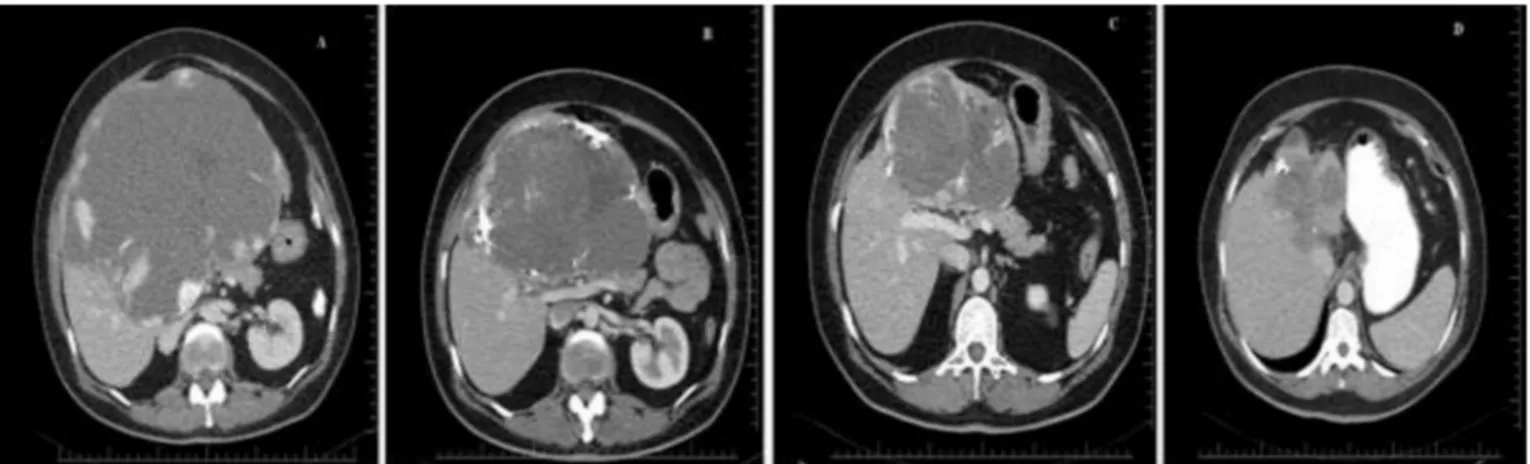
and hepatic artery is the main source of vascular supply[11]. The treatment options for symptomatic liver hemangiomas include steroids, radiotherapy, surgical resection, hepatic artery ligation, and transarterial embolization (TACE) [12]. Asymptomatic liver hemangiomas, on the other hand, have no definitive indications for Fig. 2. One-year follow-up of a giant liver hemangioma; A: computerized tomographic (CT) examination of the giant liver hemangioma before bleomycin embolization, B: CT examination 1 month after bleomycin embolization, C: CT examination 6 months after bleomycin embolization, D: CT examination 12 months after bleomycin embolization.
Fig. 3. A; Comparison of giant hemangioma size at 1, 6, and 12 months after embolization with bleomycin with pre-procedural size, B; Comparison of the size of the second non-intervened hemangiomas at 1, 6, and 12 months with pre-procedural size.
Table 1
Demographic properties of patients with giant liver hemangiomas.
Patients Ages Sex Complaint Diagnosis Localization Number Volume-1 Volume-2 Volume-3 Complication 1 42 F pain/enlargement CT bilateral multiple 89.6 190.4 64 no 2 42 M pain/enlargement USG bilateral multiple 880.4 797.94 39.9 no
3 59 F Feeling of fullness/growth USG bilateral 2 4576 405 low hemoglobin 4 47 F Feeling of fullness/growth USG bilateral multiple 510.3 fever and vomiting 5 48 M Feeling of fullness/growth USG bilateral 3 946 470.7 no
6 54 F Feeling of fullness/growth CT Right lob 1 600 no
7 39 F Feeling of fullness/growth USG Right lob 1 101 no
8 48 F pain/enlargement USG Bilateral multiple 600 no
9 67 F pain/enlargement CT Right lob 1 1340.9 no
10 49 F pain/enlargement USG Right lob 2 3021 400 no
11 47 F Feeling of fullness/growth CT Right lob 1 770 no 12 35 M Feeling of fullness/growth USG Right lob 1 658 no 13 52 M Feeling of fullness/growth USG Left lob multiple 831.6 no 14 40 M growth/Liver failure CT bilateral multiple 3705 no 15 37 M Feeling of fullness/growth USG bilateral multiple 144 229 108 no 16 45 F Feeling of fullness/growth USG Left lob multiple 1510.2 no 17 38 M Feeling of fullness/growth CT Right lob 1 810 no F; Female, M; Male, USG; ultrasonography, CT; computerized tomography.
surgery, although they must be closely monitored due to the risk of growth[3].
The decision for performing surgery for liver hemangiomas should be carefully weighed against the complication risk. A number of series have reported serious blood loss (10e27%) and death at a rate of 0e2% after the enucleation or resection of hepatic hemangiomas[11]. Surgical resection is controversial even in the case of symptomatic hemangiomas owing to the risk of severe complications. The definitive indications for surgery include intraperitoneal bleeding, intratumoral bleeding, and consumption coagulopathy (Kasabach-Merritt syndrome) and spontaneous or traumatic rupture. Nevertheless, spontaneous rupture is an extremely rare complication even with giant hemangiomas[9]. As reported previously, other definitive indications for surgery, such as Kasabach-Merritt syndrome, can also be corrected by embolization
[13]. Surgery should be reserved for patients with giant liver hemangiomas in whom the non-surgical treatment fails. Although our patients had no contraindication for surgery, they were suc-cessfully treated in a minimally invasive manner, i. e by emboliza-tion with bleomycin. By this way, they were protected against the complications of surgery.
Recent studies have emphasized the importance of the role of TACE to treat symptomatic hemangiomas as an effective treatment method that is less invasive than surgery [5]. To date, several different embolic agents like gelating sponge, steel coils, polyvinyl alcohol (PVA), ethanol, sodium morphate, and isobutyl cyano-acrylate have been used to treat hemangiomas [14]. However, liquid embolic agents like PVA and sodium morphate or ethanol may lead to serious complications such as severe pain, and may cause reflux embolization. Gelatin sponge alone does not destroy blood sinuses[4]. Bleomycin is an antimycotic and antimicrobial agent used against squamous cell carcinomas, testis cancers, and malignant lymphomas. Its antineoplastic activity stems from its inhibition of DNA biosynthesis[15]. Oikawa et al.[16]first reported the antiangiogenic action of bleomycin in 1990. Bleomycin exerts a local sclerosing action on endothelial cells. Bleomycin, alone or mixed with iodized oil (lipiodol), can be used as a sclerosing agent to treat vascular anomalies[17]. The mechanism of the antiangiogenic effects of bleomycin is unknown. Animal studies have suggested that injury starts in capillaries in the form of endothelial cell pyk-nosis, thrombocyte aggregation, and intraluminal thrombus for-mation, ultimately inducing fibroblast proliferation and collagen deposition[18]. Pulmonaryfibrosis is the most severe pulmonary complication of bleomycin. Bleomycin-induced pulmonaryfibrosis has not been reported for doses used to treat hemangiomas, how-ever[18]. Pulmonerfibrosis is seen in 30% of patients in whom the cumulative bleomycin dose exceeds 450 mg[15]. In this study, the maximum bleomycin dose was substantially lower than the toxic dose; we therefore encountered no case of pulmonaryfibrosis.
Lipiodol (iodized oil) is used as an embolic agent and anticancer drug carrier. It is selectively accumulated in vascular sinuses[19]. Lipiodol not only invades small arteries supplying a tumor, but it also carries bleomycin to it. Arteriovenous shunts inside hepatic cavernous hemangiomas are extremely rare[20]. Studies on animals and humans have demonstrated that lipiodol remains in small ar-teries and sinusoids of a tumor[19]. The mechanism of effect of bleomycin mixed with lipiodol is thought to be the destruction of endothelial cells and formation of microthrombin in sinuses, ulti-mately resulting in the atrophy andfibrosis[4]. Additionally, bleo-mycin mixed with lipiodol does not harm normal blood vessels. This study demonstrated a variable reduction of hemangioma size at 3e12 months after embolization. The difference between heman-giomas size post-and pre-embolization was statistically significant. We suggest that bleomycin and lipiodol have a superior effect on embolization than solid embolic agents. Bleomycin mixed with
lipiodol acts not only as an embolization agent, but also as a chronic sclerozing agent that inducesfibrosis. The fibrotic effect of bleo-mycin mixed with lipiodol as well as the reduction in hemangioma size and blood supply of hemangiomas were radiologically showed during 12-month follow-up of the patients.
Thanks to its relatively modest embolization effect, bleomycin mixed with lipiodol is safer than liquid embolic agents like sodium morphate and ethanol. The most common side effects of emboli-zation are pain, fever, leucocytosis, and nausea. The postemboliza-tion syndrome lasts several days and serious complicapostemboliza-tions are rare
[21]. Our patients suffered mild-moderate postembolization syn-drome, and none of them developed liver failure. Even a patient with preexisting liver failure tolerated the procedure well. This may be due to lipiodol being not taken by a normal hepatic tissue but rapidly cleared through hemangiomatous sinusoids.
5. Conclusions
This prospectively study showed that the embolization of giant liver hemangiomas with bleomycin and lipiodol mixture elimi-nated symptoms and reduced lesion size. Embolization with bleo-mycin mixed with lipiodol is an effective alternative to surgery in these patients.
Ethical approval
All patients gave written informed consent, and the study was approved by the local ethics committee of Bas¸kent University. Judgement’s reference number:D18/05
Funding None.
Author contribution
Mahir Kirnap : study design, data analysis, writing. Fatih Boyvat : data collections.
Sedat Boyacioglu: data analysis, writing. Fatih Hilmioglu : data collections. Gokhan Moray : study design. Mehmet Haberal : study design. Conflict of interest
The authors declare no conflict of interest. Guarantor
Baskent University Faculty of Medicine; Turkey. Research registration number
researchregistry3850. References
[1] Choi BY, Nguyen MH. The diagnosis and management of benign hepatic tu-mors. J Clin Gastroenterol 2005;39:401e12.
[2] Weimann A, Ringe B, Klempnauer J, et al. Benign liver tumors: differential diagnosis and indications for surgery. World J Surg 1997;21:983e90. [3] Schnelldorfer T, Ware AL, Smoot R, et al. Management of giant hemangioma of
the liver: resection versus observation. J Am Coll Surg 2010;211(6):724e30. [4] Zeng Q, Li Y, Chen Y, et al. Gigantic cavernous hemangioma of the liver treated
by intra-arterial embolization with pingyangmycin-lipiodol emulsion: a multi-center study. Cardiovasc Intervent Radiol 2004;27(5):481e2.
[5] Giavroglou C, Economou H, Ioannidis I. Arterial embolization of giant hepatic hemangiomas. Cardiovasc Intervent Radiol 2003;26:92e6.
[6] Sun JH, Nie CH, Zhang YL, Zhou GH, Ai J, Zhou TY, et al. Transcatheter arterial embolization alone for giant hepatic hemangioma. PLoS One 2015 Aug 19;10(8).
[7] Erdogan D, Busch OR, van Delden OM, et al. Management of liver hemangi-omas according to size and symptoms. J Gastroenterol Hepatol 2007;22(11): 1953e8.
[8] Ribeiro Jr MAF, Papaiordanou F, Gonçalves JM, et al. Spontaneous rupture of hepatic hemangiomas: a review of the literature. World J Hepatol 2010;2(12): 428e33.
[9] Jain V, Ramachandran V, Garg R, et al. Spontaneous rupture of a giant hepatic hemangiomadsequential management with transcatheter arterial embolization and resection. Saudi J Gastroenterol 2010;16(2): 116e9.
[10] Glinkova V, Shevah O, Boaz M, et al. Hepatic haemangiomas: possible asso-ciation with female sex hormones. Gut 2004;53(9):1352e5.
[11] Ho HY, Wu TH, Yu MC, et al. Surgical management of giant hepatic heman-giomas: complications and review of the literature. Chang Gung Med J 2012;35(1):70e8.
[12] Herman P, Costa ML, Machado MA, et al. Management of hepatic hemangi-omas: a 14-year experience. J Gastrointest Surg 2005;9:853e9.
[13] Malagari K, Alexopoulou E, Dourakis S, et al. Transarterial embolization of giant liver hemangiomas associated with Kasabache Merritt syndrome: a case report. Acta Radiol 2007;48(6):608e12.
[14] Soyer P, Levesque M. Hemoperitoneum due to spontaneous rupture of hepatic haemangiomatosis: treatment by superselective arterial embolization and partial hepatectomy. Australas Radiol 1995;39:90e2.
[15] Bennett JM, Reich SD. Bleomycin. Ann Intern Med 1979;90:945e8. [16] Oikawa T, Hirotani K, Ogasawara H, et al. Inhibition of angiogenesis by
bleomycin and its copper complex. Chem Pharm Bull 1990;38:1790e2. [17] Chen Y, Li Y, Zhu Q, et al. Fluoroscopic intralesional injection with
pin-gyangmycin lipiodol emulsion for the treatment of orbital venous malfor-mations. AJR Am J Roentgenol 2008;190(4):966e71.
[18] Duncan IC, Van Der Nest L. Intralesional bleomycin injections for the palliation of epistaxis in hereditary hemorrhagic telangiectasia. AJNR Am J Neuroradiol 2004;25(7):1144e6.
[19] Ohishi H, Uchida H, Yoshimura H, et al. Hepatocellular carcinoma detected by iodized oil. Use of anticancer agents. Radiology 1985;154(1):25e9. [20] Bozkaya H, Cinar C, Besir FB, et al. Minimally invasive treatment of giant
haemangiomas of the liver: embolisation with bleomycin. Cardiovasc Inter-vent Radiol 2014;37:101e7.
[21] Leung DA, Goin JE, Sickles C, et al. Determinants of postembolization syndrome after hepatic chemoembolization. J Vasc Interv Radiol 2001;12(3):321e6.