139
Ankara Üniversitesi Tıp Fakültesi Mecmuası 2005; 58:139-141
DAHİLİ BİLİMLER / MEDICAL SCIENCES Olgu Bildirisi / Case Report
Infected mesenteric cyst: a case report
Enfekte mesenterik kist: bir olgu sunumuNazan Dalgıç
1, Erdal İnce
1, Ergin Çiftçi
1, Selim Öncel
1, Zehra Serap Arıcı
1,
Meltem Bingöl Koloğlu
2, Ülker Doğru
11Ankara University, School of Medicine, Department of Pediatric Infectious Diseases, Ankara
2 Ankara University, School of Medicine, Department of Pediatric Surgery , Ankara
Mesenteric cysts are rare intraabdominal lesions of childhood. They may also present with acute symptoms secondary to complications such as obstruction (volvulus, extrinsic compression or entrapment in pelvis), rupture, and hemorrhage into cyst, infection or abscess formation. We report the case of a nine-month-old boy admitted with fever that resulted from an abscess of mesenteric cyst.
Key words: mesenteric cyst, intra-abdominal abscess
Mezenterik kistler çocukluk çağının nadir intraabdominal lezyonlardır. Kistin apseleşmesi veya enfeksiyonu, kistin rupture olması veya kanaması veya kistin neden olduğu barsak tıkanmaları gibi komplikasyonlara sekonder akut semptomlarla karşımıza çıkabilirler. Burada mezenterik kist apsesinin sebep olduğu ateş nedeniyle başvuran dokuz yaşında bir erkek hasta sunduk.
Anahtar kelimeler: mezenterik kist, intraabdominal apse
Received: 04.30.2004 • Accepted: 10.11.2004
Corresponding author
Nazan Dalgıç
705. Sokak, Güvengir 42 Sitesi, No: 49/Dublex, Çayyolu, Ankara, 06530, Turkey.
Tel : (312) 2410710 E-mail : [email protected]
M
esenteric cysts are rare intraabdominal lesions of childhood (1). Symp-toms are extremely variable and are related to the size and position of the cyst, with no pathognomonic signs in the uncomplicated patients (2). Mesenteric cysts may cause abdominal distension or pain. It may also pres-ent with acute symptoms secondary to complications such as obstruction (vol-vulus, extrinsic compression or entrapment in pelvis), rupture, and hemorrhage into cyst, infection or abscess formation (3). The most common acute presenta-tion in children is small bowel obstrucpresenta-tion with a possible volvulus and an as-sociated intestinal infarction (4). Infected mesenteric cyst and/or perforation are very rare complications of a very unusual lesion (1, 5). Ultrasonography (USG) and computed tomography (CT) are the best preoperative diagnostic tools. The treatment of choice is an urgent laparotomy and complete cyst removal (5).We report the case of a nine-month-old boy admitted with fever that resulted from an abscess of mesenteric cyst.
Case report
A nine-month-old boy was admitted to our hospital with fever and restlessness that had started four days before hospitalization. His medical history was unremarkable. Physical examination showed a body temperature of 38.9 oC, a heart rate of 118/ minute, a respiratory rate of 36/minute and a blood pressure of 100/60 mmHg.
On admission, he appeared ill and the oropharynx was hyperaemic. Laboratory tests were as follows: haemoglobin, 10.5 mg/dL; white blood cell count, 9100/mm3; platelets, 401 000/mm3; erythrocyte sedimentation rate 96 mm/h, serum C-reactive
140 Infected mesenteric CYST: a case report Ankara Üniversitesi Tıp Fakültesi Mecmuası 2005; 58 (3)
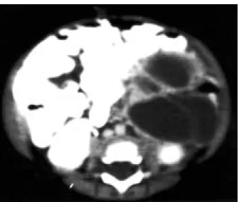
protein level 38.9 mg/dL (0-5 mg/dL). Urinanalysis and plain radiographs of the chest were normal. After blood, urine and throat cultures were obtained, intravenous ceftriaxone was ini-tiated for suspected occult bacteraemia. Fever continued for several days and then diarrhea started. Stool culture, Gram-Wright staining of stool and abdominal x-ray were normal. On the third day of hospitalization, body temperature rose to 39 °C and the child became restless. At this time, abdominal USG showed an anechoic multiloculer mass (10x5 cm) on the left side of the abdomen. A CT scan of the abdomen revealed a 6x5 cm (transverse anteroposterior diameter) fluid collec-tion in the left upper and left lower quadrants, anterior to the pancreatic tail and at the left side of the stomach (Figure 1).
The patient underwent surgery with preoperative diagnosis of infected intraabdominal cyst. At laparotomy, the patient was found to have on infected bilobular cyst (large component 10x6 cm, small component 6x4 cm) located on both sides of sigmoid colon mesentery. Enucleation of the cyst from mesentery was tried but it was not possible to eradicate the mass without com-promising blood supply to bowel. Therefore 6 cm2 sigmoid
re-section and end to end anostomosis was performed. A Penrose drain was placed at the dissected mesenterium site. The exami-nation of the histopathologic specimen showed the presence of a purulent material inside a mesenteric cyst, however, cultures of the material were sterile. On the fourth post operative day, the drain was removed because no complication, such as postopera-tive bleeding, had occurred. The patient recovered uneventfully and was discharged on the 13th day postoperatively.
Discussion
Mesenteric cyst is one of the rarest abdominal tumours. Since the first report of a mesenteric cyst by Bebevienal in 1507, only 820 cases have been reported (6).
The precise cause of mesenteric cysts is unclear. Some authors have theorized a continued growth of congenitally misplaced lymphatic tissue, whereas others have proposed that the initiating event was a failure of mesentery to fuse during development. Trauma and inflammation are ad-ditional factors implicated in the formation of these rare entities (7). Mesenteric cysts have been reported from the duodenal to the rectal mesentery but are most commonly located in the ileal mesentery (8). The cyst of our patient was located in the mesentery of the sigmoid colon. The age distribution trends toward younger patients, 70% of which are younger than 30 years. All ages are affected, however, with cases documented in utero and in the elderly (7).
The lack of characteristic clinical features and radio-logical signs may present great diagnostic difficulties. The symptoms depend on the size and location of the cyst (6). Although mesenteric cysts are often asymptomatic, they present with abdominal pain, distension, nausea, vomit-ing, diarrhea, constipation, weight loss and an abdominal mass (7). Fever and diarrhea were the first symptoms of mesenteric cyst in our patient.
In some cases, mesenteric cysts are detected incidentally by routine abdominal examination. On the other hand, emergency cases resulting from rupture or bleeding of the cyst, shock, intestinal obstruction, volvulus, or torsion of the cyst have been reported; surgical excision is the reliable treatment in such cases (9).
Acute presentation is particularly seen in children. Per-foration of a mesenteric cyst occurs following an abdom-inal trauma (sometimes minimal) or in an infected cyst (spontaneous) and is very rare (5).
A variety of diagnostic modalities can be used to con-firm the presence of a mesenteric cyst, but USG and CT tend to be the favoured methods (7). According to Seno-cak et al, abdominal USG is the most reliable diagnostic study in experienced hands. In suspected cases, the diag-nosis should be confirmed with CT (3). Our patient was diagnosed with USG and CT scan. Differential diagnosis includes abdominal cyst and tumour (10).
Surgical treatment is recommended on the basis of his-topathologic malignant features and other complications, including infection of the cyst, obstruction of adjacent bowel segment, and volvulus (7). The prognosis of patients operated in a timely manner is good with no perioperative mortality and a very low morbidity and recurrence rate (5). We suggest that infected mesenteric cyst be kept in mind as a source of fever, abdominal pain, vomiting and diarrhea attributable to no apparent cause.
Figure 1. Computed tomography scan of abdomen shows a large
141
Nazan Dalgıç, Erdal İnce, Ergin Çiftçi et al.
Journal of Ankara University Faculty of Medicine 2005; 58 (3)
References
1. Chung MA, Brandt ML, St-Vil D et al. Mesenteric cysts in children. J Pediatr Surg 1991; 26: 1306-1308.
2. Kovalivker M, Motovic A. Obstruction and gangrene of bowel with perforation due to a mesenteric cyst in a newborn. J Pediatr Surg 1987; 22: 377-378.
3. Şenocak ME, Gündoğdu H, Büyükpamukçu N et al. Mesenteric and omental cysts in children: analysis of nineteen cases. Turk J Pediatr 1994; 36: 295-302.
4. Egozı EI, Rıcketts RR. Mesenteric and omental cyst in children. Am Surg 1997; 63: 287-290.
5. Iuchtman M, Soimu U, Amar M. Peritonitis caused by a ruptured infected mesenteric cyst. J Clin Gastroenterol 2001; 32: 452-453. 6. Dequanter D, Lefebvre JC, Belva P et al. Mesenteric cysts. A case treated by laparoscopy and a review of the literature. Surg Endosc 2002; 16: 1493.
7. Mason JE, Soper NJ, Brunt LM. Laparoscopic excision of mesenteric cysts: Areport of two cases. Surg Laparosc Endosc 2001; 11: 382-384.
8. Hebra A, Brown MF, McGeehin KM et al. Mesenteric, omental, and retroperitoneal cysts in children: a clinical study of 22 cases. South Med J 1993; 86: 173-176.
9. Hideo S, Junji U, Yoshiaki O et al. Total excision of mesenteric cysts by laparoscopic surgery: Report of two cases. Surg Laparosc Endosc 1997; 7: 173-176.
10. Loreno B, Puricelli FF, Oderich C et al. Laparoscopic resection of a mesenteric cyst. Surg Laparosc Endosc 1998; 8: 402-403.