A"kara Üııiı'. Vet. Fak. Derg.
43: 281-285,1996
COMPARISON OF AgNORs STAlNING AND PCNA
IMMUNOSTAlNING METHODS, AND MITOTlC INDEX
SCORES IN INTRACUTANEOUS CORNIFYING
EPITHELIOMA AND SQUAMOUS CELL CARCINOMA
NALAN KARADEMİR' MEHMETM. ORMAN'"
TOLGA GÜVENÇ" MURA T YARIM" M. yAVUZGÜLBAHAR .... Yassı hücreli kanser ve intrakutan kornifiye epitelyoma'da AgNORs ile
PCNA boyama metodları ve mitotik indeks sonuçlarının karşılaştırılması Özet: Deri tümörleri arasında bulunan intrakutan kornifiye epitelyama ve iyi differensiye yassı hücreli kanser birbirine benzer tümörlerdir. Bu çalışmamn amacı, pralifere hücre nüklear antijen (PCNA), klon PCJO immunhistokimyasal boyama ve argirajil nükleolar organize bölgeler (AgNORs) boyama metodlarım ve mitotik indeksi bu iki tümörde karşılaştırmaktır. Çalışma için köpeklerden alınan 5 adet intrakutan kornifiye epitelyama ve çeşitli hayvan türlerinden temin edilen 20 adet iyi differensiye ya.s'sı hücreli kanser seçildi. Bütün örnekler formalinde jikze edilip parajinde bloklandı. Ortalama AgNOR sayısı her lezyon için 50 bazal ve 50 suprabazal hücrede immersiyon ile JOOObüyütmede saptandı. peNA işaretli kısımlar JO değişik büyütme alanında hesaplandı. PCNA indeksi. peNA pozitif' boyanan tümör nukleuslarımn, seçilen alanlardaki toplam hücre adedine bölünüp yüzde cinsinden ifadesi ile hesaplandı. Tümörlerdeki mitotik indeks 10 değişik büyütme alanında te.spit edildi. Intrakutan ko rnifiye epitelyoma ve iyi differensiye yassı hücreli kanser bu üç yöntemle karşılaştmldığında Mann-Whitney U testinde AgNOR sayısı önemli (p < 0,01), fakat PCNA indeksi ve mitotik indeks önemli bulunmadı.
Anahtar Kelimeler: AgNOR boyama, intrakutan kornijiye epitelyoma, mitotik indeks. PCNA, yassı hücreli kanser
Summary: Among the skin tumors, intracutaneous cornifYing epithelioma and well-differentiated squamous cell carcinoma are .s'imilar. The purpose of this study was to compare the immunostainingfor proliferating cell nuclear antigen (PCNA) clone PC i O, interphase nucleolar organizer regions staining (AgNORs) methods and mitotic index in these tumorso JıIve intracutaneous cornifYing epitheliomas in the dogs and 20 well-differentiated squamous cell carcinomas in various species were chosen. All sample.~ were .fixed in formalin and embedded in parapin. The mean number ofAgNOR was determined in 50 basal and 50 suprabasal cells of each tumor under an oil immersion lens at a magnification of 10()O.PCNA-Iabeledfractions and mitotic index were estimated in the same areas of these tumors in JO high power fields. AgNOR count (p <O,OJ) was significant, but peNA index and mitotic index were not significant in Mann-Whitney U test to compare intracutaneous cornifYing epithelioma and well-differentiated squamous cell carcinoma.
Key words: AgNOR staining, intracutaneous cornifYing epithelioma, mitotic index, PCNA, squamous cell carcinoma
•• Ara~. Gör. Dr. Universityol' Ankara, Faculty ofVeterinary Medicine, Depannent ofPathology, Ankara . ••• Araş. Gör. University of Ankara, Faculty ofVeterinary Medicine, Depannent ofPathology, Ankara . •••• Araş. Gör. University of Ankara, Faculty ofVeterinary !\1edicine, Depannent of Biostatistics, Ankara.
282 NALAl" KARADEMİR _ TOLGA GÜVENÇ - MURAT YARIM - MEHMET M. ORMAN - M. YAVUZ GÜLBAB
Introduction
Nucleolar organizer regions (NORs) are specific nucleolar components. They represent the sitcs for ribosomal RNA transcription. NOR-associated proteins bind silver (S).
111e scoring of interphase NOR numbers using the argyrophil technique has bcen shown to be of use in assessing the degree of malignancy in neoplasms (4, 6, LO).
The proliferating cell nuclcar antigen (PCNA), is a 36-kD nonhistone nuclear protein which is directly involved in DNA synthesis (16). PCNA levels increase rapidly in mid-G I, remain elevated throughout S-phase and begin to decrease from G2/M to GI-phase of cell cycle (1). Several recent papers suggest that anti-PCNA antibodies could be employed to assess the proliferative activity oftumors (13, 22, 2S).
Intracutaneous cornifying epithelioma is a distinct neoplasm observed only in the dog (17). Intracutaneous cornifying epithelioma and keratoacanthoma in humans are similar in many respect, but theyare not identical entities (2 I, 26). Keratoacanthoma in humans is a skin tumor, often demonstrates a clinical and histologic resemblance to differentiated-squamous cell carcinoma. Although a variety of histological criteria have been applied to differentiate keratoacanthoma from squamous ccll carcinoma in human s (12, 14, IS), some ofthem have proved entirely reliable.
Although there is no doubt of the diagnosis of these tumors in animals, wc want to compare nucleolar organizer region (NOR) counts, PCNA and mitotic index in well-differentiated squamous cell carcinoma and intracutaneous cornifying epithelioma.
There are very few reports in the veterinary oncology on these techniques.The PCNA immunostaining method and AgNORs staining method applied in this study was used first time in veterinary oncology in Turkey.
Materials and Methods
Four Ilm thick sections from biopsy specimens were included in this study. After routine staining with hematoxylin-eosin of the first sections, the specimens were identified as being either intracutaneous cornifying epithelioma or well-differentiated squamous cell carcinoma.
Paraffin sections from 20 cases of histologically typical differentiated squamous cell carcinoma and five cases of intracutaneous comifying epithelioma were stained by a modification of the silver NOR technique and immunostaining for PCNA, clone
pcıo.
111e second sections (four Ilm ) from the same blocks were stained by a modification of the silver NOR technique described by Ploton et al. (19).We prefer this modified technique because
overstaining produce stain deposit. The section were taken to water via xylene and grade alcohols. The sections were submitted to th AgNOR procedure at 37DC for 7S minutes Th
reaction mixture comprised 2 % gelatin in i % aqueous formic acid. This was mixed in proportion of 1:2 volumes with 2S% aqueou silver nitrate. The reaction was stopped b washing in ultrapure water for 2xS minutes an placed in S% thiosulfate solution for 2x minutes. Tissues were washed again wit distilled water. No counterstain was used. Al eounts were performed by direct observation Sections were examined under an oil immersio lens at a magnifieation of i000 and 100 nucle were studied. A variation in NOR eounts wa apparent in different areas of the tumors therefore fields were randomly selected SO basa and SO suprabasal nucleus for AgNOR countin in eaeh tumor.
All AgNOR dots, either in nucleolus o scattered in the nucleus were counted accordin to Croeker et aL's method (3) The me number of AgNORs per nucleus was calculate for each speeimen.
The third sections (four Ilm) from th same blocks were stained by immunostaining fo PCNA, clone PC
ıo.
The seetions were drie overnight at room temperature an immunostained with PC iO. The tissues wer deparaffinized in two changes of xylene for i minutes each, rehydrate in 100 %, 9S% an 70% alcohol and placed in distilled water for i minutes. Endogenous peroxidase was blocke with 3% HıOı for five minutes, then the slide were washed with tris-buffer. Sections were the slides were washed with tris-buffer. Section were then incubated with Enhanced Polyme One-Step Staining (EPOS) anti-proliferatin cell nuclear antigenIHRP (DAKO, clon PCı
O, Glostrup, Denmark) for 60 minute~ı at room temperature, after a wash in tris buffer, the sections then were incubate with diaminobenzidine/H202 for fivminutes. The slides were washed İn tris buffer and counterstained with Mayer' haematoxylin for 30 seconds. The section were washed in tap water, taken throug alcohol to xylene and mounted in syntheti medium. Negative control sections wer incubated in buffer instead of anti-PCN antibody dilution. All immunostaine sections were examined under an ai immersion lens at a magnification of 1000 T otally
ı
O high power fields were chosen The value of PCı
O index was defined as th number of positive tumor nuclei divided bOMPARISON OF AgNORs STAlNING Aı"lD PCNA IMMUNOSTAlNING METHODS, AND MITOTIC INDEX SCORES 283
e total number of cells expressed as a
ercentage were considered.
Mitotic figures were counted in anti-PCNA ained sections in a total of 1000 ceııs presents in
e same tumor area where PCNA indexes were stimated.Mann-Whitney U test was used to
atistical analysis.
Results
The mean values of the kinetic parameters, the AgNOR counts, PCNA and mitotic indexes for aıı the cas es counted, are summarized in table-I.
able ı.Mean values (:t Standart deviations), minimum and maximum values of the AgNOR counts, PCNA index and mitotic index in traeutaneous eomiıying epithelioma and squamous eell careinomas.
'voes o turnors 'lVI e oBest ean Ttanoa riDev. m. ax.
lntracutaneous !NUK .6)
Comifying ";NA 'TT
Epithelionıa ıtosıs .IT
Squamous P ,NOR .m
Cell :NA 6.R5
Carcinoma ıtosıs LO. S ılS.ı~
Figure ı. 1>. PCNA-positive nuclei in squamous eell careinorna (arrows). A lew PCNA-positive nuelei are seen. There is a little diflerenee Irorn Fig iC. Immunoperoxidase staining, Mayer's hernatoxylin counterstain, xl 000 rnagnilication.
Figure ı.B.AgNOR dots in squamous eel! carcinoma eells (arrows). In contrast to intracutaneous eomilying epithelioma eells in Fig lA, these nuelei eontain multip!e dots. Silver eolloid, no counterstain, xı000 magnification.
Figure ı. c. PC~A-positı\'e nucki in intracutaneous eonıilying epithelioma (arrows). A few PCNA-positive nuclei are seen. Immunoperoxidase staining, Mayer's hematm,')'!in eounterstain. x350 magnilieation.
In sections, AgNORs were easily entified as brown or black dots (fig.I.A, B). he mean for the number of AgNORs per ucleus was 13,02 and the range 4,23-25,84 in ifferentiated squamous ceıı carcinomas.
tracutaneous comifying epithelioma had a ean number of 4,65 AgNORs with range of ,49-5,46 (fig. 2).
'gure ı.A. AgNOR dots in intraeutaneous comiıying epithelioma fls (arrows). A few dots are visible in each nueleus. Silver colloid, ) eounterstain, x 1000 magnilication.
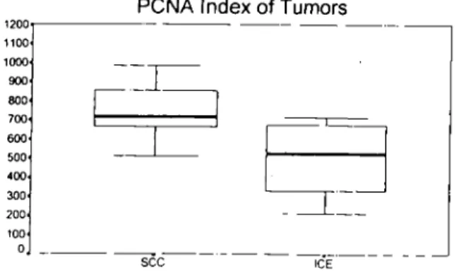
The nucleus of tumor ceııs positive for CNA stained light to dark brown (fig.l.C, D) nd PCNA reactivity was expressed by diffi.ıse uclear stai staining. Both strongly labeled uclei and less intensely labeled nuclei were ounted. Negative nuclei were light blue. The alue afthis activity, for PCNA ranged between ,88% and 9,86% in differentiated squamous eıı carcinoma and 2,13% and 7,16% in
tracutaneous comifying epithelioma (fig.3). The number of mitotic index ranged etween 1,62 and 28,29/1 000 ceııs in ifferentiated squamous cell carcinoma and 2,83
d 13,29/1000 ceııs in intracutaneous mifying epithelioma (fig. 4).
Statistic analysis of AgNOR counts (p< ,OL) was found significant but PCNA and itatic indexes were not statistically significant ol for these tumors in our study ..
284 NALAN KARADEMİR - TOLGA GÜVENÇ - MURAT YARIM - MEHMET M. ORMA.N - M. YAVUZ GeLBAHAR
Figure 4. Distribution of mitotic indexes of the tumors. As shown in box-plots: there is no difference between squamous cell careinomas (SCC) and intracutaneous comitying epithelioma (ICE).
Fiıı:ure 3. Distribution of PCI\A indexes of the tumors in box-ploK There is not a signilicant dillerence between squamous cell careinomas (SCC) and intracutaneous comifying epithelioma (ICE).
Dİscussİon
Various methods has been reported to compare squamous cell carcmoma and keratoacanthoma in humans. it has been shown that flow cytometry measurements of DNA-index and proliferative index did not heIp m separating keratoacanthoma and well-differentiated squamous cell carcinoma of skin in humans (20, 27). Ho et ai. (14), suggested that transfonning growth factor a expressıon may be a marker of epithelial differentiation and may hdp distinguish between these two tumors. lt has been reported that electron microscopy has revealed decreased numbers oftonofilaments and intercellular desmosomes m well-differentiated squamous cell carcmoma as compared to keratoacanthoma (I 2). However, the same authors did not bdieve that the subjeetive nature of those findings does yield reliable criteria for the individual case.
TIıe estimation of tumor proliferative activity by means of antibodies to cell cycle related antigen is widely employed in human and animal pathology (7, 13, 23). In our study, we counted both strongly labded and more weakly labekd nuclei which belong to different phases of cell cycle, m anti-PCNA immunostaining method. However, we could not find a corrdation between intracutaneous comifying epithelioma and well-differentiated squamous cell earcmoma and the anti-PCNA immunostaining method and also mitotic index ean not be considered as a statistically significant tool m these lesions. To date no reports are available on comparıson of the difference in values of these methods for the two tumors. However, Ehlcrs et aL. (11) and Piscioli et aL. (18), clearly demonstrated that keratoacanthomas are a heterogeneous group of lesions, including pseudocareinomatous, precancerous and cancerous neoplasms by histophotometric investigation of content of
nuclear DNA. They claimed that
keratoaeanthoma is not by definition a benign lcsion in every instance, because its components' cells have a content of malignant DNA, though rarely. However, intracutaneous comifying epithelioma is different from keratoacanthoma (2
ı,
26), but this situation may be important also for intracutaneous comifying epitheliomas Although the mvasıon of tumor cells of intracutaneous comifying epithelioma into the stroma from basement membran is not seen. it is thought that in this study this tumor may' be a precancerous tumor according to the PCNA immunostaining method.L
i _:J ıeE---
------
ICEJ
seePCNA Index of Tumors
-- --- --
---Mitotic Index of Tumors AgNor Counts of Tumors
30 ----20 25 15 10 6 8
Figure 2. Box-plots showing the mean number of AgNORs per nueleus for speeimens examined. As seen in diagram, squamous cell carcinomas (SCC) can easily distinguish trom intracutaneous comifying epithelioma (ICE).
16 14 1200 1100 1000 900 800 700 600 500 400 300 200 100 O .----
----sce--- -
-
IcE" -12 LO:)
OL - - ---sce----COMPARISO]\; OF Ag:"ORs STAINING AND PC)'.;A IMMUNOSTAI:"E\G METHODS, AND MITOTIC INDEX SCORES 285
The counting AgNORs is used for assessing the degree of malignancy (6, X, 9). In same cases this staining method are ShOWllfor the prognosis and treatment of the patient (2,24, 28). in our study, only AgNOR counts were the predictor according to the other assessment of proliferative activity methods. The squamous cell careinomas have higher AgNOR scores per nucleus than the intracutaneous comifying epithel iomas.
AgNOR indices proved to be useful for determining the proliferative activity of squamous cell carcinoma and intracutaneous comifying epithelioma. The results in this study confırm only the value AgNORs as the markers for proliferative activity
REFERENCES
I. Bolton, W. E., :-'likulka, W. R., Healey, C. G., Selunittling, R. .1., Kenyon, M. S. (i 992) Expression of prolıferatlOn assocIQted anl/gens in the cel! cyele of synchronized mammalıan cells. C)10melry, 13, i 17-126.
2. Bostoek, D. E., Croeker, .1., Harris, K., Smith, P. (1989)
Nueleolar organıser reglOns as ındıcators of post-surg/cal prognosıs ın canıne .ıpontaneous mast cel! rumours. Br. J.
Cancer, 59, 9i5-918.
3. Croeker, .1., Boldy, D. A. R., Egan, M . .1. (1989) How
should we count AgNORs? Proposals for a standard/zed approach. J. Pathol., 158, 185-188.
4. Croker, .1., Mc. Govenı, .1. (I 988). :\ucIeolar organiser regions in normal, cirrhoıic and carcinomalous livers. J. Clin. Pathol.,41,1044-1048.
5. Derenzini, M., lIenıandez-Verdwı, D., Pession, A., NoveUn, F. (1983) Stmctural organızation of chromarın ın nl/cleolar organızer reglOns o[nueleolı wıth a nucleolonema-lıke and compact nbonueleoproteın dlstnbutlOn. J. Cltraslruct. Res., 84, i6i-172.
6. Derenzini, M., Pession, A., Trere, D. (1990) Quanıııy of nucleolar sılver-staıned proteıns ıs related to proliferatıng actıv/ey ın cancer ce LLS. Lah. Invest., 63,137-140.
7. Dervan, P. A., Magee, M. H., Buekı ey, C, Canıey, D. N. (I 992) Proliferatıng ce/I nuclear anı/gen counts ın jimııalın /ixed paraffin-embedded I/ssue correlate with K/-67 ın fresh
tıssııe. Am..r. CIin. Palhol., 97. Supp!. 1,21.28.
8. Destexhe, E., VanınaIL~hoven, P., Coignoul, F. (1995)
Companson of argyrophilic nucleolar organızer region.l' by coımrıng and ımage analy,ı/.ı' ın canıne mammary tumors.
Am. 1. Yet. Res., 56,185-187.
9. De Vico, G., Papparella, S., Di Guardo, G. (i 994) Nıımber
and sıze ofsılver-stOlned nueleolı (AgNOR elusters) ın canıne semınomas: correlatlOn w/th h/stological features and tumour behaviour. J. Comp. PathoL., 110,267.273.
10. Egan, M. ,J., Raafat, F., Crocker, .1, Smith, K. (1988)
Nucleolar organıser region.l' ın /ibrous prolıferatlOns of chıldhood aııd ınfantılejibrosarcoma. 1. Clin. Pathol., 41, 31-33.
i i Elılers, G., Knoth, W., Sandritter, W. (1974)
Feıılgen-cytophotome/nsche untenuchungen an keraroacanthomen.
Haularlt. 25, 144-148.
12. Fisher, E. R., "'leCoy, M. M., Weehsher, H. Lo (1972)
Analy.I'ls of h/sropathologıc and electron mıcroscop/c determınatlOns of keratoacanthoma and squamous cell carcınoma. Cancer. 29, 1387-1397.
13. Hall, P. A., Le\'ison, D. A., Woods, A. L., Yu, C C-W.,
Kellock, D. B., Watkins, .1. A. (1990) Proliferarıng cel! nuelear ant/gen (PCNA) /mmunolocalızatlOn ın paraffin seclions: an ındex of cell proliferation wıth evidence of deregıdated expre.\SIon in some neoplasms. J. Patho!., 162, 285-294.
14. 110, T., Hom, T., Finzi, E. (1991) Transformıng growth jactor a expression helps to d/stinguısh keratoacanthomas from squamoııs cel! carcinomas. Arch. Dermato!., 127,
1167-i 171.
15. Korenberg, R., Peıuıeys, N. S., KowaIczyk, A., Nadji, M.
(1988) Quantıtation oL S-IOO protein-positıve cel/s ın ınflamed and non-inflamed keratoacanthoma and squamous cel/ carcmoma. J. Cul<ıneous PathoL.,IS, 104- 108.
16. Linden, M. D., Torres, F. X., Kuhus, .T., Zarbo, R .T. (1992)
Climcal applicatlOn o[ morphologıc and 11/1ıııunocytochel1/1calassessıııents of cel/ proliferatlOn. Am. J. Clin. PathoL.,Sııpp!. i, 4-13.
17. Moultolı,.1. E. (I 990) rumors of Doıııe.H/C Ammals. 3rd cd. London, University ofCalilomia Press. 1'.54-57.
18. Piscioli, F., Boi, S., Zwniani, G., CristofoIini, M., Chiara, S. (I 984) Metasta.ı'/zmg keratoacanthoma vs. squamous cel/ carcınoma. Am. 1. Dermatopathol., 6, 123-129.
i9. Ploton, D., Menager, M., .leanesson, P., I1imber, G., Pig('on, F., Adnett, .T•• J. (1986) lmprovement ın the staımng and m the visllOlizatlOn of the argyrophilic protein.I of the nucleolar orgamzer regıon at the opııcallevel. Histochem. J., 18,5-14.
20. Randall, M. 8., Geisinger, K. R., Kute, T. E., Buss, D. II.
(1990) DNA content and proliferative mdex m cutaneous sqııamous cel/ carcinoma and keratoacanthoma. Am.J.Clin. Paıhol., 93, 259-262.
2ı.Rudolph, R, Gnıy, A. P., Leipold, II. W. (ı 977)
Intracutaneous cormiYing epıthelioma ('Keratoacanthoma 'J
of dogs and keratoacanthoma ofman. CornelI. Yet., 67, 254-264.
22. Sarıi, G., Benazzi, c., Preziosi, R., Mareato, P. S. (I 995)
Assessment of prolıjerative act/v/ey by antı-PCNA monoclonal antıbodies ın jormalın-flxed, parajJin-embedded samples and correlatıon wıth ııı/totıc ındex. Yet. PathoL.,32,
93-96.
23. Sarıi, G., Benazzi, c.,Preziosi, R., Mareato, P. S. (1994)
Proliferallve actlvıey assessed by anıı-PCNA and KI-67 monoclonal antıbodıes ın canine testicular !Umoun. J. Comp.
Path,,!.. 110,357-368.
24. Simoes,.1. 1'.c.,Sehoııiııg, 1'., Butine, M. (1994) f'rogno.l'/s
of canıne mast cel/ rumors: a companson of three method.~.
Yet. Pathol., 31, 637-647.
25. Smith, F. G., :\1urmy, P. G., Crocker, .1. (I 993) CorrelatlOn
berween J'CNA and AgNOR scores m non-Hodgkin 's Iymphomas using sequentlOl staıning techmque. J. CIin. Paıhol., 46, 28.31.
26. StalUıard, A. A., Pıılley, L. T. (1975) lntracutaneous cormiYing epilhelioma (J:.:eratoacanthoma) m dog: A retrospective stııdy of25 cases. JAYMA, 167,385-388.
27. Stephensoıı, T . .1., Cotton, D. W. K. (1988) Flow cytometnc
companson of keratoacanrhoma and squamous cel! carcmoma. Br. J. Dermato!., 1lll, 1582.
28. Yu, C. CO -W., Fletcher, C. D. M., Nemnan, P. L., Goodlad, .J. R., Burton, .1.c.,Levison, D. A. (1992) A comparison of proliferatmg cel/ nuclear antıgen (J'CNA) /mmunostaımng. nucleolar orgamzer regıon (AgNOR) staining, and histolog/cal gradıng ın gastrointestinal stromal tumoun. J.