Ankara Üniv Vet Fak Derg, 55, 61-64, 2008
Histochemical characterization of glycoproteins in the gills of the carp
(Cyprinus carpio)
Kenan ÇINAR, Nurgül ŞENOL, M. Rüştü ÖZEN
Department of Biology, Faculty of Arts and Sciences, Süleyman Demirel University, Isparta, Turkey.
Summary:
The characteristics of the mucous cells located in gills of the fish Cyprinus carpio were investigated. Mucous cells were determined from gill arc, gill body, primary and secondary lamellae. Mostly mucous cells had oval-globular shape throughout all regions. Histochemical analysis of the gill of Cyprinus carpio showed that mucous content included glycogene and/or oxidizable dioles (PAS +), neutral or acid-rich (PAS/AB pH 2.5 +), sialic acid residues (KOH/PAS +) and strong acid sulphated (AF +) glycoproteins (GPs). Except these mucosubstances, carboxyl groups and/or with sulphate esters (AB pH 2.5 +) strong sulphate (AB pH 0.5 +) (AF/AB pH 2.5 +), O-sulphate esters (AB pH 1+) glycoproteins were also determined.Key words: Cyprinus carpio, density, gill, glycoproteins, mucous cells
Sazan (Cyprinus carpio) solungaç glikoproteinlerinin histokimyasal özellikleri
Özet:
Cyprinus carpio solungaçlarında yer alan karakteristik mukus hücreleri incelendi. Mukus hücreleri solungaç arklarında, solungaç lamellerinde, primer ve sekonder lamellerde tespit edildi. Tüm bölgeler boyunca mukus hücrelerinin çoğu oval-yuvarlak bir şekle sahipti. Histokimyasal analizler solungaçlardaki mukus hücrelerinin glikojen ya da oksidizable diolleri (PAS +), nötral ya da asitce zengin (PAS/AB pH 2.5+), siyalik asid rezidulleri (KOH/PAS +) ve asitce zengin sulfatlı (AF +) glikoproteinleri içerdiği gösterildi. Bu mukosubstanslar dışında karboksil gruplu ya da sülfat esterli (AB pH 2.5 +) güçlü sülfatlı (AB pH 0.5 +) (AF/AB pH 2.5 +), O-sülfat esterli (AB pH 1+) glikoproteinler tespit edildi.Anahtar sözcükler: Cyprinus carpio, glikoproteinler, mukus hücreleri, solungaç, yoğunluk.
Introduction
The fish gill epithelium consists of several cell
types. Many studies of fish gills have described the
morphological and functional characteristics of gill
epithelial cells: respiratory cells, chloride or
mitochondria-rich cells, pavement cells and mucous cells
(4, 9, 19, 22). As well as some studies is reported the
branchial lamellae epithelium consists of granular cells,
ciliated cells, leydig cells, bazal cells (17),
undifferentiated cells (4), and accessory cells (1). Mucus
producing cells integrate a variety of critical functions.
Physiologically, mucus is important for protection and
inhibition of micro-organisms (5, 27, 36). This function
is occurred by mucous glycoproteins (GPs) (5, 15, 24,
35), as well as the GPs are identified with O-sulphate
esters are responsible for the lubrication (8). In addition,
mucus membran is engaged important functions, such as
osmoregulation, diffusion and protection of dehydration
(8,19, 24, 36).
Mucus producing cells are numerically and
morphologically affected by different conditions as other
cells localized in gill epithelium. Increase of mucous
cells number is reported with different conditions such as
bacterial gill disease (12), amoebic gill disease (26, 30,
33), high concentrations of ammonia (12, 16), salinity (2,
13), acidity (6, 20), high pressure and low temparature
(10). On the other hand, in conditions of high concentrations
of ammonia (16), low pH (37), high concentrations of
aluminum (29), heavy metals (18), substrat of diazinon
(11) and acid plus aluminum (6), mucous cells size is
increased. It is claimed that, numerically and
morphologically the difference in mucous cells probably
relates to period of secretion. In this study, our aim was
to determine the mucous cells of the gills of the Carp
(Cyprinus carpio) with histochemical technique
Materials and Methods
In this study we choosed the omnivorous fish
species, Cyprinus carpio. We obtained these fish at
Eğirdir lake. As material, twenty five uninfected
Cyprinus carpio, length between 25 – 30 cm and weight
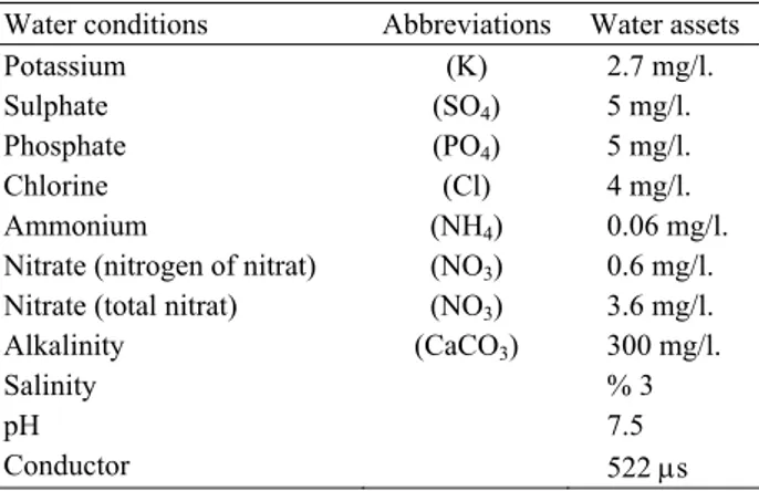
between 320 – 350 g, were used. Water conditions were
showed in table 1. The gills were rapidly excised and
fixed by immersion in 10% buffered formalin for light
microscopic studies. The samples were routinely
processed and embedded in parafin. Histochemical
techniques were performed for the density and
differentiation of carbohydrate moieties (Table 2).
Kenan Çınar - Nurgül Şenol - M. Rüştü Özen 62
Table 1. Water conditions (Photometer method, soil pool) Tablo 1. Su değerleri (Fotometre yöntemi, toprak havuzu)
Water conditions Abbreviations Water assets
Potassium (K) 2.7 mg/l.
Sulphate (SO4) 5 mg/l.
Phosphate (PO4) 5 mg/l.
Chlorine (Cl) 4 mg/l.
Ammonium (NH4) 0.06 mg/l.
Nitrate (nitrogen of nitrat) (NO3) 0.6 mg/l.
Nitrate (total nitrat) (NO3) 3.6 mg/l.
Alkalinity (CaCO3) 300 mg/l.
Salinity % 3
pH 7.5
Conductor 522 μs
Table 2. Performed the histochemical techniques in the gill epithelium of Cyprinus carpio;AB, Alcian blue; KOH, saponification; PAS, periodic acid/Schiff; AF, Aldehyde fuchsin; GPs, glycoproteins.
Tablo 2. Cyprinus carpio solungaç epitelinde uygulanan histokimyasal teknikler; AB, Alsiyan mavisi; KOH, saponifikasyon; PAS, periyodik asit/Shif; AF, Aldehit fuksin; GPs, glikoproteinler.
Procedures References 1. PAS
GPs with oxidizable vicinal diols and/or glycogen
Mc Manus (1948)
2. PAS/AB pH 2.5
Neutral and/or acid rich GPs Mowry (1956) 3. AB pH 2.5
GPs with carboxyl groups (sialic acid or uranic acid) and/or with sulphate esters
Lev and Spicer (1964)
4. AB pH 1.0
GPs with O-sulphate esters
Lev and Spicer (1964) 5. AB pH 0.5
Very sulphated GPs
Lev and Spicer (1964) 8. KOH/PAS
GPs with sialic acid residues Culling et al. (1976) 9. AF
GPs with sulphate
Gomari (1952)
10. AF/AB pH 2.5
Strong sulphated GPs Spicer and Meyer (1960)
Results
As a result of histochemical studies, mucous cells
were determined from gill arc, gill body, primary and
secondary lamellae. Density in gill arc and body was
higher. Respectively primary and secondary lamellae
followed these (Table 3). At the bases of secondary
lamellae, such cells were detected as single cells or
sometimes in groups. Mucous cells were observed to
occur towards the outer surface, but sometimes in deeper
layers. Mostly mucous cells had oval-globular shape
throughout in all regions. Exceptionally some were in
goblet shape, rather commonly in the gill arc. Despite
foamy or cloudy appearance at mucous cell cytoplasms,
seldomly cells had granular cytoplasms.
Table 3. Histochemical reactions of glycoproteins in the gills of
Cyprinus carpio; AB, Alcian blue; KOH, saponification; PAS,
periodic acid/Schiff; AF, Aldehyde fuchsin; GPs, glycoproteins. Tablo 3. Cyprinus carpio solungaç epitelinde histokimyasal reaksiyonlar; AB, Alsiyan mavisi; KOH, saponifikasyon; PAS, periyodik asit/Shif; AF, Aldehit fuksin; GPs, glikoproteinler.
Staining reactions Procedures
Gill arc Gill body Primer lamellae Sekonder lamellae PAS +++ ++ ++ + PAS/AB pH 2.5 +++ +++ ++ + AB pH 2.5 ++ +++ ++ + AB pH 1.0 ++ +++ ++ + AB pH 0.5 ++ +++ ++ + KOH/PAS +++ ++ ++ + AF ++ +++ ++ + AF/AB pH 2.5 ++ +++ ++ +
Alcian blue (AB) pH 1 (Figure 1), AB pH 2.5
(Figure 2), periodic acid/schiff (PAS) and aldehyde
fuchsin (AF) applications were intense in all regions,
while AB pH 0.5 (Figure 4, 5) was somewhat weaker.
AB (+), PAS (+) and AF (+) mucosubstances were
dominant in PAS/AB pH 2.5 (Figure 3), saponification /
periodic acid/schiff (KOH/PAS), AF/AB applications
respectively. In gill arc of some cells only AB (+)
mucosubstances were observed in PAS/AB pH 2.5.
similar results were obtained in AF/AB application. But
in AF/AB application, cells away from lumen surface
also showed AF (+) reaction at secondary lamella.
Discussion and Conclusion
Studies with different fish species shows that gill
mucous cells distributed in different areas and different
densities. In Poecilia vivipara (31) these were observed
in apical of gill filaments only in interlamellar region and
gill arc. In Micropogonias furnieri (9), mucous cells
were observed in primary and secondary lamellae, while
in Acipenser naccarii (4) along the filaments, rarely
between pavement cells. In this study, mucous cells were
identified to be broadly distributed in gills, rather in gill
arc. Mucous cells were mostly in oval-globular in shape
less commonly in goblet or pear-shaped, similar to
Morone saxatilis or M. chrysops (28).
In accordance with Diaz et al (8), except for primary
and secondary lamellae, mucous cell glycoprotein
characterization was almost the same. Also in PAS/AB
pH 2.5 applications similar results were obtained (1, 3).
The AB dominance also has been obtained in Solea
senegalensis (1), as in Cyprinus carpio in this study.
In AB pH 2.5 applications glycoprotein with
carboxyl groups are more common, while freshwater
adapter Salmo species (33) they are found less
Ankara Üniv Vet Fak Derg, 55, 2008 63
Figure 1. Secondary lamella, GPs with O-sulphate esters in mucous cells. AB pH 1.0. X 200
Şekil 1. Sekonder lamel, O-sülfat esterli glikoprotein içeren mukus hücreleri AB pH 1.0. X 200
Figure 3. Secondary lamella, GPs with carboxyl groups and/or with sulphate esters in mucous cells PAS/AB PH 2.5 (AB dominant) X 200
Şekil 3. Sekonder lamel, karboksil gruplu sülfat esterli glikoprotein içeren mukus hücreleri PAS/AB pH 2.5 X 200 Figure 2. Secondary lamella, GPs with
carboxyl groups (sialic acid and/or with sulphate esters in mucous cells. AB pH 2.5. X 200
Şekil 2. Sekonder lamel, karboksil gruplu sülfat esterli glikoprotein içeren mukus hücreleri AB pH 2.5. X 200
Figure 4. Secondary lamella, very sulphated GPs in mucous cells AB pH 0.5 X 200
Şekil 4. Sekonder lamel, güçlü sülfatlı glikoprotein içeren mukus hücreleri AB pH 0.5 X 200
Figure 5. Gill arc, very sulphated GPs in mucous cells AB pH 0.5 X 200
Şekil 5. Solungaç yayı, güçlü sülfatlı glikoprotein içeren mukus hücreleri AB pH 0.5 X 200
frequently. Similar is case for Solea senegalensis (1),
differing in that there were also some cells equally
comprising AB pH 2.5 and PAS (+) mucosubstances.
Similar to study, in Acipenser naccarii (8)
acclimated to sea, great portion of mucous cells are
observed to react with PAS (+). Also similar to M.
furnieri (8), sialic acid residues, glycoproteins with
oxidizable vicinal diols are observed. Similar to Solea
senegalensis (32) adults, in Cyprinus carpio strong
sulphated GPs were encountered with AB pH 0.5 and
AF/AB applications. Likewise glycoprotein with
sulphate groups seen in M. furnieri (8) and Salmo salar
(33) acclimated to sea water were also observed in
Cyprinus carpio in this study.
References
1. Arellano JM, Storch V, Sarasquete C (2004):
Ultrastructural and histochemical study on gills and skin of the Senegal sole, Solea senegalensis. J Appl Ichthyol,
20, 452-460.
2. Bordas MA, Balebona MC, Chabrillon M, Rodriguez-Maroto JM, Morinigo MA (2003): Influence of
temperature and salinity on the adhesion to mucous surfaces of gilt-head seabream (Sparus aurata L.) of pathogenic strains of Vibrio alginolyticus and Listonella anguillarum. B Eur Assoc Fish Pat, 23, 273-280.
3. Calabro C, Albanese MP, Lauriano ER, Martella S, Licata A (2005): Morphological, histochemical and
immunohistochemical study of the gill epithelium in the abyssal teleost fish Coelorhynchus . Folia Histochem
Cytobiol, 43, 51-56.
4. Carmona R, Garcia-Gallego M, Sanz A, Domezain A and Ostos-Garrido MV (2004): Chloride cells and
pavement cells in gill epithelia of Acipenser naccarii: ultrastructural modifications in seawater-acclimated specimens. J Fish Biol, 64, 553-566.
5. Chabrillon M, Bordas MA, Morimigo MA, Balebona MC (2004): Kinetics of adhesion of Listonella anguillarum
to the mucus of gilt-head sea bream, and the implication of surface components. Aquac Res, 35, 403-409.
6. Charles HJ, Terry AH (1997): Changes in gill
Kenan Çınar - Nurgül Şenol - M. Rüştü Özen 64
addition of acid and aluminium to stream water. Environ
Pollut, 97, 137-146.
7. Culling CFA, Reid PE, Dunn WL (1976): A new
histochemical method for the identification and visualization of both side chain acylated and non-acylated sialic acids. J Histochem Cytochem, 24, 1225-1230.
8. Diaz AO, Garcia AM, Devincenti CV, Goldemberg AL (2001): Mucous cells in Micropogonias furnieri gills:
histochemistry and ultrastructure. Anat Histol Embryol ,
30, 135-139.
9. Diaz AO, Garcia AM, Devincenti CV, Goldemberg AL (2005): Ultrastructure and histochemical study of
glycoconjugates in the gills of the White Croaker (Micropogonias furnieri). Anat Histol Embriyol, 34, 117-122.
10. Dunel EB, Sebest P, Chevalier C, Simon B, Bart HL (1996): Morphological changes indiced by acclimation
high pressure in the gill epithelium of the freshwater Yellow Eel. J Fish Biol, 48, 1018-1022.
11. Dutar HM, Richmands CR, Zeno T (1993): Effect of
diazinon on the gills of Bluegill Sunfish, Lepomis macrochirus J Environ Pathol Toxicol Oncol, 12, 219-227.
12. Ferguson HW, Morrison D, Ostland VE, Lumsden J, Byme P (1992) : Response of mucus-producing cell in gill
disease of Rainbow trout (Oncorhynchus mykiss). J Comp
Pathol, 106, 255-265.
13. Franklin GC (1990): Surface ultrastructural changes in
the gills of Sockeye salmon (Teleostei: Oncorhynhus nerka) during seawater transfer: comparison of successful seawater adoptation. J Morphol, 206, 13-23.
14. Gomari G (1952): Gomari’ s aldehyde fuchsin stain. 238. In: Cellular Pathology Technique, CFA Culling, RT Allison, WT Barr (eds). Butterworths, London.
15. Herrler G, Rott R, Klenk HD, Muller HP, Shukla AK and Schauer R (1985): The receptor destroying enzyme of
influenza C virus is neuraminate- O- acetyl esterase. Embo
J, 4, 1503-1506.
16. Hilary M, Lease-Hansen-James A, Bergman-Harold L, Meyer-Joseph S (2003): Structural changes in gills of
Lost River suckers exposed to elevated pH and ammonia concentrations. Comparative Biochemistry and Physiology
Part C: Toxicol Pharmacol, 134, 491-500.
17. Jarial MS, Wilkins JH (2003): Ultrastructure of the
external gill epithelium of the axolotl, Ambystoma mexicanum with reference to ionic transport. J Submicrosc
Cytol Pathol, 35,445-455.
18. Jezierska B, Witeska M (2004): The effect of metals on
fish gill functions-Gas and ion change (review). Fresenius
Environ Bullet, 13, 1370-1378.
19. Laurent P, Perry SF (1990): Effect of cortisol on gill
chloride cell morphology and ionic uptake in the freshwater trout, Salmo gairdneri. Cell Tissue Res, 259,
429-442.
20. Ledy K, Giamberini L, Pihan PC (2003): Mucous cell
responces in gill and skin of brown trout Salmo trutta fario in acidic, aluminium containing stream water. Dis Aquat
Organ, 56, 235-240.
21. Lev R, Spicer SS (1964): Specific staining of sulphate
groups with alcian blue at low pH. J Histochem Cytochem,
12, 309.
22. Matei VE (1993): Changes in the cellular ultrastructure of
the gill epithelium in Tilapia under the action of cadmium on the fish. Tsitol, 35, 34-41.
23. McManus JFA (1948): Histological and histochemical
uses of periodic acid. Stain Technol, 23, 99-108.
24. Mittal AK, Fujimori O, Ueda T, Yamada K (1995):
Carbohydrates in the epidermal mucous cells of a fresh-water fish Mastacembelus (Mastacembelidae, Pisces) as studied by electron-microscopic cytochemical methods.
Cell Tissue Res, 280, 531-539.
25. Mowry RW (1956): Alcian blue techniques for the
histochemical study of acidic carbohydrates. J Histochem
Cytochem, 4, 407-408.
26. Munday BL, Zilberg D, Findlay V (2001): Gill disease
of morine fish caused by infection with Neoporamoeba pemaguidensis. J Fish Dis, 24, 497-507.
27. Park CM, Reid PE, Owen DA, Volz D, Dunn WL (1987): Histochemical studies of epithelial cell
glycoproteins in normal rat colon. Histochem J, 19, 546-554.
28. Pfeiffer CJ, Smith BJ, Smith SA (1999): Ultrastructural
morphology of the gill of the hybrid striped bass (Morone saxatilis X M. chrysops). Anat Histol Embryol 28,
337-344.
29. Playle RC, Wood CM (1991): Mechanisms of aluminium
extraction and accumilation at the gills of rainbow trout, Oncorhynchus mykiss (Walbaum), in acidic soft water. J
Fish Biol, 38, 791-805.
30. Powell MD, Porsons HJ, Nowak BF (2001): Physiological effects of freshwater bathing of Atlantic salmon (Salmo salar) as a treatment for amoebic gill disease. Aquaculture,199,259-266.
31. Sabaoia-Moraes SMT, Hernandez-Blazquez FJ, Mota DL, Bittencourt AM (1996): Mucous cell rypes in
branchial epithelium of the euryhaline fish Poecilia vivipara. J Fish Biol, 49, 549-558.
32. Sarasquete C, Canales MLG, Arellano J, Cueto JAM, Ribeiro L, Dinis MT (1998): Histochemical study of skin
and gills of Senegal solea, Solea senegalensis larvae and adults. Histol Histopathol, 13, 727-735.
33. Shane DR, Powell MD (2003): Comparative ionic flux
and gill mucous cell histochemistry: effects of salinitiy and disease status in Atlantic salmon (Salmo salar L.). Comp
Biochem Physiol Part A: Molecular and Integrative Physiology, 134, 525-537.
34. Spicer SS, Meyer DB (1960): Aldehyde fuchsin / alcian
blue. 233. In: Cellular Pathology Technique, CFA Culling,
RT Allison, WT Barr (eds). Butterworths, London. 35. Suprasert A, Fujioka T, Yamada K (1987): The
histochemistry of glycoconjugates in the colonic epithelium of the chicken. Histochemistry, 86, 491-497.
36. Whitear M (1986): The skin of fishes including
cyslostomes epidermis. 8-38. In: Biology of the
Integument, Vol. 2. Vertebrates, J Bereiter-Hahn, AG Matoltsy, KS Richards (eds). Springer-Verlag, Berlin. 37. Wilson RW, Bergman HL, Wood CM (1994): Metabolic
costs and physiological consequences of acclimation to aluminium in juvenile rainbow trout, Oncorhynchus mykiss (Walbaum), gill morphology, swimming performance and aerobic scope. Can Fish Aquat Sci, 51, 536-544.
Geliş tarihi: 05.02.2007 / Kabul tarihi: 16.05.2007
Address for correspondance
Dr. Kenan Çınar Department of Biology Faculty of Arts and Sciences Süleyman Demirel University 32260, Isparta, Turkey e-mail: [email protected]