ORIGINAL ARTICLE
The improvement of biocompatibility of adhesives
The effects of resveratrol on biocompatibility and dentin micro-tensile bond strengths
of self-etch adhesives
Cigdem Atalayin1 &Huseyin Tezel1&Zeynep Ergucu1&Nimet Unlu2&Guliz Armagan3&Taner Dagci4&Timur Kose5 Received: 15 August 2018 / Accepted: 6 November 2018 / Published online: 10 November 2018
# Springer-Verlag GmbH Germany, part of Springer Nature 2018
Keywords Adhesive . Antioxidant . Cell viability . Microtensile bond strength . Resveratrol
Introduction
The clinical success of adhesives are related with their phys-ical and chemphys-ical properties and the biocompatibility [1]. Antioxidant addition to adhesives has been known to decrease the hydrolysis and degradation caused by free radicals [2]. Thus, the effect of different antioxidants was investigated to protect cells from the cytotoxicity of resin-based materials. Resveratrol (RES) is a polyphenolic antioxidant found in a large variety of foods such as grapes and berries. RES has protective potential against oxidative damage at lower dose wheras it has apoptotic actions at higher dose. Therefore, the dose-dependent profile is a crucial point for the effects of RES in health benefits [3]. RES has been reported to protect oral fibroblasts from reactive oxygen species (ROS)-inducing * Cigdem Atalayin
1
Department of Restorative Dentistry, Ege University School of Dentistry, 35100 Izmir, Turkey
2
Department of Restorative Dentistry, Selcuk University School of Dentistry, Konya, Turkey
3 Department of Biochemistry, Ege University Faculty of Pharmacy,
Izmir, Turkey
4
Department of Physiology, Ege University School of Medicine, Izmir, Turkey
5
Department of Biostatistics and Medical Informatics, Ege University School of Medicine, Izmir, Turkey
Abstract
Objective The aim of this in vitro study is to evaluate the effects of resveratrol (RES) addition on the cytotoxicity and microtensile bond strength (μTBS) of different adhesives.
Materials and methods Five self-etching adhesives (G-aenial Bond-GC, Optibond All in One-Kerr, Gluma Self Etch-Kulzer, Clearfil S3Bond-Kuraray, and Nova Compo-B Plus-Imicryl) were tested. They were applied to L-929 cell culture by the extract method. In the test groups, 0.5μM RES (Sigma-Aldrich) was added into the medium. Cell viability was assessed by MTT assay after 24 h. Human extracted third molars were used forμTBS test (n = 7). The adhesives with or without 0.5 μM RES addition were applied on dentin surfaces. A composite build-up was constructed. Then, the specimens were sectioned into multiple beams with the non-trimming version of the microtensile test and subjected to microtensile forces. Statistical analysis was performed using ANOVA and post hoc Tukey test (p ˂ 0.05).
Results The extracts of all adhesives decreased the cell viability. However, RES addition increased the cell viability in all groups (p ˂ 0.05). RES addition did not cause any decrease in μTBS values of the adhesives compared to baseline. Optibond All in One showed the highestμTBS after RES addition. It was followed by Clerafil S3Bond and Nova Compo-B Plus. No difference was determined between the Optibond All in One and Clearfil S3Bond. There was difference between Optibond All in One and Nova Compo-B Plus (p ˂ 0.05).
Conclusion RES addition may improve the biocompatibility without causing negative influence onμTBS of the adhesives. Clinical relevance RES addition has clinical applicable potential to overcome the adverse biocompatibility of adhesives.
agents [4]. In a previous study that investigated the cytotoxic-ity induced by adhesives, RES was found to have generally positive effects on cell viability and reduced oxidative stress, ROS production, and DNA damage [5]. The results of the study depicted that RES addition might contribute to the bio-compatibility of the adhesives. However, the protective effect of RES against cytotoxicity varied according to the content of the adhesives. This has been attributed to the interaction be-tween RES and the structural components of the adhesives. The effectiveness of adhesive systems is crucial for the clinical success in adhesive dentistry. Therefore, the critical point to achieve clinical applicable potential is to improve the biocom-patibility without causing a negative influence on the adhesion properties of the agents. Bond strength tests are known as useful methods to investigate the effect of an experimental procedure on a product or analyze a new product [6]. In addi-tion, microtensile bond strength (μTBS) test is one of the most preferred and advantageous method to examine the strength of dentin adhesive sytems [7].
From this point of view, the aim of this in vitro study is to investigate the effect of RES addition on the cytotoxicity and μTBS of five different one-step, self-etching adhesives. The hypothesis to be tested was that RES addition does not nega-tively affect the bonding performance of the adhesives.
Methodology
Cell viability test
Five different one-step, self-etching adhesives [G-aenial-Bond (GC), Optibond All in One (Kerr), Gluma Self Etch (Kulzer), Clearfil S3 Bond (Kuraray), and Nova Compo-B Plus (Imicryl)] were used in the study (Table1). The adhesives were applied to L-929 mouse fibroblast cells (HUKUK, Foot and Mouth Disease Institute, Animal Cell Culture
Collection, Ankara, Turkey) by extract method applied in pre-vious studies [5,8]. The cells (5 × 10³cells/well) were cul-tured in medium containing RPMI 1640 (Sigma-Aldrich), 10% fetal bovine serum (Gibco Invitrogen), 1%L-glutamine (Sigma-Aldrich), and 100 units/mL penicillin/streptomycin (Gibco Invitrogen) at 37 °C in 5% CO2. Then, the cells were
exposed to extracts of the adhesives with and without RES addition. For the preparation of the extracts; 5μL adhesive was dropped in 10 mL vial and shaked gently to provide the diffusion of the drop at the base of the vial (2 cm diameter). Then the light source was kept 2 mm away from the base of vial during curing to simulate the clinical application. The extended polymerization time is assumed for high degree con-version of resin structures [9,10]; therefore, the adhesive was light cured by LED (Elipar Freelight, 3M ESPE) for 20 s. Then 5 mL medium was added per vial and incubated at 37 °C in 5% CO2for 24 h. The incubated extract medium
was filtered through a sterile 0.22μm syringe filter. The ex-tract medium was added to the wells with 1:10 concentration (100μL extract medium in 1 mL medium). In the experimen-tal groups, RES was added into the medium prior to the ad-hesive extract exposure. Considering the dose-dependent ef-fect of RES [3] the most effective dose of RES that signifi-cantly increased the cell viability [5], 0.5μM was added. The fresh medium and medium with 0.1 mM H2O2was used as
control groups. The layout of the well-plates were constituted randomly. The cell viability was assessed using the 3-(4,5-dimethylthiazol-2-yl)-2,5-diphenyl tetrazolium bromide (MTT) assay after 24 h. The absorbance was measured at 570 nm and 630 nm using a microplate reader (VersaMax, Molecular Devices, USA).
μTBS test
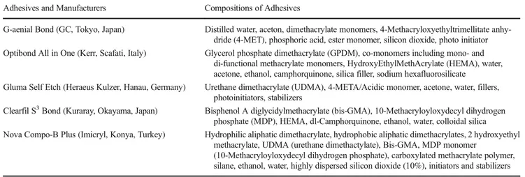
The study protocol was approved by the Human Ethical Committee of Ege University (Research no: 16-10.1/1) and Table 1 The adhesives and product details. Data were provided from manufacturers as declared
Adhesives and Manufacturers Compositions of Adhesives
G-aenial Bond (GC, Tokyo, Japan) Distilled water, aceton, dimethacrylate monomers, 4-Methacryloxyethyltrimellitate anhy-dride (4-MET), phosphoric acid, ester monomer, silicon dioxide, photo initiator Optibond All in One (Kerr, Scafati, Italy) Glycerol phosphate dimethacrylate (GPDM), co-monomers including mono- and
di-functional methacrylate monomers, HydroxyEthylMethAcrylate (HEMA), water, acetone, ethanol, camphorquinone, silica filler, sodium hexafluorosilicate
Gluma Self Etch (Heraeus Kulzer, Hanau, Germany) Urethane dimethacrylate (UDMA), 4-META/Acidic monomer, acetone, water, fillers, photoinitiators, stabilizers
Clearfil S3Bond (Kuraray, Okayama, Japan) Bisphenol A diglycidylmethacrylate (bis-GMA), 10-Methacryloyloxydecyl dihydrogen phosphate (MDP), HEMA, dl-Camphorquinone, ethanol, water, colloidal silica Nova Compo-B Plus (Imicryl, Konya, Turkey) Hydrophilic aliphatic dimethacrylate, hydrophobic aliphatic dimethacrylates, 2 hydroxyethyl
methacrylate, UDMA (urethane dimethactylate), Bis-GMA, MDP monomer
(10-Methacryloyloxydecyl dihydrogen phosphate), carboxylated methacrylate polymer, silane, ethanol, water, highly dispersed silicon dioxide (10%), initiators and stabilizers
informed consent was obtained from patients before tooth extraction. Human extracted third molar teeth stored in 0.1% thymol and distiled water were used for the study. The roots of all teeth were removed 2 mm beneath the cemento-enamel junction. Than the occlusal enamel were cut 2 mm above the cemento-enamel junction using a slow-speed water-cooled di-amond saw and exposing a flat surface of dentin (Isomet, Buehler; Lake Bluff, IL, USA). The pulpal tissue was re-moved. The exposed dentin surface was abraded with a 600-grit SiC paper for 60 s under running water to obtain a purely dentin surface and create a standardized smear layer. Prepared teeth were randomly divided into ten groups (n = 7).
The adhesives were applied on the flattened dentin sur-faces with and without RES addition. The most effective dose of RES (0.5μM) that significantly increased the cell viability [5] was added into the adhesives before bonding procedure. RES (Sigma-Aldrich, Saint Louis, USA) was added into the bottle of the adhesive at the specified con-centration and then homogenized by shaking. RES addi-tion was performed just prior to bonding applicaaddi-tion in order to prevent the inactivation of the antioxidant. The adhesives were applied according to the manufacturer’s instructions and then polymerized with LED light source for 20 s (Elipar Freelight, 3M ESPE, USA).
A composite build up in two layers (each layer 2 mm thick) was constructed with Filtek Z250 (3M ESPE, St. Paul, USA) for each tooth and then each layer polymer-ized with LED light source for 40 s. The teeth were stored in distiled water for 24 h at 37 °C. Matchsticks (1.00 ± 0.003 × 1.00 ± 0.003 mm2) were obtained from each tooth by sectioning the bonded teeth. The central matchsticks were evaluated and periphery matchsticks including enamel were eliminated. Twenty central matchsticks se-lected randomly were tested for each group. The match-sticks were fixed to a jig using a cyanoacrylate adhesive and tested to microtensile forces in a microtensile testing machine (Microtensile Tester, BISCO, Inc., Schaumburg, IL, USA) at 1.0 mm/min. The exact cross-sectional area was measured after failure with a digital caliper. The microtensile bond strengths (μTBS) of the sticks from the same bonded tooth were averaged and used for the statistical analysis. Means and standard deviations were calculated and expressed in MPa.
The specimen surfaces were evaluated under a stereomi-croscope (LG-P52, Olympus Co, Tokyo, Japan) at × 50 mag-nification to determine the apparent failure modes. The fail-ures were classified as adhesive (interfacial failure), cohesive in dentin, cohesive in resin, and mix.
IBM SPSS was used for the statistical analysis. The vari-ables were summarized by means of mean ± standard devia-tion. Statistical analysis was performed by analysis of variance (ANOVA) and followed by post hoc Tukey test for the com-parison between the groups (p < 0.05).
Results
The MTT assay results are presented in Table 2. The tested adhesives caused reduction in cell viability, although not as much as hydrogen peroxide. The lowest cell viability was observed in the G-aenial Bond and Optibond All in One groups and the highest cell viability was observed in the Gluma Self Etch, Nova Compo-B Plus, and Clerafil S3 Bond groups. After the RES addition, cell viability was in-creased in all groups.
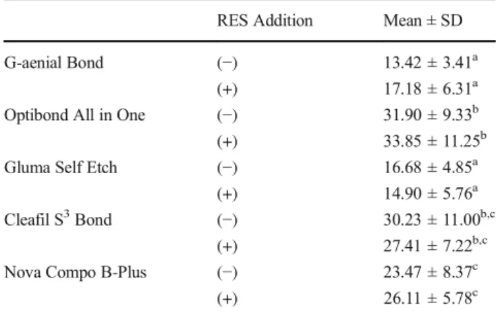
The mean μTBS values of the groups are presented in Table3. Among the groups without RES addition, the highest μTBS values were determined in Optibond All in One Clearfil S3Bond and Nova Compo-B Plus, while the lowest values were obtained in G-aenial Bond and Gluma Self Etch. No significant differences were found between theμTBS values of G-aenial Bond and Gluma Self Etch. RES addition did not cause reduction in theμTBS values of adhesives. The highest μTBS values were also determined in Optibond All in One, Clearfil S3 Bond, and Nova Compo-B Plus after RES addition.
The fracture modes of the groups are shown in Fig.1. The adhesive failure was the most frequent pattern of failure for all test groups.
Discussion
In this in vitro study, the effect of RES addition on the cyto-toxicity andμTBS of the adhesives was investigated. RES is a food-derived antioxidant existing in grapes, berries, nuts, and red wine [11,12]. This polyphenolic compound has been re-ported to have chemopreventive, cardioprotective, and Table 2 Cell viability (%) (mean ± SD). The different superscripts indicate statistical difference (p < 0.05)
RES addition Mean ± SD
Control (−) 99.92 ± 8.78a (+) 131.75 ± 16.69d H2O2 (−) 58.69 ± 7.52b (+) 128.49 ± 14.82d G-aenial Bond (−) 78.11 ± 0.88c (+) 124.12 ± 17.07d Optibond All in One (−) 79.38 ± 4.30c
(+) 116.56 ± 12.70d Gluma Self Etch (−) 84.60 ± 5.06a,c
(+) 120.91 ± 12.47d
Cleafil S3Bond (−) 88.58 ± 5.31a,c
(+) 116.64 ± 13.53d
Nova Compo-B Plus (−) 87.78 ± 7.66a,c
neuroprotective effects [13–15]. RES is an antioxidant which has also potential positive effects on adhesive biocompatibil-ity [5]. In the current study, based on the previous findings, RES was added into different self-etching adhesives in order to improve their biocompatibility without causing any nega-tive effect on their adhesive potentials. The results indicated that RES addition increased cell viability in L929 cell culture exposed to adhesive extracts and furthermore RES addition did not cause any negative influence on theμTBS of the tested adhesives.
The monomers such as Bis-GMA and UDMA have been reported to have more cytotoxicity compared to TEG-DMA and HEMA [16–18]. In this in vitro study, the cytotoxicity of adhesives including different monomer combinations was in-vestigated instead of the analysis of a single monomer. The least cytotoxic effect was depicted in Bis-GMA and UDMA incorporated adhesives. The adhesives including HEMA showed different cell viabilty rates in different monomer com-binations. These results suggest that adhesives may exhibit cytotoxic effects more or less than the monomer alone, con-sistent with the previous report [19].
The antioxidants have been reported to protect cells against cytotoxicity caused by resin-based materials [20, 21]. In
accordance with previous reports, in this study, it was deter-mined that RES, a strong antioxidant increased the cell viabil-ity. On the other hand, the innovative aspect of this study is that the analysis was not limited to cytotoxicity test alone, and μTBS test was also applied to determine the effect of RES addition on the bonding performance of the materials.
TheμTBS values obtained in this study were consistent with the previous reports [22–25]. However, there were dif-ferences between the groups considering the adhesive type as variable. The variability between the performances of the dif-ferent self-etch adhesives may be attributed to their difdif-ferent functional monomers (Table1). Optibond All in One, Clearfil S3Bond, and Nova Compo-B Plus including hydroxyethyl methacrylate (HEMA) had the highestμTBS values with or without RES addition among the all groups. The remarkably lowerμTBS values were depicted in G-aenial Bond including HEMA-free formulation and Gluma Self Etch including ure-thane dimethacrylate (UDMA). These findings support that the absence of HEMA leads to the contact of water with hy-drophobic groups and creates unfavorable condition causing water separation [25].
The monomer 10-methacryloxydecyl dihydrogen phos-phate (10-MDP) has been reported to be stable due to the adherence to hydroxyapatite [25, 26]. The higher μTBS values of Clearfil S3Bond and Nova Compo-B Plus in this study may be correlated with 10-MDP content of the materials consistent with a previous report [25]. On the other hand, the bonding potential of 4-methacryloxyethyl trimellitic acid (4-MET) was noticed to be lower [26]. In accordance with the previous findings, G-aenial Bond including 4-MET showed lowerμTBS values. Furthermore, the rapid convertion poten-tial of 4-META to 4-MET has been reported [27]. Thus, the lowerμTBS values of Gluma Self Etch may also related with 4-META content.
It is known that some special antioxidants are added into dental adhesives to eliminate the free radicals and inhibit spontaneous polymerization [28]. Antioxidants have also been reported to reduce hydrolysis [2]. In a previous study, the antioxidants vitamin C, vitamin E and quercetin were Fig. 1 The fracture modes of the
tested groups
Table 3 μTBS values (mean ± SD) in MPa. The different superscripts indicate statistical difference (p < 0.05)
RES Addition Mean ± SD
G-aenial Bond (−) 13.42 ± 3.41a
(+) 17.18 ± 6.31a
Optibond All in One (−) 31.90 ± 9.33b (+) 33.85 ± 11.25b Gluma Self Etch (−) 16.68 ± 4.85a
(+) 14.90 ± 5.76a
Cleafil S3Bond (−) 30.23 ± 11.00b,c (+) 27.41 ± 7.22b,c
Nova Compo B-Plus (−) 23.47 ± 8.37c
added into dentin adhesives and the bond strengths of the antioxidant-doped adhesives were determined to be main-tained or increased over the time [29]. It has been reported that ascorbic acid and ferric chloride might improve the micro-tensile bond strength between resin and dentin [30]. In the present study, RES a potential protective agent against cyto-toxicity, was used as antioxidant. RES addition did not cause any adverse effect onμTBS of the adhesives and adhesive performances were determined to be maintained after antiox-idant addition, where this finding was consistent with the pre-vious antioxidant reports. Although RES addition into the adhesives did not cause statistically significant differences re-garding theμTBS values, it caused an upward trend in μTBS values of G-aenial Bond, Optibond All in One, and Nova Compo-B Plus. On the contrary, it induced tendency to de-crease inμTBS values of Gluma Self Etch and Clearfil S3 Bond. Although Optibond All in One, Clearfil S3Bond, and Nova Compo-B Plus include HEMA, the tendency to de-crease inμTBS values was remarkable in Clearfil S3Bond after RES addition. This finding suggests that the interaction of RES with different combinations of monomers may cause diverse effects. On the other hand, it is also noteworthy that despite the tendency to decrease, the bonding strength of Clearfil S3Bond after RES addition was higher compared to the G-aenial Bond and Gluma Self Etch.
In this study, theμTBS of the specimens were investi-gated 24 h later without any aging procedure, as we could not predict how RES addition would affect the perfor-mance of the adhesives. However, it has been reported that aging procedure is of great importance since it may affect the initial bond strength in the long term [29]. Therefore, further studies are necessary to determine the long-term effect of RES addition on μTBS of adhesives. The diffusion of monomers into the dentin and polymer-ization of the diffused monomer are the key points for adhesion. The antioxidants may display further reaction with degradation products after the initial polymerization and this reaction has the potential to increase the bond strength in long term [29]. But the realization of this po-tential needs to be investigated for RES addition.
Different antioxidants were tested to restore dentin bond strength after dental bleaching in previous studies. Vitamins C and E have been reported to improve dentin bond strength after dental bleaching, as they reduce oxidative compounds and free radicals [31,32]. Considering the results of this study, the investigation of the effect of RES on bleached enamel may be recommended for further studies. The adhesives included in this study have different monomers with different diffusion properties. The bond strength of adhesives might be depen-dent on both chemical composition and pH of adhesives [22]. Therefore, pH measurement may be included in further stud-ies to obtain more evidence about the effect of RES addition on adhesive durability.
Conclusion
Within the limitation of this in vitro study, it can be concluded that the addition of RES, a polyphenolic antioxidant, to adhe-sives increases the cell viability and it does not adversely affect the dentinμTBS strength. Thus, RES seems to have a potential to improve the biocompatibility without causing any adverse effect on the adhesive properties of the material. But, further studies are necessary to investigate the stability of ad-hesives after RES addition for further understanding.
Compliance with ethical standards
Conflict of interest The authors declare that they have no conflict of interest.
Ethical approval All procedures performed in studies involving human participants were in accordance with the ethical standards of the institu-tional and/or nainstitu-tional research committee and with the 1964 Helsinki declaration and its later amendments or comparable ethical standards. Informed consent Informed consent was obtained from all individual participants included in the study.
References
1. Schweikl H, Spagnuolo G, Schmalz G (2006) Genetic and cellular toxicology of dental resin monomers. J Dent Res 85(10):870–877.
https://doi.org/10.1177/154405910608501001
2. Erhardt MC, Osorio R, Viseras C, Toledano M (2011) Adjunctive use of an antioxidant agent to improve resistance of hybrid layers to degradation. J Dent 39(1):80–87.https://doi.org/10.1016/j.jdent. 2010.10.013
3. Mukherjee S, Dudley JI, Das DK (2010) Dose-dependency of res-veratrol in providing health benefits. Dose Response 8:478–500.
https://doi.org/10.2203/dose-response.09-015.Mukherjee
4. San Miguel SM, Opperman LA, Allen EP, Zielinski J, Svoboda KK (2012) Bioactive polyphenol antioxidants protect oral fibroblasts from ROS-inducing agents. Arch Oral Biol 57(12):1657–1667.
https://doi.org/10.1016/j.archoralbio.2012.04.021
5. Atalayin C, Armagan G, Konyalioglu S, Kemaloglu H, Tezel H, Ergucu Z et al (2015) The protective effect of resveratrol against dentin bonding agents induced cytotoxicity. Dent Mater J 34(6): 766–773.https://doi.org/10.4012/dmj.2015-079
6. Armstrong S, Geraldeli S, Maia R, Raposo LH, Soares CJ, Yamagawa J (2010) Adhesion to tooth structure: a critical review ofBmicro^ bond strength test methods. Dent Mater 26(2):50–62.
https://doi.org/10.1016/j.dental.2009.11.155
7. Sano H, Shono T, Sonoda H, Takatsu T, Ciucchi B, Carvalho R, Pashley DH (1994) Relationship between surface area for adhesion and tensile bond strengthevaluation of a microtensile bond test. Dent Mater 10(4):236–240. https://doi.org/10.1016/0109-5641(94)90067-1
8. El-kholany NR, Abielhassan MH, Elembaby AE, Maria OM (2012) Apoptotic effect of different self-etch dental adhesives on odontoblasts in cell cultures. Arch Oral Biol 57(6):775–783.https:// doi.org/10.1016/j.archoralbio.2011.11.019
9. Amato PA, Martins RP, dos Santos Cruz CA, Capella MV, Martins LP (2014) Time reduction of light curing: influence on conversion degree and microhardness of orthodontic composites. Am J Orthod
Dentofac Orthop 146(1):40–46.https://doi.org/10.1016/j.ajodo. 2014.03.022
10. Schubert A, Ziegler C, Bernhard A, Bürgers R, Miosge N (2018, 2018) Cytotoxic effects to mouse and human gingival fibroblasts of a nanohybrid ormocer versus dimethacrylate-based composites. Clin Oral Investig.https://doi.org/10.1007/s00784-018-2419-9
11. Gescher AJ, Steward WP (2003) Relationship between mecha-nisms, bioavailibility, and preclinical chemopreventive efficacy of resveratrol: a conundrum. Cancer Epidemiol Biomark Prev 12(10): 953–957
12. Wenzel E, Somoza V (2005) Metabolism and bioavailability of transresveratrol. Mol Nutr Food Res 49(5):472–481.https://doi. org/10.1002/mnfr.200500010
13. Nawaz W, Zhou Z, Deng S, Ma X, Ma X, Li C, Shu X (2017) Therapeutic versatility of resveratrol derivatives. Nutrients 9(11): E1188.https://doi.org/10.3390/nu9111188
14. Delmas D, Lançon A, Colin D, Jannin B, Latruffe N (2006) Resveratrol as a chemopreventive agent: a promising molecule for fighting cancer. Curr Drug Targets 7(4):423–442.https://doi. org/10.2174/138945006776359331
15. Marambaud P, Zhao H, Davies P (2005) Resveratrol promotes clearance of Alzheimer’s disease amyloid-beta peptides. J Biol Chem 280(45):37377–37382. https://doi.org/10.1074/jbc. M508246200
16. Kusdemir M, Gunal S, Ozer F, Imazato S, Izutani N, Ebisu S, Blatz MB (2011) Evaluation of cytotoxic effects of six self-etching adhe-sives with direct and indirect tests. Dent Mater J 30(6):799–805.
https://doi.org/10.4012/dmj.2011-046
17. Spagnuolo G, Mauro C, Leonardi A, Santillo M, Paterno R, Schweikl H et al (2004) NF-κ protection against apoptosis induced by HEMA. J Dent Res 83(11):837–842.https://doi.org/10.1177/ 154405910408301103
18. Janke V, von Neuhoff N, Schlegelberger B, Leyhausen G, Geurtsen W (2003) TEGDMA causes apoptosis in primary human gingival fibroblasts. J Dent Res 82(10):814–818.https://doi.org/10.1177/ 154405910308201010
19. Ratanasathien S, Wataha JC, Hanks CT, Dennison JB (1995) Cytotoxic interactive effects of dentin bonding components on mouse fibroblasts. J Dent Res 74(9):1602–1606.https://doi.org/ 10.1177/00220345950740091601
20. Kim NR, Park HC, Kim I, Lim BS, Yang HC (2010) In vitro cytocompatibility of N-acetylcysteine-supplemented dentin bond-ing agents. J Endod 36(11):1844–1850.https://doi.org/10.1016/j. joen.2010.08.005
21. Krifka S, Spagnuolo G, Schmalz G, Schweikl H (2013) A review of adaptive mechanisms in cell responses towards oxidative stress
caused by dental resin monomers. Biomaterials 34(19):4555– 4563.https://doi.org/10.1016/j.biomaterials.2013.03.019
22. Ahn J, Jung KH, Son SA, Hur B, Kwon YH, Park JK (2015) Effect of additional etching and ethanol-wet bonding on the dentin bond strength of one-step self-etch adhesives. Restor Dent Endod 40(1): 68–74.https://doi.org/10.5395/rde.2015.40.1.68
23. Zilberman U, Lasilla L (2014) The use of glass-fibers ribbon and composite for prosthetic restoration of missing primary teeth-laboratory and clinical research. Open Dent J 8:220–228.https:// doi.org/10.2174/1874210601408010220
24. Pinzon LM, Watanabe LG, Reis AF, Powers JM, Marshall SJ, Marshall GW (2013) Analysis of interfacial structure and bond strength of self-etch adhesives. Am J Dent 26(6):335–340 25. Poptani B, Gohil KS, Ganjiwale J, Shukla M (2012) Microtensile
dentin bond strength of fifth with five seventh-generation dentin bonding agents after thermocycling: an in vitro study. Contemp Clin Dent 3(2):167–171.https://doi.org/10.4103/0976-237X. 101079
26. Yoshida Y, Nagakane K, Fukuda R, Nakayama Y, Okazaki M, Shintani H, Inoue S, Tagawa Y, Suzuki K, de Munck J, van Meerbeek B (2004) Comparative study on adhesive performance of functional monomers. J Dent Res 83(6):454–458.https://doi.org/ 10.1177/154405910408300604
27. Fujisawa S, Ito S (1999) 1H-NMR studies of the interaction of dental adhesive monomer, 4-META with calcium. Dent Mater J 18(1):54–62.https://doi.org/10.4012/dmj.18.54
28. Van Landuyt KL, Snauwaert J, De Munck J, Peumans M, Yoshida Y, Poitevin A et al (2007) Systematic review of the chemical com-position of contemporary dental adhesives. Biomaterials 28(26): 3757–3785.https://doi.org/10.1016/j.biomaterials.2007.04.044
29. Gotti VB, Feitosa VP, Sauro S, Correr-Sobrinho L, Leal FB, Stansbury JW et al (2015) Effect of antioxidants on the dentin interface bond stability of adhesives exposed to hydrolytic degra-dation. J Adhes Dent 17(1):35–44.https://doi.org/10.3290/j.jad. a33515
30. Soeno K, Taira Y, Jimbo R, Sawase T (2008) Surface treatment with ascorbic acid and ferric chloride improves the micro-tensile bond strength of 4-META/MMA-TBB resin to dentin. J Dent 36(11): 940–944.https://doi.org/10.1016/j.jdent.2008.07.010
31. Güler E, Gönülol N, Özyilmaz ÖY, Yücel AÇ (2013) Effect of sodium ascorbate on the bond strength of silorane and methacrylate composites after vital bleaching. Braz Oral Res 27(4):299–304.
https://doi.org/10.1590/S1806-83242013000400002
32. Whang HJ, Shin DH (2015) Effects of applying antioxidants on bond strength of bleached bovine dentin. Restor Dent Endod 40(1):37–43.https://doi.org/10.5395/rde.2015.40.1.37