Elasto
fibroma dorsi management and outcomes: review of 16 cases
Ozgur Karakurt
a, Tevfik Kaplan
b,*, Nesimi Gunal
c, Gultekin Gulbahar
d, Bulent Kocer
a, Serdar Han
b,
Koray Dural
cand Unal Sakinci
aa
Department of Thoracic Surgery, Ankara Numune Teaching and Research Hospital, Ankara, Turkey
b Department of Thoracic Surgery, Ufuk University School of Medicine, Ankara, Turkey c
Department of Thoracic Surgery, Kirikkale University School of Medicine, Kirikkale, Turkey
d Department of Thoracic Surgery, Sincan State Hospital, Ankara, Turkey
* Corresponding author. Department of Thoracic Surgery, Ufuk University School of Medicine, Dr. Ridvan Ege Teaching and Research Hospital, Mevlana Bulvarı (Konya Yolu) No: 86-88, 06520 Balgat - Ankara, Turkey. Tel: +90-312-2044201; fax: +90-312-2872390; e-mail: tevfi[email protected] (T. Kaplan).
Received 15 July 2013; received in revised form 12 September 2013; accepted 12 September 2013
Abstract
OBJECTIVES: Elastofibroma dorsi (ED) is a rare, benign lesion arising from connective tissue, usually found at the inferior pole of the scapula.
To date, only a few small series have been reported in the English literature and there are few data about the long-term outcomes after surgery. Our goal is to contribute a better understanding of this tumour and to determine the long-term outcomes after surgery.
METHODS: Sixteen patients with a diagnosis of ED were identified from the unit’s database. The clinical presentation, diagnosis, pathological evidences and long-term outcomes were evaluated.
RESULTS: There were 11 females and 5 males with a mean age of 61.1 years (range 38–78 years). The tumour was located on the right in 5 (31.2%) patients, on the left in 6 (37.5%) patients and bilaterally in 5 (31.2%). Six patients had painful scapular swelling resulting in restriction of movement of the shoulder whereas 10 reported only painful scapular mass. All 16 patients underwent complete resections. The tumour size ranged from 3 to 15 cm. The mean hospital stay was 3.1 ± 1.4 days with a morbidity of 18.75% (seroma observed in 3 patients). The mean follow-up was 58.4 ± 29.5 months (range 11–92 months). In 2 patients (12.5%) a new occurrence on the contralateral side was observed at the follow-follow-up.
CONCLUSIONS: Elastofibroma dorsi is a rare, ill-defined, pseudotumoural lesion of the soft tissues. Surgical treatment can be proposed if the
lesion is symptomatic. Furthermore, at the follow-up, the possibility of new occurrences on the contralateral side should be kept in mind.
Keywords:Chest wall• Benign tumour • Treatment • Outcomes
INTRODUCTION
Elastofibroma dorsi (ED) is a benign tumour of the thoracic wall,
which is characterized by proliferation of the elastin component of
the encapsulatedfibrous tissue [1]. The lesion usually arises beneath
the rhomboid major and latissimus dorsi muscles subjacent to the inferior angle of the scapula [2]. The pathogenesis of these lesions remains unclear. Some authors suggested that elastofibroma is a re-active process initiated by mechanical friction of the scapula to the
chest wall and hyperproliferation offibroelastic tissue caused by
re-current microtraumas [1]. Since itsfirst description, this neoplasm
has received little attention in the modern literature and only a few studies have analysed the aetiology and the clinical behaviour of this tumour. In this study, we describe a series of 16 patients with ED and on the basis of our data we discuss the main features and the long-term outcomes of this entity.
PATIENTS AND METHODS
All ED patients who underwent surgery in Ankara Numune Teaching and Research Hospital between January 2003 and
December 2012 were retrospectively reviewed. The clinical fea-tures including patients’ diagnostic methods, treatments, patho-logical characteristics of ED and the follow-up records of the outpatient clinic were evaluated. Ultrasound, computed tomog-raphy (CT) of the thorax and magnetic resonance imaging (MRI) were performed alone or in combination during the preoperative diagnostic work-up evaluation. This study protocol was approved by the Medical Ethics Committee of Ankara Numune Teaching and Research Hospital.
RESULTS
A total of 16 patients were treated within the study period. There were 11 females and 5 males (F/M ratio = 2.2). The mean age at
presentation was 61.1 years (range 38–78 years). Six patients
(37.5%) were manual labourers and the remaining 10 (62.5%)
patients did not report any significant information on heavy
manual labour or sport activity. No genetic tendency that could be an aetiological factor was determined.
Elastofibroma dorsi appeared in the subscapular region in 13 (81.2%) patients and parascapular in 3 (18.7%). The tumour was
© The Author 2013. Published by Oxford University Press on behalf of the European Association for Cardio-Thoracic Surgery. All rights reserved.
ORIG
IN
AL
ARTI
CLE
Interactive CardioVascular and Thoracic Surgery 18 (2014) 197–201
ORIGINAL ARTICLE
– THORACIC
doi:10.1093/icvts/ivt442 Advance Access publication 11 November 2013located on the right in 5 (31.2%) patients, on the left in 6 (37.5%) patients and bilaterally in 5 (31.2%).
In all patients, the major symptoms were pain and swelling but in 6 patients, painful scapular swelling resulted in a restriction of shoulder movement. The time from the onset of the pain to dis-covery of the lesions ranged between 3 and 42 months. First line
radiology comprised ultrasound scan and thorax CT (Fig. 1).
Magnetic resonance imaging was performed only in second
in-stance to add information relevant to diagnostic clarification. The
patients’ data are summarized in Table1.
In all patients, complete excision was performed following frozen-section examination demonstrating benign soft tissue tumours. A complete surgical excision was done via muscle sparing technique, which requires preparation of latissimus dorsi and
ser-ratus anterior muscleflaps. The level of incision varied between
the sixth and eighth ribs, depending on the size and location of
the tumour. Tumours were usuallyfixed to the periostium of the
ribs and tip of scapula. Our resection does not include resection of muscle and bone structures of the chest wall. As a result, after excision of the tumour the integrity and rigidity of the chest wall is protected. After complete excision of the uncapsulated tumour, a
suction drain was placed and muscleflaps were proximated with
running sutures. The surgical wounds were closed and we used
compression bandage. The drain was removed after 24–48 h. The
patients with bilateral ED underwent complete excision of both
sides during the same session (Fig. 2). The resected specimen
ranged from 3 to 15 cm in diameter. A postoperative seroma developed in 3 patients (18.75%) requiring needle aspiration. Mean
hospitalization was 3.1 ± 1.4 days (range 1–7 days).
In general, the lesions macroscopically appeared as irregular masses with indistinct borders and hard elastic consistence
(Fig.2). Microscopically, the tumours were composed of dense
collagenfibre bundles and elastic fibres (Fig.3).
The mean follow-up was 58.4 ± 29.5 months (range 11–92
months). At the end of the follow-up, all the patients were alive and well with no sign of recurrent disease. Surgery achieved excel-lent pain relief as well as a good range of shoulder movement. Interestingly, 2 patients (12.5%) developed a new ED on the op-posite site of the operation. The time for the new occurrences
after the operation was 13 months for the first patient and
19 months for the second one. The 2 patients were manual labourers. Re-evaluation of the initial thorax CT of the patients revealed no lesion on the other site. These 2 patients underwent surgery again and they are well at the follow-up.
DISCUSSION
Elastofibroma dorsi is a rare, benign tumour of the thoracic wall.
It wasfirst defined by Jarvi and Saxen [1] in 1961. Elastofibroma
dorsi could be defined as a subscapular tumour clinically and
it is also characterized by distinct elastosis histopathologically. Pathophysiological determinants are still not clear and several hy-potheses have been put forward. Repeated microinjuries between the chest wall and the scapula, the source of excess elastin produc-tion and collagen degeneraproduc-tion could play a physiopathological
role in this rare lesion [1,3]. This view has been supported by the
higher ED prevalence particularly among individuals who work at hard manual labours. However, patients who have never been
involved in hard manual work, as well as those with elastofibromas
in different locations, have undermined this view. In this study 6 patients (37.5%) were manual labourers. A new ED on the contra-lateral side developed in 2 of these patients at the follow-up. So this supports the hypothesis of microinjuries between the chest wall and the scapula in the pathogenesis of this rare proliferative lesion.
Elastofibroma dorsi is more commonly seen in older women,
with a reported female:male ratio of 5/1 and a mean age at
diag-nosis of 65–70 years and there is one case report of a 6-year old
child with ED [4–6]. In this study, the female:male ratio was 2.2 and
the mean age at presentation was 61.1 years (range 38–78 years).
Ninety-nine percent of EDs are located in the subscapular
region and more commonly on the right; however, 10–66% of
cases are bilateral [4,7]. In our study, the tumour was located on
the right in 5 (31.2%) patients, on the left in 6 (37.5%) patients and bilaterally in 5 (31.2%). Also ED appeared in the subscapular region in 13 (81.2%) patients and parascapular in 3 (18.7%).
In the physical examination, ED is in the form of well-defined
lesions that do not adhere to the skin and can easily be palpated on the thoracic wall. The scapula may sometimes mask the lesion. When both arms of the patient are strongly pulled forward and
the body is bent forward at an angle of 10–15°, the lesion
becomes more evident. Pain is the most common clinical com-plaint; however, it is mild in almost all patients. There may be lim-itations in the motions of the upper extremities. While some
authors [4,8] have reported that in most patients, ED progresses
asymptomatically, in this study, 6 patients had painful scapular swelling resulting in a movement restriction of the shoulder and 10 reported only painful scapular mass.
Elastofibroma dorsi tumours were initially considered a rare
and slowly growing lesion; however, this lesion should be known to differentiate it from malignant tumour and to avoid wide or
radical surgery [8,9]. In this study, the strategy was to use
frozen-section histological evaluation during the operation, and in the case of benign histology, to perform complete resection.
Figure 1: Thorax CT of a case with bilateral elastofibroma dorsi. CT scan demonstrates an 8 × 8 cm on the left and 9.5 × 7 cm on the right side regular soft-tissue masses (arrowheads) posterior to both scapulas. The masses have at-tenuation similar to that of the adjacent skeletal muscle.
On ultrasound ED usually appears as a hypoechoic mass with
interspersed linear echogenicity resembling muscle [4,10]. On
com-puted tomography ED appears as a heterogeneous soft tissue mass with muscle-like density and, depending on the fat tissue, contains
areas of low density [8,11]. This appearance on CT is diagnostic for
ED. On MRI, a typical feature of ED is that the interposed areas of decreased signal intensity also appear as low signal intensity on
T2-weighted sequences [9,11]. Computed tomography and MRI
images are not pathognomic for ED, but the image of a lesion
located in the subscapular region of a middle-aged or older
individ-ual is highly suggestive of ED [5,12].
Some researchers believe that imaging features are not
sufficient to diagnose ED and believe that biopsy is indicated to
exclude more aggressive tumours [7]. Butfine needle aspiration is
Table 1: Characteristics of the patients
Case Age Sex Heavy manual labour
Location Size (cm) Symptom Complication Follow-up
1 68 F No Right subscapular 3 × 2 Painful mass No No recurrence
2 53 F No Right subscapular 8 × 6 Painful mass No No recurrence
3 48 M Yes Right subscapular 13 × 7 Painful mass
Movement restriction
No No recurrence New occurrence Contralateral side
4 57 M No Left parascapular 5 × 3 Painful mass No No recurrence
5 64 F Yes Bilateral subscapular 10 × 6 8 × 5 Painful mass Movement restriction Seroma No recurrence 6 73 F No Bilateral subscapular 7 × 6 8 × 5
Painful mass No No recurrence
7 78 F No Left subscapular 5 × 3 Painful mass No No recurrence
8 54 M Yes Bilateral subscapular 11 × 8 15 × 9 Painful mass Movement restriction No No recurrence 9 67 F No Bilateral parascapular 8 × 5 7 × 4
Painful mass No No recurrence 10 41 M Yes Right parascapular 12 × 8 Painful mass
Movement restriction
Seroma No recurrence New occurrence Contralateral side
11 38 F No Left subscapular 7 × 6 Painful mass No No recurrence
12 62 M Yes Bilateral subscapular 9 × 8 11 × 6
Painful mass Movement restriction
No No recurrence
13 59 F Yes Left subscapular 15 × 9 Painful mass
Movement restriction
Seroma No recurrence
14 76 F No Left subscapular 8 × 7 Painful mass No No recurrence
15 62 F No Right subscapular 5 × 4 Painful mass No No recurrence
16 78 F No Left subscapular 6 × 5 Painful mass No No recurrence
Figure 2:Macroscopic view of excised material from a patient with bilateral elastofibroma dorsi. Regularly shaped, non-encapsulated dense masses.
Figure 3:Histopathological view of elastofibroma dorsi. Original magnification, ×100; haematoxylin-eosin stain reveals thefibrous bands to be composed of a mixture of collagen bundles and dense, eosinophilic elasticfibres (arrowheads). Mature adipose tissue is also seen.
ORIG
IN
AL
ARTI
not recommended due to the hypocellular structure of the
tumour [13,14]. Open biopsy or, at least, core needle biopsy is
needed to obtain sufficient tissue samples [14]. In this study
we did not performfine needle aspiration biopsy or core needle
biopsy because a complete tumour resection was proposed due to persisting symptoms and absence of malignancy evidences.
Complete excision is the recommended treatment for ED. Nevertheless, some studies advocate conservative treatment of patients whose diagnoses of ED are established radiologically and recommend surgical excision only when there are functional motion limitations, compression symptoms, pain or tumour
size of 5 cm or larger [8,15]. Surgical treatment is effective in
elimination of ED-related complaints. In our study, all the pat-ients had painful scapular swelling and in 7 of these patpat-ients the mass restricted movement of the shoulder. Surgery has achieved excellent pain relief as well as a good range of shoulder movement.
Post-surgical complications after the excision of ED include
seroma, postoperative haematoma and wound infections [16,17].
Seroma and haematoma were common in one series, which was
attributed to insufficient immobilization [14]. In this study, we use
postoperative wound drainage and compression bandage to reduce these complications. But even then a postoperative seroma developed in 3 patients requiring needle aspiration.
Pathologically, ED is in the form of a fibrous lesion of dirty
white colour, is non-encapsulated and contains streaks of fat
tissue. Some elastofibromas may have cystic degeneration. The
histological appearance of the lesion is typical. In large areas, it contains hyalinized collagenous stroma and little amount of
fat tissue in between. In hypocellular collagenous stroma,fibrils
and globules that show eosinophilic staining are striking. In
sec-tions of haemotoxylen-eosin, the presence offibrils and globules
is important for determining the location of the lesion and its
diagnosis [4,10].
Nagamineet al. [7], however, reported 1 case with recurrence in
their 170-patient series. Briccoliet al. [8] reported 1 patient with
ED who had a second lesion on the other side 2 years after
surgi-cal resection. Marinoet al. [18] reported 10% recurrence rate after
a mean post-surgical period of 47 months. We followed up these patients 58.4 ± 29.5 months. We observed no recurrences on the operation site but interestingly 2 patients developed a new ED on the contralateral site of the operation and these 2 patients were manual labourers.
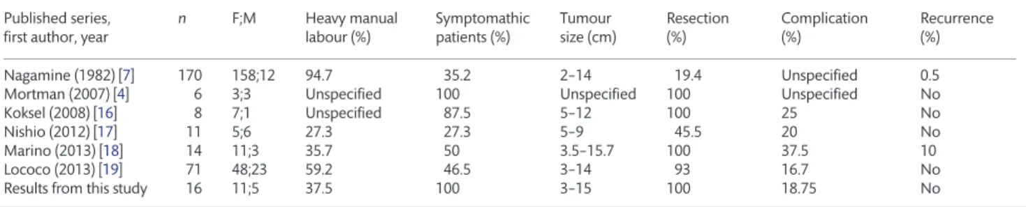
Only a few studies [4–19] have reported on the outcomes of
surgical treatment of ED (Table2). In a series of 71 patients who
underwent complete excision of ED, with similar outcomes, with a tumour size between 3 and 14 cm, complications of 16.7% and no recurrence at follow-up, have been reported [19].
In conclusion for symptomatic ED, surgery should be the choice of treatment because it is easy, has few complications, and there are no recurrences after complete excision. Furthermore, at follow-up, the possibility of new occurrences on the contralateral side should be kept in mind especially in manual labourer patients.
Funding
This research received no specific grant from any funding agency
in the public, commercial or not-for-profit sectors.
Conflict of interest: none declared.
REFERENCES
[1] Jarvi OH, Saxen AE. Elastofibroma dorsi. Acta Pathol Microbiol Scand 1961;51:83–4.
[2] Freixinet J, Rodrigez P, Hussein M. Elastofibroma of the thoracic wall. Interact CardioVasc Thorac Surg 2008;7:626–8.
[3] Fibla J, Molins L, Marco V, Pérez J, Vidal G. Bilateral elastofibroma dorsi. Joint Bone Spine 2007;74:194–6.
[4] Mortman KD, Hochheiser GM, Giblin EM, Matos YM, Frankel KM. Elastofibroma dorsi: clinocopathological review of 6 cases. Ann Thorac Surg 2007;83:1894–7.
[5] Naylor MF, Nascimento AG, Sherrick AD, McLeod RA. Elastofibroma dorsi: radiologicalfindings in 12 patients. AJR Am J Roentgenol 1996;167: 683–7.
[6] Marin ML, Perzin KH, Markowitz AM. Elastofibroma dorsi: benign chest wall tumor. J Thorac Cardiovasc Surg 1989;98:234–8.
[7] Nagamine N, Nohara Y, Ito E. Elastofibroma in Okinawa. A clinicopatholo-gic study of 170 cases. Cancer 1982;50:1794–805.
[8] Briccoli A, Casadai R, Di Renzo M, Favale L, Bacchini P, Bertoni F. Elastofibroma dorsi. Surg Today 2000;30:147–52.
[9] Kransdorf MJ, Meis JM, Montgomery E. Elastofibroma: MR and CT appear-ance with radiolgic-pathologic correlation. AJR Am J Roentgenol 1992; 159:575–9.
[10] Kara M, Dikmen E, Kara SA, Atasoy P. Bilateral elastofibroma dorsi: proper positioning for an accurate diagnosis. Eur J Cardiothorac Surg 2002;22: 839–41.
[11] Brandser EA, Goree JC, El-Khoury GY. Elastofibroma dorsi: prevalance in an elderly patient population as revealed by CT. AJR Am J Roentgenol 1998;171:977–80.
[12] Yu JS, Weis LD, Vaughan LM, Resnick D. MRI of elastofibroma dorsi. J Comput Assist Tomogr 1995;19:601–3.
[13] Kourda J, Ayadi-Kaddour A, Meria S, Hantous S, Miled KB, Mezni FE. Bilateral elastofibroma dorsi. A case report and review of the literature. Orthop Traumatol Surg Res 2009;95:383–7.
[14] Daigeler A, Vogt PM, Busch K, Pennekamp W, Weyhe D, Lehnhardt M et al. Elastofibroma dorsi-differential diagnosis in chest wall tumours. World J Surg Oncol 2007;5:5–15.
Table 2: Outcomes of selected series of elastofibroma dorsi
Published series, first author, year
n F;M Heavy manual labour (%) Symptomathic patients (%) Tumour size (cm) Resection (%) Complication (%) Recurrence (%) Nagamine (1982) [7] 170 158;12 94.7 35.2 2–14 19.4 Unspecified 0.5 Mortman (2007) [4] 6 3;3 Unspecified 100 Unspecified 100 Unspecified No
Koksel (2008) [16] 8 7;1 Unspecified 87.5 5–12 100 25 No
Nishio (2012) [17] 11 5;6 27.3 27.3 5–9 45.5 20 No
Marino (2013) [18] 14 11;3 35.7 50 3.5–15.7 100 37.5 10
Lococo (2013) [19] 71 48;23 59.2 46.5 3–14 93 16.7 No
[15] Schafmayer C, Kahlke V, Leuschner I, Pai M, Tepel J. Elastofibroma dorsi as differential diagnosis in tumors of the thoracic wall. Ann Thorac Surg 2006;82:1501–4.
[16] Koksel O, Apaydin FD, Ayhan E, Demir M, Ozdulger A. Elastofibroma dorsi: review of eight cases. Surg Today 2010;40:423–7.
[17] Nishio J, Isayama T, Iwasaki H, Naito M. Elastofibroma dorsi; diagnostic and therapeutic algorithm. J Shoulder Elbow Surg 2012;21:77–81.
[18] Marino IT, Solis PS, Lara AP, Malo JM, Vazquez ML, Tamimi F. Sensitivity and positive predictive value of magnetic resonance imaging in the diag-nosis of elastofibroma dorsi: review of fourteen cases. J Shoulder Elbow Surg 2013;22:57–63.
[19] Lococo F, Cesario A, Mattei F, Petrone G, Vita LM, Petracca-Ciavarelle L et al. Elastofibroma dorsi; clinicopathological analysis of 71 cases. Thorac Cardiovasc Surg 2013;61:215–22. ORIG IN AL ARTI CLE