Journal of Experimental and Clinical Medicine
Case Report doi: 10.5835/jecm.omu.31.04.011
Solitary plasmacytoma of the femur: A case report
Ferhat Saya*, Deniz Gürlerb, Murat Bülbülc
a Department of Orthopaedics and Traumatology, Faculty of Medicine, Ondokuz Mayıs University, Samsun, Turkey b Department of Orthopaedics and Traumatology, Samsun Training and Research Hospital, Samsun, Turkey c Department of Orthopaedics and Traumatology, Faculty of Medicine, Medipol University, Istanbul, Turkey
ARTICLE INFO ABSTRACT
Article History
Received 09 / 11 / 2013 Accepted 29 / 12 / 2013
Solitary plasmacytoma (SP) is a rare type of myeloma which is a hematologic malign tumour originated from plasma cells. It is an isolated tumour which myeloma signs are not observed. SP primarily affects axial skeleton and involvement of extremity is seen rarely. We report a case of SP of proximal femur origin in a 52-year-old woman. The patient was treated with cemented modüler tumour prosthesis following tumour excision. In a one year follow up period, no recurrens or multiple myeloma signs were observed and the patient was able to walk without support and without pain so the treatment resulted successfully.
J. Exp. Clin. Med., 2014; 31:251-253
© 2014 OMU * Correspondence to:
Ferhat Say
Department of Orthopaedics and Traumatology,
Faculty of Medicine, Ondokuz Mayıs University, Samsun, Turkey e-mail : [email protected] Keywords: Multiple myeloma Plasma cells Proximal femur Solitary plasmacytoma 1. Introduction
Plasma cell neoplasms are malignant tumours of the immune system. The most commonly seen plasma cell neoplasm showing diffuse involvement is multiple myeloma (MM). Solitary plasmacytoma (SP) and extramedullary plasmacytoma, which do not show findings of systemic myeloma are isolated tumours formed from malignant plasma cells. The World Health Organisation has classified plasmacytoma as SP of the bone and extrameduallary/outside the bone plasmacytoma (Jaffe, 2009).
SP is often found in the axial skeleton, particularly bone-marrow dense bones such as vertebra. SP is seen at a rate of less than 10% in plasma cell neoplasms. SP has been reported in the femur (Kivioja et al., 1992), tibia (Kumar et al., 2011), jawbone (Agostini et al., 2011) and patella (Lebon et al., 2011). Only one percent of SP are located in an extremity
(Homann et al., 2002). A case is presented here of SP in the upper end of the femur which was causing destructive changes and was therefore treated surgically.
2. Case report
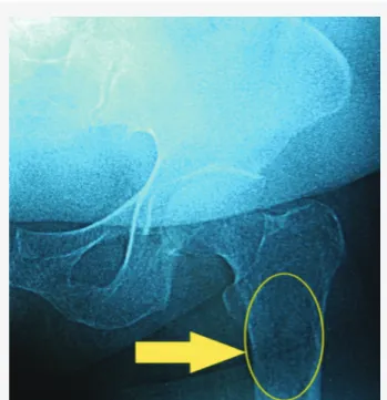
A fifty two year-old female patient presented at our clinic with pain in the left hip. The history revealed that the pain had been continuing for several months. There was nothing remarkable complaint apart from the pain on physical examination. On a direct radiograph, a regular edged, multilocular destructive lesion, 11 cm x 4 cm in size, was determined at the upper end of the femur (Fig. 1).
Magnetic resonance imaging (MRI) of the left femur showed destruction of the bone marrow in subtrochanteric region (Fig. 2).
252
tomography (PET) and 3-phase scintigraphy of the skeletal system. There were no pathological findings in the lung and abdominal examination. After evaluation of these results, rather than metastatic disease, primary bone pathology was considered. So bone marrow biopsy, complete blood count, serum electrophoresis, 24-hour urine electrophoresis and C-reactive protein tests were ordered. No pathology was found except a high sedimentation level and a low level of albumin in the protein electrophoresis. A biopsy was performed because of the mass at the upper end of the left femur. The biopsy revealed monoclonal plasma cell infiltration. It was decided to treat the patient surgically. After excision of the mass and femoral head, femoral shaft was evaluated with frozen to limit surgery. Reconstruction was achieved with a cemented resection type prosthesis (Fig.
3). The patient was mobilised with support postoperatively. The patient was monitored by the haemotology and radiation oncology clinic. At the postoperative one-year follow-up, the patient was able to walk without support and without pain. There was no recurrence and no findings of MM.
3. Discussion
SP is a tumour seen twice as frequently in males, at average age 55 years and often showing vertebral involvement (Dimopoulos et al., 2000), which is thought to be a low-grade, precursor of MM. The most common symptom may be pain caused by bone destruction and there may be findings of spinal cord or nerve root compression in vertebral involvement (Terada, 2011). In some cases, detection may be coincidental in radiological tests carried out for a different reason. The case reported here was in the appropriate age range and symptoms were given as complaints of hip pain.
Fig. 1. A regular edged, multilocular destructive lesion located
at the subtrochanteric region of femur
Fig. 3. Direct radiograph after resection and reconstruction
with cemented moduler tumour prosthesis.
Fig. 2A. Coronal magnetic resonance imaging view of the
lesion
Journal of Experimental and Clinical Medicine 31 (2014) 251-253
253 Diagnostic criteria for SP have been defined as bone
destruction in a single area by clonal plasma cells, normal bone-marrow aspiration, no involvement of another bone radiologically, no findings of anaemia, hypercalcemia or renal insufficiency, no M-protein in the serum or urine (Soutar et al., 2004). The case reported here was determined to have involvement in a single area and no additional lesions were detected radiologically. Bone marrow aspiration and laboratory tests were determined as normal. Diagnosis was made on pathological examination of SP.
Radiotherapy is the preferred treatment. Radiotherapy can achieve local control in more than 80% of patients and increases survival in approximately 35% of patients (Weber, 2005). There is a greater possibility of local recurrence with radiotherapy on lesions greater than 5 cm (Tsang et al., 2001). Patients who do not respond to radiotherapy should be treated with chemotherapy. In young patients, this includes high-dosage and autologous hematopoietic stem cell transplantation (Soutar et al., 2004).
In extremity involvement of SP, surgery is recommended
when there is structural instability (Soutar et al., 2004; Kumar, 2008). In the case reported here, on evaluation of the mass located in the left subtrochanteric femur, 10 points were scored according to the Mirel criteria (Mirel, 1989) so surgical treatment was applied. With the aim of regaining a pain-free extremity and an early return to daily activities, surgery was applied as extensive resection of the tumour and reconstruction with a cemented modular tumour prosthethis.
In SP cases, although progression to myeloma is seen at mean 2-3 years, survival is 7-12 years longer compared to myeloma early phase patients (Soutar et al., 2004; Weber, 2005; Kilciksiz et al., 2008). The prognosis of SP is worse than that of the extramedullary plasmacytoma (Kilciksiz et al., 2008). Due to the possibility of SP becoming multiple myeloma, patients require life-long follow-up of symptoms, findings and laboratory tests (Soutar et al., 2004). In the case reported here, the treatment was determined to have been sucessful as the one-year follow-up showed no recurrence or findings of MM and the patient was able to walk without support and without pain.
REFERENCES
Agostini, T., Sacco, R., Bertolai, R., Acocella, A., Lazzeri, D., 2011. Solitary plasmacytoma of the jaw. J. Craniofac. Surg. 22, 2-10. doi: 10.1097/ SCS.0b013e31822ec79a.
Dimopoulos, M.A., Moulopoulos, L.A., Maniatis, A., Alexanian, R., 2000. Solitary plasmacytoma of bone and asymptomatic multiple myeloma. Blood. 96, 2037-2044.
Homann, H.H., Lehnhardt, M., Kuhnen, C., Tilkorn, D., Steinau, H.U., 2002. Solitary plasmacytoma of the shoulder. Diagnosis and therapeutic methods. Chirurg. 73, 736-738.
Jaffe, E.S., 2009. The 2008 WHO classification of lymphomas: Implications for clinical practice and translational research. Hematology Am. Soc. Hematol. Educ. Program. 523-531. doi: 10.1182/asheducation-2009.1.523.
Kilciksiz, S., Celik, O.K., Pak, Y., Demiral, A.N., Pehlivan, M., Orhan, O., Tokatli, F., Agaoglu, F., Zinciroglu, B., Atasoy, B.M., Ozseker, N., Yersal, O., Niamg, U., Haydaroglu, A., 2008. Turkish Oncology Group-Sarcoma Working Party. Clinical and prognostic features of plasma-cytomas: A multicenter study of Turkish Oncology Group-Sarcoma Working Party. Am. J. Hematol. 83, 702-707. doi: 10.1002/ajh.21211. Kivioja, A., Sundell, B., Karaharju, E., 1992. Treatment of solitary plasmacytoma of the femur. A case report. Eur. J. Surg. Oncol. 18, 195-198. Kumar, S., 2008. Solitary plasmacytoma: Is radiation therapy sufficient? Am. J. Hematol. 83, 695-696. doi: 10.1002/ajh.21248.
Kumar, P., Sharma, S.C., Saikia, U.N., Kumar, N., Vyas, S., Angurana, S.L., 2011. Solitary plasmacytoma of the proximal tibia in an adolescent. Pediatr. Blood Cancer. 56, 158-160. doi: 10.1002/pbc.22687.
Lebon, D., Saidi, L., Merlusca, L., Leduc, F., Royer, B., 2011. Patella plasmacytoma: An unusual localization. Am. J. Hematol. 86, 504. doi: 10.1002/ajh.21961.
Mirel, H., 1989. Metastatic disease in long bones: A proposed scoring system for diagnosing impending pathologic fractures. Clin. Orthop. 249, 256-264.
Soutar, R., Lucraft, H., Jackson, G., Reece, A., Bird, J., Low, E., Samson, D., 2004. Guidelines Working Group of the UK Myeloma Forum; British Committee for Standards in Haematology; British Society for Haematology. Guidelines on the diagnosis and management of solitary plasmacytoma of bone and solitary extramedullary plasmacytoma. Br. J. Haematol. 124, 717-726.
Terada, T., 2011. Solitary plasmacytoma of the thoracic vertebra presenting with sudden paraplegia and back pain: A pathologic case report. Pathol. Oncol. Res. 17, 167-169. doi: 10.1007/s12253-010-9292-4.
Tsang, R.W., Gospodarowicz, M.K., Pintilie, M., Bezjak, A., Wells, W., Hodgson, D.C., Stewart, A.K., 2001. Solitary plasmacytoma treated with radiotherapy: Impact of tumor size on outcome. Int. J. Radiat. Oncol. Biol. Phys. 50, 113-120.
Weber, D.M., 2005. Solitary bone and extramedullary plasmacytoma. Hematology Am. Soc. Hematol. Educ. Program. 373-376.