O
riginala
rticle1 Arq Bras Oftalmol. 2017;80(1):1-3 http://dx.doi.org/10.5935/0004-2749.20170002
INTRODUCTION
Psoriasis is a skin disease characterized by chronic relapsing T-cell-mediated autoimmune inflammation that causes excessive proliferation of the underlying epidermis which ends with scaling. The diagnosis is based on the clinical appearance, which frequently presents as characteristic chronic plaques (Psoriasis vulgaris) that are well-demarcated, bright red, and covered with adherent silvery-white scales. Psoriasis is very common, with a reported prevalence in adults ranging from 0.91% (United States) to 8.5% (Norway)(1). The etiology of this disease remains unknown, but it has the highest prevalence in white males and has strong genetic and environmental associa-tions(2). Psoriasis can manifest at any age but is most common in two age groups: 20-30 years and 50-60 years.
The ocular effects of psoriasis are often disregarded because they are also observed in many other ophthalmic diseases and underlying psoriasis may be missed by a physician. The most recent American Academy of Dermatology guidelines for the management of pso-riasis and psoriatic arthritis contain no recommendations for ocular screening(3,4).
Previous studies of patients with psoriasis have reported inflamma-tory ocular changes, such as blepharitis, conjunctivitis, uveitis, and ocular ABSTRACT
Purpose: The aim of this study was to evaluate tear osmolarity, tear film function,
and ocular surface changes in patients with psoriasis.
Methods: At a single center, 30 eyes of 30 patients with psoriasis (group 1) and 30
eyes of 30 healthy individuals (group 2) were evaluated using the Ocular Surface Disease Index (OSDI) questionnaire, Schirmer I test, tear film break-up time (TBUT) test, scoring of ocular surface fluorescein staining using a modified Oxford scale, and tear osmolarity measurement.
Results: Tear osmolarity values, OSDI, and Oxford scale scores were significantly
higher in group 1 (309.8 ± 9.4 mOsm, 38.9 ± 1.1, and 0.7 ± 1.1, respectively) than in group 2 (292.7 ± 7.7 mOsm, 4.2 ± 0.3, and 0.1 ± 0.3, respectively; p<0.01 for all). TBUT was significantly lower in group 1 (8.7 ± 3.6 s) than in group 2 (18.1 ± 2.8 s; p<0.001). No significant differences were detected in Schirmer I test values between the groups (16.2 ± 2.5 mm in group 1 and 16.6 ± 2.3 mm in group 2; p=0.629).
Conclusions: The results of this study showed that psoriasis may influence tear
osmolarity and tear film function. Patients with psoriasis showed tear hyperos-molarity and tear film dysfunction.
Keywords: Dry eye syndromes; Tears; Osmolar; Psoriasis; Tear film
RESUMO
Objetivo: O objetivo deste estudo foi avaliar a osmolaridade da lágrima, função do
filme lacrimal e alterações da superfície ocular em pacientes com psoríase.
Método: Em um único centro, 30 olhos de 30 pacientes com psoríase (grupo 1) e 30
olhos de 30 indivíduos saudáveis (grupo 2) foram avaliados pelo questionário do Índice de Doença da Superfície Ocular (OSDI), teste de Schirmer tipo I, tempo de ruptura do filme lacrimal (TBUT), coloração por fluoresceína da superfície ocular utilizando a escala de Oxford modificada e osmolaridade lacrimal.
Resultados: Os valores de osmolaridade lacrimal, OSDI e escores da escala de Oxford
foram significativamente maiores no grupo 1 (309,8 ± 9,4 mOsm, 38,9 ± 1,1 e 0,7 ± 1,1, respectivamente) em comparação com o grupo 2 (292,7 ± 7,7 mOsm, 4,2 ± 0,3 e 0,1 ± 0,3, respectivamente) (p<0,01 para todos). TBUT no grupo 1 (8,7 ± 3,6 s) foi significativamente menor em comparação com o grupo 2 (18,1 ± 2,8 s) (p<0,001). Não foram detectadas diferenças significativas nos valores de teste de Schirmer (16,2 ± 2,5 mm no grupo 1 e 16,6 ± 2,3 mm no grupo 2, p=0,629).
Conclusões: Este estudo mostrou que a psoríase pode influenciar osmolaridade
lágrima e função do filme lacrimal. Os pacientes com psoríase apresentaram hipe-rosmolaridade lágrima e disfunção do filme lacrimal.
Descritores: Síndromes do olho seco; Lágrima; Concentração osmolar; Psoríase;
Fil me lacrimal
surface diseases, such as dry eye, which includes ectropion, trichiasis, conjunctivitis, conjunctival hyperemia, and corneal dryness with punctate keratitis and corneal melting(5). Her et al.(6) showed that dry eye is common in patients with psoriasis and that the tear film in these patients is relatively unstable because of ocular surface damage. In this study, we investigated the osmolarity of the tear film in patients with psoriasis and healthy individuals.
METHODS
A total of 30 eyes of 30 patients with psoriasis (group 1) and 30 eyes of 30 healthy individuals without signs or symptoms of dry eye disease or other ocular pathology (group 2) were included in this single-center, cross-sectional observational study. Right eye data for each patient were assessed.
Patients in group 1 were diagnosed with psoriasis by a dermato-logy specialist (A.B.) based on the results of dermatological and his-to pathological evaluations. Subjects were excluded if they had a history of smoking, current or recent drug use that could affect the lacrimal functional unit, active ocular infection or allergy, ocular surface scarring, previous eye surgery, or current contact lens use. Systemic
Tear osmolarity and ocular surface parameters in patients with psoriasis
Parâmetros de osmolaridade lacrimal e da superfície ocular em pacientes com psoríase
GoktuG Demirci1, Sevil karaman erDur1, rukiye ayDin1, ali Balevi1, muStafa eliacik1, muStafa ozSutcu1, oktay olmuScelik2Submitted for publication: April 29, 2016 Accepted for publication: October 23, 2016
1 Department of Ophthalmology, Istanbul Medipol University, Istanbul, Turkey. 2 Department of Internal Medicine, Istanbul Medipol University, Istanbul, Turkey.
Funding: No specific financial support was used for this study.
Disclosure of potential conflicts of interest: None of the authors have any potential conflict of interest to disclose.
Corresponding author: Sevil Karaman Erdur. Department of Ophthalmology. Istanbul Medipol University - Istanbul 34214 - Turkey - E-mail: [email protected]
Approved by the following research ethics committee: Medipol University Ethical Committee (#10840098-604.01.01-E.2734).
Te a ro s m o l a r i T ya n do c u l a rs u r fac epa r a m e T e r si npaT i e n T sw i T hp s o r i a s i s
2 Arq Bras Oftalmol. 2017;80(1):1-3
disease other than psoriasis was the other exclusion criterion. All pa-tients were examined by an internal specialist (O.O). Healthy control subjects were also examined to exclude psoriasis or other dermato-logical diseases.
The study was reviewed and approved by the Istanbul Medipol University Ethics Committee, and written informed consent was obtained from each patient before enrollment. The study was con-ducted in accordance with the tenets of the Declaration of Helsinki. Initially, patients completed the International Ocular Surface Disease Index (OSDI) survey. All subjects then underwent a full ophthal-mological examination in the same order, including visual acuity assessment and standardized slit-lamp and fundus examinations. The ophthalmologic examination and evaluation of tear osmolarity were performed on the same day but at different times.
Tear osmolarity measurements were evaluated using a TearLab osmometer (TearLab Co., San Diego, CA, USA). Tears were collected from the inferior lateral tear meniscus. Three consecutive measurements were obtained, and the mean was used for statistical analysis. All eyes underwent corneal fluorescein staining scoring using a modified Oxford scheme.
Tear film break-up time (TBUT) was assessed after the instillation of 2% fluorescein under a cobalt blue filter. The time interval between the last complete blink and the appearance of the first dry spot was recorded. The mean of three consecutive measurements was calculated. The Schirmer I test was performed with topical anesthesia using a standardized filter strip (Bio-Tech Vision Care, Ahmedabad, Gujarat, India). The amount of wetting was measured after 5 min.
The normality of the distribution of each of the parameters was checked using the Kolmogorov-Smirnov normality test. The tear osmo-larity measurements, results of the Schirmer I test with anesthesia, TBUT values, and OSDI scores were compared between the groups using an independent t-test. A p value of less than 0.05 was considered statistically significant.
RESULTS
The mean age of the subjects was 33.9 ± 5.1 years (range: 25-42 years) in group 1 (15 women and 15 men) and 33.8 ± 5 years (range: 25-43 years) in group 2 (16 women and 14 men). No significant diffe-rences were observed between the groups with respect to age or sex (p=0.960 and p=0.796, respectively).
A summary of the parameter comparisons between groups 1 and 2 is shown in table 1. Mean tear osmolarity was significantly higher in group 1 than in group 2 (p<0.001; Figure 1). The TBUT measurements for group 1 were significantly lower than those for group 2 (p<0.001; Figure 2). Mean superficial punctate staining, measured using the Oxford scale, differed significantly between the groups (p<0.001). Mean OSDI scores were significantly higher in group 1 than in group 2 (p<0.001). No statistically significant difference in Schirmer test values was observed between the groups (p=0.605).
DISCUSSION
Psoriasis is a complex, chronic, multifactorial, inflammatory, non-con tagious autoimmune disease that involves the hyperproliferation of keratinocytes in the epidermis with increased epidermal cell tur-nover. Psoriasis affects 1%-3% of the adult population with multiple extracutaneous manifestations(7,8). Recently published population-based studies examined the prevalence and incidence of psoriasis worldwide and found that the prevalence varied from 0.91% in the United States to 8.5% in Norway, with higher frequencies observed in countries at higher latitudes. Major advances in immunologic and genetic research have resulted in considerable progress in understan-ding the pathogenesis of psoriasis, which in turn has facilitated the identification of new therapeutic targets and development of more selective biologic agents.
The ophthalmic complications of psoriasis are numerous and affect almost every part of the eye; however, they remain clinically underappreciated(9). These findings are more common in men and are almost always preceded by cutaneous findings. Psoriasis may also affect the eyelids in several ways. Cram(10) has suggested that blepharitis, a common inflammatory condition of the eyelids, is the most prevalent ocular finding in patients with psoriasis. Ectropion, trichiasis, conjunctivitis with conjunctival hyperemia, and corneal dryness with punctate keratitis and corneal melting are the most frequently reported symptoms of ocular involvement in psoriasis(11,12).
Table 1. Comparison of the mean tear film parameters and OSDI scores between the two groups
Parameter psoriasis (mean ± SD)Patients with (mean ± SD) p value*Controls
Tear osmolarity (mOsm/L) 309.8 ± 9.4 292.7 ± 7.7 <0.001
Schirmer test (mm) 016.2 ± 2.5 016.6 ± 2.3 <0.605
Corneal staining 000.7 ± 1.1 000.1 ± 0.3 <0.002
TBUT (s) 008.7 ± 3.6 018.1 ± 2.8 <0.001
OSDI 038.9 ± 1.1 004.2 ± 0.3 <0.001
OSDI = ocular surface disease index; SD = standard deviation; TBUT = tear film break-up time. *= independent sample t-test.
Figure 1. Tear osmolarity in each group.
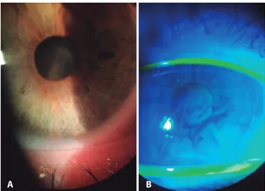
Figure 2. A) Eyelid margin examination reveals inspissated glands. B) Tear break-up time measurement showing breaks in the fluorescein after 8 s.
De m i rc i G, e t a l.
3 Arq Bras Oftalmol. 2017;80(1):1-3 The pathogenesis of both dry eye and psoriasis is not fully
understood. Similar to dry eye, psoriatic skin shows evidence of immune-mediated inflammation induced by T-cells in the kerati no-cytes(13,14). A valuable study by Her et al.(6) suggested that a common denominator exists between dry eye and psoriasis. This commonality may be immune-mediated inflammation or L-arginine deficiency and increased β-defensin production, which are key factors in both psoriasis and systemic illnesses associated with dry eye. Her et al.(6) reported that patients with psoriasis have higher OSDI and corneal fluorescein staining scores but lower TBUT values and goblet cell den-sity than controls. They found no significant differences in Schirmer test values between patients with psoriasis and controls. These results are compatible with those of the present study.
Zengin et al.(15) found that mean Schirmer I test values in patients with psoriasis were within normal limits, whereas TBUT was signifi-cantly lower than normal limits. These results are similar to those of the present study. Zengin et al.(15) also evaluated meibomian gland function and found that patients with psoriasis had higher plugging and thickness indices but normal volumes of meibomian gland secretion. These findings suggest an obstructive type of meibomian gland dysfunction in patients with psoriasis, which might result from increased turnover of the epithelial lining of the meibomian gland duct. Given these observations and hypotheses, the authors recommend meibomian gland expression therapy, lid hygiene, and systemic tetracyclines for the management of MGD with psoriasis(15). A letter from Friedlaender, MD, in response to the study advises that “There is a useful take-home message here. Ask your external disease patients about ongoing skin conditions, like psoriasis and rosacea. Attention to lid disease may be helpful in the management of these individuals’’(16).
Celik et al.(17) investigated dry eye and corneal biomechanical properties in psoriasis patients and found that the results of dry eye tests were statistically significant in the psoriasis group; however, significant corneal biomechanical changes were also found, contrary to the results of previous studies. Celik et al.(17) also found lower TBUT and Schirmer test values in patients with psoriasis than the controls(17) and concluded that dry eye assessment based on these measure-ments might be performed more accurately using an OcuSense TearLab osmometer (OcuSense Inc., San Diego, CA), which would allow patients to be grouped according to the severity of the disease. We also found lower TBUT values in patients with psoriasis than the controls in the present study, but there was no significant difference in Schirmer test values between the groups. This difference may be explained by patient age as our patients were younger than those in the study by Celik et al.(17)
Campanati et al.(18,19) also conducted comprehensive studies showing a significant association among dry eye syndromes, con-junctival hyperemia, and psoriasis. The authors observed lower TBUT and Schirmer test values in patients with psoriasis than the controls. Although their results in Schirmer test differed from our results, as we noted no difference in Schirmer test values between patients and controls, we again attribute this difference to our study patients’ being younger.
The most important finding of the studies by Campanati et al.(18,19) is the improvement in clinical disease severity indexes (DLQI and PASI) and inflammatory parameters (CRP) observed after treatment with tumor necrosis factor alpha inhibitors. Their findings suggest that tumor necrosis factor alpha plays a crucial role in the development of psoriasis. This finding agrees with those of numerous studies confirming the significant role of immunosuppressive treatments in improving the systemic and inflammatory symptoms of psoriasis.
These findings may also explain why tear film osmolarity is higher in psoriasis patients. Eye findings in conjunction with psoriatic arthritis were reported in 1976 by Lambert and Wright(20), who noted
the presence of ocular inflammation in 31.2% of 112 patients with psoriatic arthritis. Conjunctivitis was the most common lesion (19.6%), followed by iritis (7.1%). The inflammation in dry eye due to psoriasis may explain the increased tear osmolarity in these patients.
The present study had some limitations. Quantitative studies of conjunctival goblet cells and meibography were not performed. Further studies will be required to obtain these data.
Clinicians should remain mindful that the most frequently en -coun tered psoriatic ocular complications of psoriasis are often vague and represent otherwise common symptoms, such as dry eye or blepharitis(9). The results of our study show that tear osmolarity in pa tients with psoriasis is increased, which contributes to the dry eye symptoms that are often underappreciated by physicians. Because psoriasis, similar to dry eye, is an inflammatory condition, we hypo-thesize that tear osmolarity is also affected by inflammatory processes. We suggest routine eye examinations for patients with psoriasis of all types for the early detection of subclinical dry eye. The noninvasive measurement of tear osmolarity can easily rule out subclinical dry eye in psoriasis patients.
REFERENCES
1. Parisi R, Symmons DP, Griffiths CE, Ashcroft DM. Identification and Management of Psoriasis and Associated ComorbidiTy (IMPACT) project team. Global epidemiology of psoriasis: a systematic review of incidence and prevalence. J Invest Dermatol. 2013; 133(2):377-85.
2. Gonzalez-Andrades M, Arias-Santiago S, García-Serrano JL, González Gallardo MD, McAlinden C. Sterile corneal infiltrates secondary to psoriasis exacerbations: topical tacrolimus as an alternative treatment option. Eye Contact Lens. 2015. doi: 10.1097/ ICL.0000000000000178.
3. Listed N. The definition and classification of dry eye disease: report of the Definition and Classification Subcommittee of the International Dry Eye WorkShop (2007). Ocul Surf. 2007;5(2):75-92.
4. Menter A, Gottlieb A, Feldman SR, Van Voorhees AS, Leonardi CL, Gordon KB, et al. J Guide-lines of care for the management of psoriasis and psoriatic arthritis: Section 1. Overview of psoriasis and guidelines of care for the treatment of psoriasis with biologics. Am Acad Dermatol. 2008;58(5):826-50.
5. Kilic B, Dogan U, Parlak AH, Goksugur N, Polat M, Serin D, Ozmen S, et al. Ocular fin-dings in patients with psoriasis. Int J Dermatol. 2013;52(5):554-9.
6. Her Y, Lim JW, Han SH. Dry eye and tear film functions in patients with psoriasis. Jpn J Ophthalmol. 2013;57(4):341-6.
7. Griffiths C, Camp R, Barker J. Rock’s textbook of dermatology. Oxford: Blackwell Science; 2004.
8. Zachariae H. Pathologic findings in internal organs in psoriasis. Int J Dermatol. 1994; 33(5):323-6.
9. Rehal B, Modjtahedi BS, Morse LS, Schwab IR, Maibach HI. Ocular psoriasis. J Am Acad Dermatol. 2011;65(6):1202-12.
10. Cram DL. Corneal melting in psoriasis. J Am Acad Dermatol. 1981;5(5):617. 11. Catsarou-Catsari A, Katambus A, Theodorpoylos P. Ophthalmological manifestations
in patients with psoriasis. Acta Derm Venereol (Stock). 1984;64(6):557-59. 12. Huynh N, Cervantes-Castaneda RA, Bhat P, Gallagher MJ, Foster CS. Biologic response
modifier therapy for psoriatic ocular inflammatory disease. Ocul Immunol Inflamm. 2008;16(3):89-93.
13. Ayroldi E, Bastianelli A, Cannarile L, Petrillo MG, Delfino DV, Fierabracci A. A pathoge-netic approach to autoimmune skin disease therapy: psoriasis and biological drugs, unresolved issues, and future directions. Curr Pharm Des. 2011;17(29):3176-90. 14. Perera GK, Di Meglio P, Nestle FO. Psoriasis. Annu Rev Pathol. 2012;7:385-422. 15. Zengin N, Tol H, Balevi S, Gündüz K, Okudan S, Endoğru H. Tear film and meibomian
gland functions in psoriasis. Acta Ophthalmol Scand. 1996;74(4):358-60.
16. Friedlaender MH. tear film and meibomian gland functions in psoriasis. Comment in Surv Ophthalmol. 1997;41(6):500-1.
17. Celik U, Aykut V, Celik B, Tas M, Yazgan S, Kaldırım H, et al. A comparison of corneal biomechanical properties in patients with psoriasis and healthy subjects. Eye Contact Lens. 2015; 41(2):127-9
18. Campanati A, Neri P, Giuliodori K, Arapi I, Carbonari G, Borioni E, et al. Psoriasis beyond the skin surface: a pilot study on the ocular involvement. Int Ophthalmol. 2015;35(3): 331-40.
19. Campanati A, Giuliodori K, Ganzetti G, Liberati G, Offidani AM. A patient with psoriasis and vitiligo treated with etanercept. Am J Clin Dermatol. 2010;11(1):46-8. 20. Lambert JR, Wright V. Eye inflammation in psoriatic arthritis. Ann Rheum Dis. 1976;35(4):