Original Article
Nilüfer Bıçakçı
1, Murat Elli
21University of Health Sciences Turkey, Samsun Training and Research Hospital, Clinic of Nuclear Medicine, Samsun, Turkey 2İstanbul Medipol University Faculty of Medicine, Department of Pediatric Oncology, İstanbul, Turkey
18
Fluorine-fluorodeoxyglucose PET/CT Imaging in Childhood
Malignancies
Çocukluk Çağı Malignitelerinde
18
Flor-florodeoksiglukoz PET/BT Görüntüleme
©Copyright 2021 by Turkish Society of Nuclear Medicine
Molecular Imaging and Radionuclide Therapy published by Galenos Yayınevi.
DOI:10.4274/mirt.galenos.2020.64436
Mol Imaging Radionucl Ther 2021;30:18-27
Address for Correspondence: Nilüfer Bıçakçı MD, University of Health Sciences Turkey, Samsun Training and Research Hospital,
Clinic of Nuclear Medicine, Samsun, Turkey
Phone: +90 362 311 15 00 E-mail: [email protected] ORCID ID: orcid.org/0000-0003-4124-1225 Received: 08.08.2020 Accepted: 13.10.2020
Abstract
Objectives: The aim of the study was to evaluate the utility of 18fluorine-fluorodeoxyglucose (18F-FDG) positron emission tomography/computed tomography (PET/CT) in the diagnosis, staging, restaging, and treatment response of childhood malignancies.
Methods: This study included 52 patients (32 boys, 20 girls) who were referred to our clinic between November 2008 and December 2018 with
the diagnosis of malignancy. The patients were evaluated retrospectively. Median age of the patients was 13 years (range 2-17). 18F-FDG was given to the patients intravenously, and time of flight with PET/16 slice CT was performed 1 hour thereafter. The lowest dose was 2 mCi (74 MBq) and the highest dose was 10 mCi (370 MBq). Fasting blood sugars of all patients were found below 200 mg/dL (11.1 mmol/L).
Results: 18F-FDG PET/CT was performed to evaluate the response to treatment in 38 of 52 children, staging in 11 patients (staging and evaluation of the response to treatment in nine of them), restaging in 2 patients, restaging, and evaluation of the response to treatment in 1 patient. 18F-FDG PET/CT examination was reported as normal in 13 patients (5 girls, 8 boys). The pathological 18F-FDG uptake was detected in 39 patients (14 girls, 25 boys), which indicated metastasis and/or recurrence of the primary disease. Total number of deaths was 30 (13 girls, 17 boys).
Conclusion: 18F-FDG PET/CT has a significant role for staging, restaging, treatment response, and detection of metastatic disease but it is limited for the early diagnosis of childhood cancers.
Keywords: 18F-FDG PET/CT, childhood malignancy, staging, restaging, response
Öz
Amaç: Çalışmamızın amacı, çocukluk çağı malignitelerinin tanı, evreleme, yeniden evreleme ve tedaviye cevabın değerlendirilmesinde 18 flor-florodeoksiglukoz (18F-FDG) pozitron emisyon tomografisi/bilgisayarlı tomografinin (PET/BT) yararını göstermektir.
Yöntem: Kasım 2008 ve Aralık 2018 tarihleri arasında, malignensi tanılı 52 hastanın (32 erkek, 20 kız) dosyaları ve görüntüleri geriye dönük olarak
incelendi. Ortalama yaş 13 (2-17) idi. 18F-FDG’nin intravenöz enjeksiyonundan 1 saat sonra, time of flight/16 kesit BT yapıldı. Çalışmamızda en düşük doz 2 mCi (74 MBq), en yüksek doz 10 mCi (370 MBq). Tüm hastaların açlık kan şekerleri 200 mg/dL’nin (11,1 mmol/L) altındaydı.
Bulgular: 18F-FDG PET/BT, 52 hastanın 38’ine tedaviye yanıt değerlendirilmesi, 11 hastaya evreleme (9 hasta evreleme ve aynı zamanda tedaviye yanıt değerlendirilmesi), 2 hastaya yeniden evreleme, 1 hastaya yeniden evreleme ve tedaviye yanıt değerlendirilmesi amacıyla yapıldı. 18F-FDG PET/ BT çalışması 13 hastada (5 kız, 8 erkek) normaldi. Otuz dokuz hastada (14 kız, 25 erkek) çalışma, metastaz ve/veya primer hastalığın nüksü ile uyumlu bulundu. Toplam ölüm sayısı 30 (13 kız, 17 erkek) idi.
Sonuç: 18F-FDG PET/BT çocukluk çağı malignensilerinin tanı, evreleme, yeniden evreleme ve tedaviye yanıt değerlendirilmesi açısından çok faydalıdır ancak erken tanıda yararı sınırlıdır.
Introduction
18
Fluorine-fluorodeoxyglucose (
18F-FDG) positron emission
tomography/computed tomography (PET/CT) plays an
important role for diagnosis, staging, restaging, response
to treatment, and evaluation of prognosis in childhood
malignancies (1,2). PET-only examinations have been
replaced by hybrid systems in the recent decades, where PET
and CT are used together in oncology (3). In this imaging
system, PET and CT are used together for functional data
and morphological information, respectively (4).
18F-FDG
PET/CT is also known to have high sensitivity and specificity
(86% and 80%, respectively) in childhood malignancies
(5,6,7).
The type of childhood malignancies varies according to
the age groups. The most common childhood malignancy
is leukemia with a rate of 30%; other malignancies are
brain tumors (20%), lymphomas (14%), neuroblastoma
(7%), soft tissue sarcomas (7%), Wilms’ tumor (6%),
bone tumors (5%), germ cell tumors (3%), melanoma
(3%), hepatic tumors (1%), etc. Lymphoma and germ cell
tumors are more common in children between the ages
of 14 and 19 years (8,9,10,11,12,13,14). The childhood
tumors in which
18F-FDG PET/CT is used frequently
include lymphomas, brain tumors, soft tissue sarcomas,
neuroblastoma, Wilms’ tumor, germ cell tumors, and
neurofibromatosis 1 (15). The most commonly used
radionuclides in nuclear medicine for the cancer imaging
are gallium-67 (
67Ga) citrate, thallium-201 chloride,
technetium-99m sestamibi, and
18F-FDG.
18F-FDG causes
lower radiation exposure due to relatively short half-life
(110 minutes), and it is also a widely available radionuclide
agent (2).
18F-FDG mimics glucose in cell uptake process
and thus acts as a marker of glucose usage.
18F-FDG is not
a tumor-specific agent and can be kept in cells in case
of many physiological and pathological conditions.
Dual-time-point imaging can help to increase the specificity of
18
F-FDG imaging (3).
We evaluated the role of
18F-FDG PET/CT in diagnosis,
staging, restaging, treatment response, and detection of
metastatic disease of childhood malignancies in this study.
Materials and Methods
Fifty-two children (32 boys, 20 girls) with tissue-confirmed
malignancies underwent
18F-FDG PET/CT examination
between November 2008 and December 2018. The median
age of the patients was 13 years (range 2-17 years). The
study was approved by the University of Health Sciences
Turkey, Samsun Training and Research Hospital of Local
Ethics Committee (protocol number: GOKA/2020/10/6).
All imaging studies were performed under at least 4
hours of total fasting. The dose of
18F-FDG was calculated
as 0.15 mCi/kg (5.55 MBq/kg) between 2008 and
2010. After 2010, it was calculated according to the
radiopharmaceutical doses published in the 2016 North
American Consensus Guidelines, which has been updated
as the whole-body
18F-FDG with 3.7-5.2 MBq/kg (0.1-0.4
mCi/kg), and the minimum dose was recommended as 37
MBq (1 mCi). In our study, the lowest dose was 2 mCi
(74 MBq), and the highest dose was 10 mCi (370 MBq).
Fasting blood sugar level of all patients was found to be
less than 200 mg/dL (11.1 mmol/L). CT parameters were
obtained with ultra-low dose (80 kVp, 5 mAs, and 1.5:1
pitch). After 45-60 minutes from application of
18F-FDG, CT
images were obtained for attenuation correction without
intravenous contrast, and then PET images were gathered.
18
F-FDG examination was performed with time of flight
PET/16 section CT (Philips Gemini TF), and the PET detector
crystal material was LYSO.
Sedation was used in 6 patients who were under 8 years of
age during the
18F-FDG PET/CT examination. We used the
oral chloral hydrate as 50-70 mg/kg for young children less
than 15 kg of body weight, according to application guide
of the American Academy of Pediatrics (16,17). This dosage
is appropriate in most nuclear medicine applications. In our
study it was sufficient for the younger age group.
Brown adipose tissue produces heat in case of exposure
to cold and causes focal increased
18F-FDG uptake and
may mimic muscle or malignancy (18,19,20). However,
diazepam was not used in any of our patients as the waiting
room temperatures were ensured to be high enough to
prevent cold exposure in our clinic.
18
F-FDG PET/CT indications and findings of the patients
were analyzed retrospectively. Patient characteristics are
listed in Table 1.
No statistical analysis was performed.
Results
18
F-FDG PET/CT was applied to 52 children for evaluation
of response to treatment in 38, staging in 11 (2 staging
and nine staging and evaluating response to treatment),
restaging in 2, evaluation of response to treatment with
restaging in 1 patient.
Twenty-three patients had the diagnosis of lymphoma [14
non-Hodgkin’s lymphoma (NHL), 9 HL], and
18F-FDG PET/
CT was performed for staging and response to treatment
in 10, for response to treatment in 11, and for restaging
in 2 patients.
18F-FDG PET/CT detected more nodal lesions
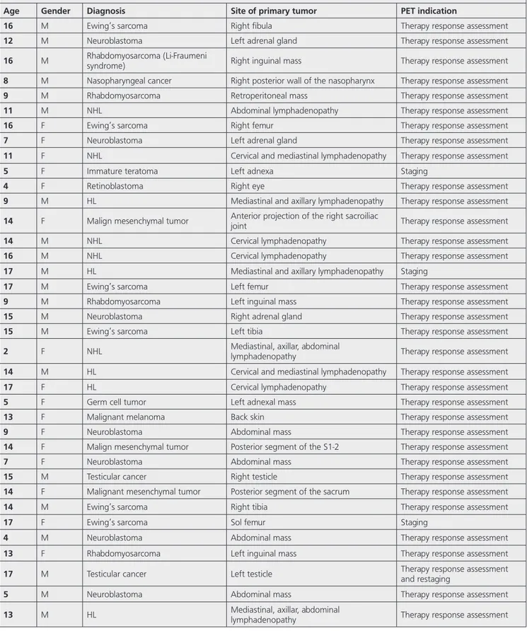
Table 1. Patient characteristics
Age Gender Diagnosis Site of primary tumor PET indication
16 M Ewing’s sarcoma Right fibula Therapy response assessment
12 M Neuroblastoma Left adrenal gland Therapy response assessment
16 M Rhabdomyosarcoma (Li-Fraumeni syndrome) Right inguinal mass Therapy response assessment
8 M Nasopharyngeal cancer Right posterior wall of the nasopharynx Therapy response assessment
9 M Rhabdomyosarcoma Retroperitoneal mass Therapy response assessment
11 M NHL Abdominal lymphadenopathy Therapy response assessment
16 F Ewing’s sarcoma Right femur Therapy response assessment
7 F Neuroblastoma Left adrenal gland Therapy response assessment
11 F NHL Cervical and mediastinal lymphadenopathy Therapy response assessment
5 F Immature teratoma Left adnexa Staging
4 F Retinoblastoma Right eye Therapy response assessment
9 M HL Mediastinal and axillary lymphadenopathy Therapy response assessment
14 F Malign mesenchymal tumor Anterior projection of the right sacroiliac joint Therapy response assessment
14 M NHL Cervical lymphadenopathy Therapy response assessment
16 M NHL Cervical lymphadenopathy Therapy response assessment
17 M HL Mediastinal and axillary lymphadenopathy Staging
17 M Ewing’s sarcoma Left femur Therapy response assessment
9 M Rhabdomyosarcoma Left inguinal mass Therapy response assessment
15 M Neuroblastoma Right adrenal gland Therapy response assessment
15 M Ewing’s sarcoma Left tibia Therapy response assessment
2 F NHL Mediastinal, axillar, abdominal lymphadenopathy Therapy response assessment
14 M HL Cervical and mediastinal lymphadenopathy Therapy response assessment
17 F HL Cervical lymphadenopathy Therapy response assessment
5 F Germ cell tumor Left adnexal mass Therapy response assessment
13 F Malignant melanoma Back skin Therapy response assessment
9 F Neuroblastoma Abdominal mass Therapy response assessment
14 F Malign mesenchymal tumor Posterior segment of the S1-2 Therapy response assessment
7 F Neuroblastoma Abdominal mass Therapy response assessment
15 M Testicular cancer Right testicle Therapy response assessment
14 F Malignant mesenchymal tumor Posterior segment of the sacrum Therapy response assessment
14 M Ewing’s sarcoma Right tibia Therapy response assessment
17 F Ewing’s sarcoma Sol femur Staging
4 M Neuroblastoma Abdominal mass Therapy response assessment
13 F Rhabdomyosarcoma Left inguinal mass Therapy response assessment
17 M Testicular cancer Left testicle Therapy response assessment and restaging
5 M Neuroblastoma Abdominal mass Therapy response assessment
in the skeletal system and bone marrow increased the
stage in these patients (Figure 1).
Patients with Ewing’s sarcoma (ES), rhabdomyosarcoma,
neuroblastoma,
malignant
melanoma,
malignant
mesenchymal tumor, retinoblastoma, nasopharynx
carcinoma, and germ cell tumors did not undergo
18F-FDG
PET/CT study before treatment, and
18F-FDG PET/CT was
performed after treatment to evaluate the response to
treatment. Metastatic disease was detected by
18F-FDG PET/
CT in the bone, liver, brain, and abdominal and mediastinal
lymph nodes of the patients with neuroblastoma (n=7)
during follow-up.
Seven patients with ES and one with peripheric primitive
neuroendocrine tumor were evaluated with
18F-FDG PET/
CT for local and systemic involvement after chemotherapy.
Three local recurrences and five abdominal/inguinal
metastatic lymph nodes were detected with the
18F-FDG
PET/CT. In patients with rhabdomyosarcoma,
18F-FDG PET/
CT detected three recurrent diseases and one metastatic
disease on follow-up after adjuvant therapy (one had
Li-Fraumeni syndrome).
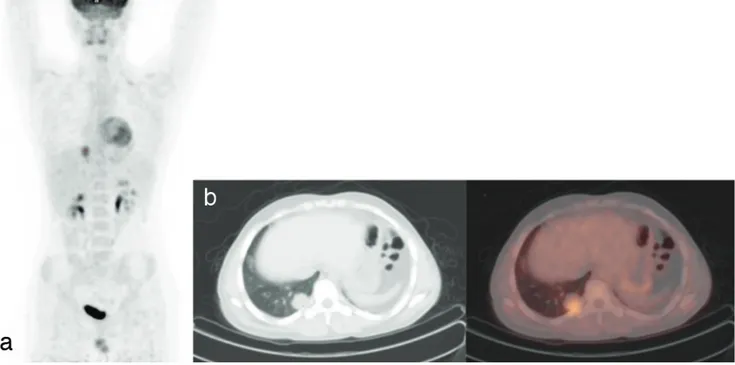
18
F-FDG PET/CT was performed for evaluation of treatment
response in 2 patients with testicular carcinoma. In the
other patient,
18F-FDG PET/CT was performed for restaging,
and a lung metastasis was detected (Figure 2).
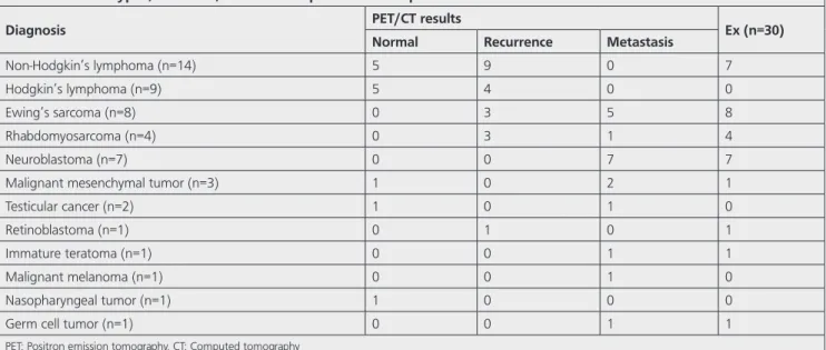
No recurrence or metastasis was identified in
18F-FDG
PET/CT of 13 patients. Thirty patients died on follow-up;
7 patients had NHL, and the other 23 patients had ES
(n=8), neuroblastoma (n=7), rhabdomyosarcoma (n=1),
malignant mesenchymal tumor (n=1), germ cell tumor
(n=1), immature teratoma (n=1), and retinoblastoma (n=1)
(Table 2).
Discussion
Our findings indicate that
18F-FDG PET/CT is an essential
imaging modality and provided important information for
diagnosis, staging, restaging, evaluation of the response to
treatment, and detection of metastatic disease. However,
Table 1. Continued
Age Gender Diagnosis Site of primary tumor PET indication
14 F NHL (Burkitt’s lymphoma) Cervical lymphadenopathy Therapy response assessment
11 M Peripheral primitive neuroectodermal tumor Left posterior mediastinum Therapy response assessment
8 F Ewing’s sarcoma Right femur Therapy response assessment
15 M NHL (Burkitt’s lymphoma) Mediastinal, abdominal, and pelvic lymphadenopathy Staging and therapy response assessment
15 M NHL Abdominal lymphadenopathy Staging and therapy response assessment
14 M NHL Abdominal lymphadenopathy Staging and therapy response assessment
14 M NHL Abdominal and pelvic lymphadenopathy Staging and therapy response assessment
16 M HL Cervical lymphadenopathy Staging and therapy response assessment
15 M HL Cervical and mediastinal lymphadenopathy Staging and therapy response assessment
13 F NHL Abdominal lymphadenopathy Staging and therapy response assessment
11 F NHL Abdominal and pelvic lymphadenopathy Restaging
16 M HL Cervical lymphadenopathy Restaging
15 M HL Cervical lymphadenopathy Therapy response assessment
12 M NHL Mediastinal, axillar, abdominal lymphadenopathy Staging and therapy response assessment
11 M NHL Abdominal lymphadenopathy Staging and therapy response assessment
this study is limited in early diagnosis of childhood
malignancies.
Although childhood malignancies are relatively rare as
compared to adults, still they are a significant cause of
mortality and constitute the second most frequent cause of
death after trauma in children (21). Leukemia accounts for
more than half of all childhood solid tumors, and the other
frequent childhood cancers are brain tumors, lymphomas,
neuroblastoma, soft tissue sarcomas, Wilms’ tumor, and
bone tumors (8,21).
Childhood cancers differ from adults in terms of
epidemiology, histological patterns, clinical behavior,
Figure 1. MIP (a), transaxial CT (b), and fusion 18F-FDG PET/CT images of a 15-year-old male patient. Abdominal lymph node biopsy revealed a
high-grade malign B-cell lymphoma (Burkitt’s lymphoma). Multiple hypermetabolic mediastinal, abdominal, pelvic lymph nodes, massive abdominal fluid, and bone marrow involvement were seen on 18F-FDG PET/CT imaging
18F-FDG: 18Fluorine-fluorodeoxyglucose, PET: Positron emission tomography, CT: Computed tomography, MIP: Maximum intensity projection
Table 2. Cancer types, numbers, and follow-up results of all patients
Diagnosis PET/CT results Ex (n=30)
Normal Recurrence Metastasis
Non-Hodgkin’s lymphoma (n=14) 5 9 0 7
Hodgkin’s lymphoma (n=9) 5 4 0 0
Ewing’s sarcoma (n=8) 0 3 5 8
Rhabdomyosarcoma (n=4) 0 3 1 4
Neuroblastoma (n=7) 0 0 7 7
Malignant mesenchymal tumor (n=3) 1 0 2 1
Testicular cancer (n=2) 1 0 1 0
Retinoblastoma (n=1) 0 1 0 1
Immature teratoma (n=1) 0 0 1 1
Malignant melanoma (n=1) 0 0 1 0
Nasopharyngeal tumor (n=1) 1 0 0 0
Germ cell tumor (n=1) 0 0 1 1
treatment response, and prognosis. Appropriate treatment
reduces the mortality rate. Early and correct diagnosis is
essential. Improved oncological results lead to an increased
incidence of late complications of childhood cancers.
18F-FDG
PET/CT as an imaging technique is well studied in adults.
18
F-FDG PET/CT is increasingly used for staging, prognosis,
determination of biopsy location, evaluation of treatment
response, radiotherapy planning, and follow-up in many
types of childhood cancers (5,22,23,24,25,26,27,28). The
role of
18F-FDG PET/CT is, however, limited for the early
diagnosis of childhood cancers but has a significant role for
staging, treatment response, and detection of metastatic
disease. Thus,
18F-FDG PET/CT has been used increasingly
in children with malignancy for these features.
18
F-FDG is the most commonly used radiopharmaceutical
in PET for oncological purposes.
18F-FDG is a cyclotron
radiopharmaceutical with a half-life of 110 minutes.
18
F-FDG is a glucose analog and is transported into the cell
by glucose transporters and often participates in the first
stage of the physiological glycolytic pathway. Therefore, the
degree of
18F-FDG uptake indicates the metabolic activity of
the cells (29). Evaluation after treatment with therapeutic
agents does not affect tumor size immediately but inhibits
tumor metabolism and proliferation. So, accumulation of
18
F-FDG in metabolically active tumor cells has revolutionized
oncological imaging. Although this discovery was made
several decades ago, the ability of
18F-FDG PET imaging for
differentiation of active/stable disease and to provide more
clinical information than the simple anatomical localization
of the disease has been appreciated recently.
New generation PET devices are faster and have higher
resolution.
18F-FDG PET reflects both the metabolic status
and the proliferative potential of the disease in patients
receiving either conventional or experimental therapy.
18
F-FDG PET can be used in the majority of childhood
cancers as convenient as CT and magnetic resonance
imaging (MRI) (30,31,32,33). Metabolic changes induced by
chemotherapy occur before morphological changes. Since
the
18F-FDG intake provides direct measurement of tumor
glucose metabolism, the tumor’s response to treatment can
be evaluated earlier before the tumor shrinks. The response
to treatment may also be predicted more accurately than
conventional techniques (34,35,36,37). In our study, we
also used
18F-FDG as imaging radiopharmaceutical in all
pediatric patients. We adjusted the radiopharmaceutical
doses in children in line with the 2016 North American
Consensus Guidelines renewed in 2010 and later (38,39).
Lymphomas are the third most common type of tumor in the
childhood group that account for 14% of all cancer cases.
While NHL is more commonly found in young children, HL
is more common in the adolescent group.
18F-FDG PET/
CT is used for staging, evaluation of treatment response,
and relapse of disease, before bone marrow or stem cell
Figure 2. MIP (a), transaxial CT (b), and fusion 18F-FDG PET/CT images of a 16-year-old male patient. Histopathologically, diagnosis was
rhabdomyosarcoma. Hypermetabolic metastatic nodule was seen in the right lung posterobasal segment on 18F-FDG PET/CT imaging
transplantation for diagnostic and prognostic information
in children (40). London et al. (41) in their study compared
conventional imaging methods (CT, ultrasonography, MRI,
and bone scintigraphy) with
18F-FDG PET/CT in pediatric
patients diagnosed with HL and NHL to differentiate
malignant lesion and to predict poor response to treatment.
The sensitivity, specificity, and accuracy (95.9%, 99.7%,
and 99.6%, respectively) of
18F-FDG PET/CT were found
to be higher than other conventional imaging methods
(70.1%, 99.0%, and 98.3%, respectively) for lymphoma
in children. In a study by Cheng et al. (6),
18F-FDG PET/
CT detected lesions that could not be detected by CT in
50% of children with HL and 42.9% of children with NHL.
In our study
18F-FDG PET/CT detected more nodal lesions
than CT in 10 patients (50% of children with HL and 50%
of children with NHL). The stage of malignancy was also
increased because of additional lesions in the skeletal
system and bone marrow in these patients.
Tumors of sympathetic nervous system constitute about
7% of all childhood tumors, and neuroblastoma is the most
common tumor in this group (42). Approximately 10% of
neuroblastomas do not uptake metaiodobenzylguanidine
(MIBG), and
18F-FDG PET/CT can be used in the evaluation
of MIBG-negative patients (42,43,44). Another study
reported that MIBG scintigraphy and
18F-FDG PET/CT
were equally effective for patients with distant disease
in demonstrating bone metastases after primary tumor
resection and chemotherapy (45). Choi et al. (46)
showed that
18F-FDG PET/CT is more sensitive than CT
for evaluation of distant lymph node metastases and can
detect recurrent lymph node metastases. Similarly, bone,
liver, brain, and widespread lymph node metastases in the
abdomen and mediastinum were detected by
18F-FDG PET/
CT in our patients with neuroblastoma after the adjuvant
therapy. Other alternative diagnostic imaging technique
in neuroblastoma without MIBG uptake has been
investigated including radiolabeled somatostatin analogs
such as octreotide and DOTA-conjugated peptides [e.g.,
68
Ga DOTATATE (DOTA0-Try3) octreotate],
68Ga DOTATOC
(DOTA0-Try3) octreotide, and
68Ga DATANOC
(DOTA0-1NaI3) octreotide. These analogs can bind selectively to
somatostatin receptors 2 (47). DOTA-peptides can also be
labeled with beta-emitting isotopes, for example,
177Lu or
90Y, to provide peptide receptor radionuclide therapy for
neuroendocrine tumors in adults (48,49,50,51,52,53,54)
and have been used in small studies with relapsed
neuroblastoma in children (55,56,57,58).
ES is a heterogenous tumor including ES of the bone,
extraosseous ES, and peripheral primitive neuroectodermal
tumor. It is the second most common bone malignancy
in the pediatric age group, and its incidence among all
childhood cancers is approximately 3% (59). Like many
other malignant tumors, ES has an increased glycolysis rate,
and as a result, it shows increased
18F-FDG accumulation.
18F-FDG PET/CT is particularly useful in detecting, staging,
and restaging of the bone metastases in musculoskeletal
tumors and often provides important additional
information that may alter the treatment plan (60). Seven
patients with ES and one patient with peripheral primitive
neuroectodermal tumor were evaluated with
18F-FDG PET/
CT for local and systemic disease after chemotherapy in
our study. Three local recurrences and five abdominal/
inguinal metastatic lymph nodes were detected with the
18
F-FDG PET/CT.
Rhabdomyosarcoma is responsible for 4%-8% of
malignant diseases in children under 15 years of age (2).
Although most of the cases are sporadic, some related
congenital and genetic diseases are reported (61). One
of our four rhabdomyosarcoma patients had Li-Fraumeni
syndrome.
18F-FDG PET/CT detected three recurrent and
one metastatic disease on follow-up after treatment
of rhabdomyosarcoma. There are few studies in the
literature on the role of
18F-FDG PET/CT in treatment
response evaluation in childhood rhabdomyosarcoma.
Eugene et al. (62) reported that
18F-FDG PET/CT predicted
the treatment response better than conventional imaging
methods in a study group of 23 patients after 3 cycles of
treatment. They also had demonstrated 69% complete
radiological response with
18F-FDG PET/CT while it was
reported as 8% in conventional methods. This finding
supports that the metabolic response of the treatment
occurred earlier than the response in tumor size.
18F-FDG
PET/CT was also performed in our clinic for evaluating
response to treatment in patients with malignant
mesenchymal tumor, testicular tumors, retinoblastoma,
immature teratoma, nasopharyngeal cancers, and germ
cell tumors.
18F-FDG PET/CT guided the treatment in
these patients by evaluating the local recurrence and
metastatic disease.
18
F-FDG PET/CT detected more nodal lesions than CT in 10
staged patients in our study.
18F-FDG PET/CT also increased
the stage in these patients by detecting multiple lesions
in the skeletal system and bone marrow. So, it has been
confirmed that
18F-FDG PET/CT has addictive effects on the
outcomes and the prognosis of patients.
Despite the above-mentioned beneficial roles of
18F-FDG
PET/CT in malignancy, it has some limitations. Level of
radiation dose is a severe problem in children. Lack of
simultaneous data acquisition causes image artifacts
because of patient movement. Another drawback is
that CT provides only limited soft tissue contrast. These
problems could be overcome by integrating the PET
detectors into MR scanner. Dose reductions of up to 73%
have been reported when performing PET/MRI instead of
18
F-FDG PET/CT because of lack of the CT component, and
decreasing the amount of PET tracer administered (because
of longer imaging times in PET/MRI) could further reduce
the radiation dose (63). Other advantage of PET/MRI is
improved soft tissue contrast. Improved soft tissue contrast
of MRI leads to improved localization of PET tracer uptake
(64). Although
18F-FDG PET/CT remains the mainstay for
functional imaging of oncologic and neurologic processes
in children, early experience shows that PET/MRI has great
potential in diagnostic algorithms of several pediatric
diseases.
The acquisition parameters for the CT portion of the scan
should be tailored to the patient’s size. CT parameters
were obtained with ultra-low dose (80 kVp, 5 mAs,
and 1.5:1 pitch) in our study. Decreasing the absorbed
radiation dose without compromising the image quality
can be provided by reducing milliamperes proportionately.
This modification results in lower exposed radiation dose
in
18F-FDG PET/CT than the diagnostic CT. Combination
of
18F-FDG PET/CT and diagnostic CT has been reported
to be used in the literature to prevent doubled radiation
exposure to the patient (65). The follow-up of the patients
can be performed reliably with
18F-FDG PET/CT in order to
further reduce the radiation exposure.
Conclusion
To conclude,
18F-FDG PET/CT provides important
information for the staging, restaging, response to
treatment, and detection of metastatic disease, but it has
limited contribution to early diagnosis in childhood tumors
particularly in lymphoma, primary bone, and soft tissue
tumors. It is a non-invasive imaging method that reflects
both the metabolic features and the structural status of
the tumors. As the preparation and image interpretation of
the pediatric patients differ from adults, these procedures
should be performed with specific information and
experience on this age group. It should also be noted
that indications of
18F-FDG PET/CT must be considered
appropriately since the exposure to radiation in children
has more severe consequences than the adults.
Ethics
Ethics Committee Approval: The study was approved by
the University of Health Sciences Turkey, Samsun Training
and Research Hospital of Local Ethics Committee (protocol
number: GOKA/2020/10/6).
Informed Consent: Consent form was filled out by all
participants.
Peer-review: Externally peer-reviewed.
Authorship Contributions
Surgical and Medical Practices: N.B., M.E., Concept: N.B.,
Design: N.B., M.E., Data Collection or Processing: N.B.,
M.E., Analysis or Interpretation: N.B., Literature Search:
N.B., Writing: N.B.
Conflict of Interest: No conflict of interest was declared
by the authors.
Financial Disclosure: The authors declared that this study
has received no financial support.
References
1. Voss SD. Pediatric oncology and the future of oncological imaging. Pediatr Radiol 2011;41:172-185.
2. Freebody J, Wegner EA, Rossleigh MA. 2-deoxy-2-((18)F) fluoro-D-glucose positron emission tomography/computed tomography imaging in paediatric oncology. World J Radiol 2014;6:741-755.
3. Costantini DL, Vali R, Chan J, McQuattie S, Charron M. Dual-time-point FDG PET/CT for the evaluation of pediatric tumors. AJR Am J Roentgenol 2013;200:408-413.
4. Shulkin BL. PET imaging in pediatric oncology. Pediatr Radiol 2004;34:199-204.
5. Uslu L, Donig J, Link M, Rosenberg J, Quon A, Daldrup-Link HE. Value of 18F-FDG PET and PET/CT for evaluation of pediatric malignancies. J Nucl Med 2015;56:274-286.
6. Cheng G, Servaes S, Zhuang H. Value of (18)F-fluoro-2-deoxy-D-glucose positron emission tomography/computed tomography scan versus diagnostic contrast computed tomography in initial staging of pediatric patients with lymphoma. Leuk Lymphoma 2013;54:737-742.
7. Miller E, Metser U, Avrahami G, Dvir R, Valdman D, Sira LB, Sayar D, Burstein Y, Toren A, Yaniv I, Even-Sapir E. Role of 18F-FDG PET/CT in staging and follow-up of lymphoma in pediatric and young adult patients. J Comput Assist Tomogr 2006;30:689-694.
8. Steliarova-Foucher E, Stiller C, Lacour B, Kaatsch P. International Classification of Childhood Cancer, third edition. Cancer 2005;103:1457-1467.
9. Percy CL, Smith MA, Linet M. Et al. Lymphomas and reticuloendothelial neoplasms.In: Ries LAG, Smith MA, Gurney, et al., eds. Cancer incidance and Survival Among children and adolescents: United States SEER Program 1975-1995. Bethesda MD: National Cancer İnstitude;1999. NIH publication 99-4649.
10. Amankwah EK, Conley AP, Reed DR. Epidemiology and therapies for metastatic sarcoma. Clin Epidemiol 2013;5:147-162.
11. Ducimetière F, Lurkin A, Ranchère-Vince D, Decouvelaere AV, Péoc’h M, Istier L, Chalabreysse P, Muller C, Alberti L, Bringuier PP, Scoazec JY, Schott AM, Bergeron C, Cellier D, Blay JY, Ray-Coquard I. Incidence of sarcoma histotypes and molecular subtypes in a prospective epidemiological study with central pathology review and molecular testing. PLoS One 2011;6:20294. 12. Arndt CA, Crist WM. Common musculoskeletal tumors of childhood and
adolescence. N Engl J Med 1999;341:342-352.
13. Burns DK, Kumar V. The musculoskeletal system. In: Kumar V, Cotran RS, Robbins SL, eds. Robbins Basic Pathology. 7th ed. Philadelphia, Pennsylvania: Saunders; 2003:769-770.
14. Bhojwani D, McCarville MB, Choi JK, Sawyer J, Metzger ML, Inaba H, Davidoff AM, Gold R, Shulkin BL, Sandlund JT. The role of FDG-PET/CT in
the evaluation of residual disease in paediatric non-Hodgkin lymphoma. Br J Haematol 2015;168:845-853.
15. Uslu-Beşli L, Atay Kapucu LÖ, Karadeniz C, Akdemir ÜÖ, Pinarli FG, Aydos U, Okur A, Kaya Z, Samanci C, Karabacak NI. Comparison of FDG PET/MRI and FDG PET/CT in Pediatric Oncology in Terms of Anatomic Correlation of FDG-positive Lesions. J Pediatr Hematol Oncol 2019;41:542-550. 16. American Academy of Pediatrics Committee on Drugs: Guidelines
for monitoring and management of pediatric patients during and after sedation for diagnostic and therapeutic procedures. Pediatrics 1992;89:1110-1115.
17. American Society of Anesthesiologists Task Force on Sedation and Analgesia by Non-Anesthesiologists. Practice guidelines for sedation and analgesia by non-anesthesiologists. Anesthesiology 2002;96:1004-1017. 18. Cohade C, Mourtzikos KA, Wahl RL. “USA-Fat”: prevalence is related to
ambient outdoor temperature-evaluation with 18F-FDG PET/CT. J Nucl Med 2003;44:1267-1270.
19. Cohade C, Osman M, Pannu HK, Wahl RL. Uptake in supraclavicular area fat (“USA-Fat”): description on 18F-FDG PET/CT. J Nucl Med 2003;44:170-176.
20. Yeung HW, Grewal RK, Gonen M, Schöder H, Larson SM. Patterns of (18)F-FDG uptake in adipose tissue and muscle: a potential source of false-positives for PET. J Nucl Med 2003;44:1789-1796.
21. Steliarova-Foucher E, Colombet M, Ries LAG, Moreno F, Dolya A, Bray F, Hesseling P, Shin HY, Stiller CA; IICC-3 contributors. International incidence of childhood cancer, 2001-10: a population-based registry study. Lancet Oncol 2017;18:719-731.
22. Nihayah S, Shammas A, Vali R, Parra D, Alexander S, Amaral J, Connolly B. Correlation of PET/CT and Image-Guided Biopsies of Pediatric Malignancies. AJR Am J Roentgenol 2017;208:656-662.
23. Dong Y, Zhang X, Wang S, Chen S, Ma C. 18F-FDG PET/CT is useful in initial staging, restaging for pediatric rhabdomyosarcoma. Q J Nucl Med Mol Imaging 2017;61:438-446.
24. Hurley C, McCarville MB, Shulkin BL, Mao S, Wu J, Navid F, Daw NC, Pappo AS, Bishop MW. Comparison of (18) F-FDG-PET-CT and Bone Scintigraphy for Evaluation of Osseous Metastases in Newly Diagnosed and Recurrent Osteosarcoma. Pediatr Blood Cancer 2016;63:1381-1386.
25. Treglia G, Taralli S, Bertagna F, Salsano M, Muoio B, Novellis P, Vita ML, Maggi F, Giordano A. Usefulness of whole-body fluorine-18-fluorodeoxyglucose positron emission tomography in patients with neurofibromatosis type 1: a systematic review. Radiol Res Pract 2012;2012:431029.
26. London K, Cross S, Onikul E, Dalla-Pozza L, Howman-Giles R. 18F-FDG PET/CT in paediatric lymphoma: comparison with conventional imaging. Eur J Nucl Med Mol Imaging 2011;38:274-284.
27. Kluge R, Kurch L, Georgi T, Metzger M. Current Role of FDG-PET in Pediatric Hodgkin’s Lymphoma. Semin Nucl Med 2017;47:242-257. 28. Flerlage JE, Kelly KM, Beishuizen A, Cho S, De Alarcon PA, Dieckmann
U, Drachtman RA, Hoppe BS, Howard SC, Kaste SC, Kluge R, Kurch L, Landman-Parker J, Lewis J, Link MP, McCarten K, Punnett A, Stoevesandt D, Voss SD, Wallace WH, Mauz-Körholz C, Metzger ML. Staging Evaluation and Response Criteria Harmonization (SEARCH) for Childhood, Adolescent and Young Adult Hodgkin Lymphoma (CAYAHL): Methodology statement. Pediatr Blood Cancer 2017;64.
29. Gatenby RA, Gillies RJ. Why do cancers have high aerobic glycolysis? Nat Rev Cancer 2004;4:891-899.
30. Portwine C, Marriott C, Barr RD. PET imaging for pediatric oncology: an assessment of the evidence. Pediatr Blood Cancer 2010;55:1048-1061. 31. McCarville MB. PET-CT imaging in pediatric oncology. Cancer Imaging
2009;9:35-43.
32. Franzius C. FDG-PET/CT in pediatric solid tumors. Q J Nucl Med Mol Imaging 2010;54:401-410.
33. Kleis M, Daldrup-Link H, Matthay K, Goldsby R, Lu Y, Schuster T, Schreck C, Chu PW, Hawkins RA, Franc BL. Diagnostic value of PET/CT for the staging and restaging of pediatric tumors. Eur J Nucl Med Mol Imaging 2009;36:23-36.
34. Furth C, Steffen IG, Amthauer H, Ruf J, Misch D, Schönberger S, Kobe C, Denecke T, Stöver B, Hautzel H, Henze G, Hundsdoerfer P. Early and late therapy response assessment with [18F] fluorodeoxyglucose positron emission tomography in pediatric Hodgkin’s lymphoma: analysis of a prospective multicenter trial. J Clin Oncol 2009;27:4385-4391. 35. Gallamini A, Hutchings M, Avigdor A, Polliack A. Early interim PET
scan in Hodgkin lymphoma: where do we stand? Leuk Lymphoma 2008;49:659-662.
36. Hawkins DS, Conrad EU 3rd, Butrynski JE, Schuetze SM, Eary JF. [F-18]-fluorodeoxy-D-glucose-positron emission tomography response is associated with outcome for extremity osteosarcoma in children and young adults. Cancer 2009;115:3519-3525.
37. Hawkins DS, Schuetze SM, Butrynski JE, Rajendran JG, Vernon CB, Conrad EU 3rd, Eary JF. [18F]Fluorodeoxyglucose positron emission tomography predicts outcome for Ewing sarcoma family of tumors. J Clin Oncol 2005;23:8828-8834.
38. Gelfand MJ, Parisi MT, Treves ST; Pediatric Nuclear Medicine Dose Reduction Workgroup. Pediatric radiopharmaceutical administered doses: 2010 North American consensus guidelines. J Nucl Med 2011;52:318-322.
39. Treves ST, Gelfand MJ, Fahey FH, Parisi MT. 2016 Update of the North American Consensus Guidelines for Pediatric Administered Radiopharmaceutical Activities. J Nucl Med 2016;57:15-18.
40. Qiu L, Chen Y, Wu J. The role of 18F-FDG PET and 18F-FDG PET/CT in the evaluation of pediatric Hodgkin’s lymphoma and non-Hodgkin’s lymphoma. Hell J Nucl Med 2013;16:230-236.
41. London K, Cross S, Onikul E, Dalla-Pozza L, Howman-Giles R. 18F-FDG PET/CT in paediatric lymphoma: comparison with conventional imaging. Eur J Nucl Med Mol Imaging 2011;38:274-284.
42. Sharp SE, Gelfand MJ, Shulkin BL. Pediatrics: diagnosis of neuroblastoma. Semin Nucl Med 2011;41:345-353.
43. Piccardo A, Lopci E, Conte M, Foppiani L, Garaventa A, Cabria M, Villavecchia G, Fanti S, Cistaro A. PET/CT imaging in neuroblastoma. Q J Nucl Med Mol Imaging 2013;57:29-39.
44. Mueller WP, Coppenrath E, Pfluger T. Nuclear medicine and multimodality imaging of pediatric neuroblastoma. Pediatr Radiol 2013;43:418-427. 45. Kushner BH, Yeung HW, Larson SM, Kramer K, Cheung NK. Extending
positron emission tomography scan utility to high-risk neuroblastoma: fluorine-18 fluorodeoxyglucose positron emission tomography as sole imaging modality in follow-up of patients. J Clin Oncol 2001;19:3397-3405.
46. Choi YJ, Hwang HS, Kim HJ, Jeong YH, Cho A, Lee JH, Yun M, Lee JD, Kang WJ. (18)F-FDG PET as a single imaging modality in pediatric neuroblastoma: comparison with abdomen CT and bone scintigraphy. Ann Nucl Med 2014;28:304-313.
47. Alexander N, Vali R, Ahmadzadehfar H, Shammas A, Baruchel S. Review: The Role of Radiolabeled DOTA-Conjugated Peptides for Imaging and Treatment of Childhood Neuroblastoma. Curr Radiopharm 2018;11:14-21.
48. Bodei L, Cremonesi M, Grana CM, Fazio N, Iodice S, Baio SM, Bartolomei M, Lombardo D, Ferrari ME, Sansovini M, Chinol M, Paganelli G. Peptide receptor radionuclide therapy with 177Lu-DOTATATE: the IEO phase I-II
study. Eur J Nucl Med Mol Imaging 2011;38:2125-2135.
49. Bodei L, Mueller-Brand J, Baum RP, Pavel ME, Hörsch D, O’Dorisio MS, O’Dorisio TM, Howe JR, Cremonesi M, Kwekkeboom DJ, Zaknun JJ. The joint IAEA, EANM, and SNMMI practical guidance on peptide receptor radionuclide therapy (PRRNT) in neuroendocrine tumours. Eur J Nucl Med Mol Imaging 2013;40:800-816.
50. Brans B, Mottaghy FM, Kessels A. 90Y/177Lu-DOTATATE therapy: survival of the fittest? Eur J Nucl Med Mol Imaging 2011;38:1785-1787. 51. Delpassand ES, Samarghandi A, Zamanian S, Wolin EM, Hamiditabar
M, Espenan GD, Erion JL, O’Dorisio TM, Kvols LK, Simon J, Wolfangel R, Camp A, Krenning EP, Mojtahedi A. Peptide receptor radionuclide therapy with 177Lu-DOTATATE for patients with somatostatin receptor-expressing neuroendocrine tumors: the first US phase 2 experience. Pancreas 2014;43:518-525.
52. Kunikowska J, Królicki L, Hubalewska-Dydejczyk A, Mikołajczak R, Sowa-Staszczak A, Pawlak D. Clinical results of radionuclide therapy of neuroendocrine tumours with 90Y-DOTATATE and tandem 90Y/177Lu-DOTATATE: which is a better therapy option? Eur J Nucl Med Mol Imaging 2011;38:1788-1797.
53. Maecke HR, Reubi JC. Somatostatin receptors as targets for nuclear medicine imaging and radionuclide treatment. J Nucl Med 2011;52:841-844.
54. Waldherr C, Pless M, Maecke HR, Haldemann A, Mueller-Brand J. The clinical value of [90Y-DOTA]-D-Phe1-Tyr3-octreotide (90Y-DOTATOC) in the treatment of neuroendocrine tumours: a clinical phase II study. Ann Oncol 2001;12:941-945.
55. Kong G, Hofman MS, Murray WK, Wilson S, Wood P, Downie P, Super L, Hogg A, Eu P, Hicks RJ. Initial Experience With Gallium-68 DOTA-Octreotate PET/CT and Peptide Receptor Radionuclide Therapy for Pediatric Patients With Refractory Metastatic Neuroblastoma. J Pediatr Hematol Oncol 2016;38:87-96.
56. Gains JE, Bomanji JB, Fersht NL, Sullivan T, D’Souza D, Sullivan KP, Aldridge M, Waddington W, Gaze MN. 177Lu-DOTATATE molecular radiotherapy for childhood neuroblastoma. J Nucl Med 2011;52:1041-1047. 57. Menda Y, O’Dorisio MS, Kao S, Khanna G, Michael S, Connolly M, Babich
J, O’Dorisio T, Bushnell D, Madsen M. Phase I trial of 90Y-DOTATOC therapy in children and young adults with refractory solid tumors that express somatostatin receptors. J Nucl Med 2010;51:1524-1531.
58. Sadowski SM, Neychev V, Millo C, Shih J, Nilubol N, Herscovitch P, Pacak K, Marx SJ, Kebebew E. Prospective Study of 68Ga-DOTATATE Positron Emission Tomography/Computed Tomography for Detecting Gastro-Entero-Pancreatic Neuroendocrine Tumors and Unknown Primary Sites. J Clin Oncol 2016;34:588-596.
59. Ludwig JA. Ewing sarcoma: historical perspectives, current state-of-the-art, and opportunities for targeted therapy in the future. Curr Opin Oncol 2008;20:412-418.
60. Bestic JM, Peterson JJ, Bancroft LW. Pediatric FDG PET/CT: Physiologic uptake, normal variants, and benign conditions [corrected]. Radiographics 2009;29:1487-1500.
61. Hartley AL, Birch JM, Blair V, Kelsey AM, Harris M, Jones PH. Patterns of cancer in the families of children with soft tissue sarcoma. Cancer 1993;72:923-930.
62. Eugene T, Corradini N, Carlier T, Dupas B, Leux C, Bodet-Milin C.
18F-FDG-PET/CT in initial staging and assessment of early response to
chemotherapy of pediatric rhabdomyosarcomas. Nucl Med Commun 2012;33:1089-1095.
63. Schäfer JF, Gatidis S, Schmidt H, Gückel B, Bezrukov I, Pfannenberg CA, Reimold M, Ebinger M, Fuchs J, Claussen CD, Schwenzer NF. Simultaneous whole-body PET/MR imaging in comparison to PET/CT in pediatric oncology: initial results. Radiology 2014;273:220-231. 64. Rausch I, Quick HH, Cal-Gonzalez J, Sattler B, Boellaard R, Beyer T.
Technical and instrumentational foundations of PET/MRI. Eur J Radiol 2017;94:3-13.
65. Qi Z, Gates EL, O’Brien MM, Trout AT. Radiation dose reduction through combining positron emission tomography/computed tomography (PET/ CT) and diagnostic CT in children and young adults with lymphoma. Pediatr Radiol 2018;48:196-203.