Turk Kardiyol Dern Ars 2016;44(5):423-426 doi: 10.5543/tkda.2015.31624
Successful ablation of coexistent Mahaim tachycardia and right
posterior accessory pathway in a patient with Ebstein’s anomaly
Ebstein anomalisi olan çocukta Mahaim taşikardi ve
sağ arka aksesuvar yol birlikteliğinin başarılı ablasyonu
Department of Cardiology, İstanbul Medipol University Faculty of Medicine, İstanbul, Turkey#Department of Pediatric Cardiology, İstanbul Medipol University Faculty of Medicine, İstanbul, Turkey
Enes Elvin Gül, M.D., Celal Akdeniz, M.D.,# Volkan Tuzcu, M.D.#
Özet– Atriyofasiküler aksesuvar yolak (AY) diğer deyimle Mahaim yolakları preeksitasyonun nadir formu olup aksesu-var yolakların %3’den daha azını oluşturmaktadır. Mahaim aksesuvar yolak dekremental, sadece önedoğru iletisi olan ve sol dal bloklu antidromik taşikardi ile kendini gösterir. Ma-haim aksesuvar yolağın Ebstein anomalisindeki prevalan-sı oldukça yüksektir. Ayrıca Wolf-Parkinson-White (WPW) sendromu ve Mahaim AY birlikteliği Ebstein anomalisinde gösterilmiştir. Bu yazıda, Ebstein anomalisi olan hastada WPW ve Mahaim AY birlikteliğinin floroskopisiz başarılı ab-lasyonunu sunuyoruz.
Summary– The atriofascicular accessory pathway (AP), known as the Mahaim pathway, is a rare form of pre-ex-citation, comprising less than 3% of all APs. Mahaim AP is characterized by decremental, anterograde-only conduc-tion, and antidromic tachycardia with left bundle branch morphology. Prevalence of Mahaim AP in Ebstein’s anom-aly is significantly high. In addition, combination of Wolff– Parkinson–White (WPW) syndrome and Mahaim AP in pa-tients with Ebstein’s anomaly has been reported. Presently described is the coexistence of Mahaim AP and manifest WPW syndrome in a patient with Ebstein’s anomaly, who was successfully ablated without fluoroscopy.
423
C
oexistence of Mahaim AP with manifest Wolff–Parkinson-White (WPW) syndrome in patients
with Ebstein’s anomaly has been reported.[1]
Treat-ment modality of multiple APs depends upon clinical significance and risk assessment of these pathways. Radiofrequency (RF) ablation can be used safely and effectively for treatment of various types of tachycar-dias in patients with Ebstein’s anomaly.[2]
CASE REPORT
A 12-year-old boy with a diagnosis of Ebstein’s anomaly was referred due to palpitations. Twelve-lead resting electrocardiography revealed short PR in-terval (110 msec) and wide QRS duration (119 msec) with delta waves consistent with manifest WPW syn-drome. No tachycardia was observed on electrocardi-ography. Echocardiogram revealed Ebstein’s anomaly with normal right and left ventricular functions. There
was no family history of syncope, sudden cardiac death, or arrhythmias. Physical examination was normal. Electro-physiological study was
planned for risk stratification of WPW syndrome. Electrophysiological study was performed un-der general anesthesia without intubation. Quadripo-lar catheters were positioned at the high atrium, His bundle area, and right ventricular apex. A steerable de-capolar catheter was inserted into the coronary sinus for positional reference. EnSite NavX system (St. Jude Medical, Inc., Little Canada, MN, USA) was used for 3-dimensional mapping and catheter navigation. Right atrial anatomy was reconstructed without fluoroscopy. Baseline electrophysiological measurements were as follows: AH interval of 32 msec, HV interval of 24
Received:August 23, 2015 Accepted:December 03, 2015
Correspondence: Dr. Enes Elvin Gül. 334 Queen Mary Rd, K7M 7E7 K7M7E7 Kingston - Canada. Tel: +1 613 328 2052 e-mail: [email protected]
© 2016 Turkish Society of Cardiology
Abbreviations:
AP Accessory pathway LBBB Left bundle branch block RF Radiofrequency
SVT Supraventricular tachycardia WPW Wolff–Parkinson–White CASE REPORT
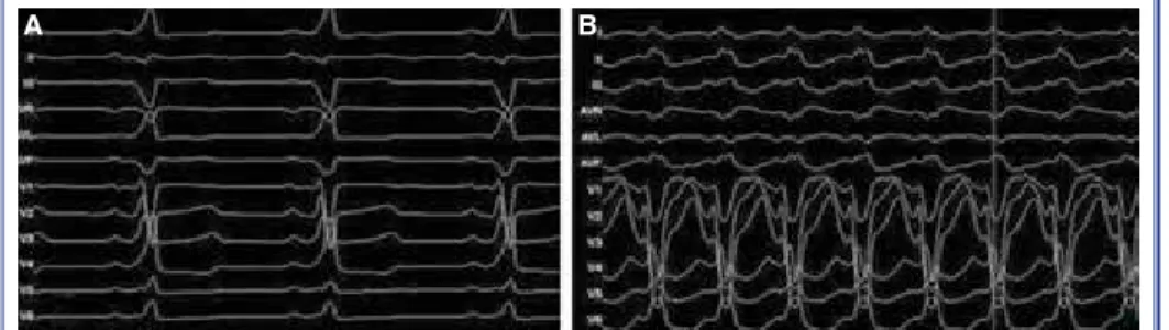
msec, and baseline cycle length of 582 msec. There was evidence of pre-excitation consistent with right-sided AP. Wide QRS tachycardia with left bundle branch block (LBBB) and inferior axis pattern was induced spontaneously due to catheter manipulation. QRS mor-phology was different from baseline pre-excited QRS morphology (Figure 1). Atrial pacing demonstrated a difference in pre-excited QRS morphology, compared to tachycardia QRS morphology (Figure 2). Tachycar-dia cycle length was 280 msec, and was not considered antidromic supraventricular tachycardia (SVT) of the pathway leading to baseline pre-excitation. Tachycar-dia was terminated with overdrive pacing due to hemo-dynamic impairment during tachycardia.
To assess risk stratification of WPW, effective refractory period of AP and shortest pre-excited RR duration during atrial fibrillation were measured (ef-fective refractory period was 320 msec and shortest pre-excited RR was 338 msec). Morphology of tachy-cardia during atrial fibrillation was different from baseline QRS morphology. However, this tachycardia was similar to the wide QRS tachycardia previously induced. Due to unchanged QRS morphology in spite of irregularity in RR intervals, this wide QRS tachy-cardia was not interpreted as aberrant SVT, but rather as Mahaim tachycardia. During the mapping around the tricuspid annulus, typical Mahaim potential was detected at the lateral tricuspid annulus (Figure 3).
Turk Kardiyol Dern Ars 424
Figure 1. (A) Twelve-lead electrocardiography showing pre-excited QRS morphology with short PR interval. (B) Wide QRS tachycardia with LBBB morphology and inferior axis, con-sistent with Mahaim tachycardia.
A B
Figure 2. Atrial pacing from high right atrium showing pre-excited and Mahaim QRS mor-phologies. The first 2 post-atrial pacing beats were consistent with pre-excited QRS, and the third beat showed left bundle branch block with inferior axis, consistent with Mahaim automatic tachycardia.
Coexistent Mahaim tachycardia and right posterior accessory pathway in a patient with Ebstein’s anomaly 425
RF energy was delivered to this region and an ac-celerated wide QRS rhythm with similar QRS mor-phology to Mahaim tachycardia was detected dur-ing ablation. This finddur-ing also supported evidence of Mahaim AP. Following successful RF and during atrial pacing, the wide QRS morphology of the Ma-haim pathway was not observed again, though pre-excitation was still noted at baseline. Antegrade and retrograde mapping located the pathway in the right posterior area of the tricuspid valve annulus. AP was
eliminated at the 14th second of RF ablation.
Follow-ing successful ablation and durFollow-ing ventricular pacFollow-ing, no retrograde conduction was observed. At the end of the post-procedural waiting period of 30 minutes, atrioventricular block was obtained with adenosine administration, and no pre-excitation was observed. It was conclusively confirmed that the anterograde limb of the wide QRS tachycardia was Mahaim tachy-cardia, using the right posterior AP as the retrograde limb, as no retrograde conduction was present at the atrioventricular node. Total procedure time was 230 min, and no fluoroscopy was used.
DISCUSSION
The most accepted and successful site of catheter ab-lation of Mahaim pathways is at the site of Mahaim potential,[3,4] though ablation at the septal insertion site near the right bundle branch can be performed. Mahaim pathway is characterized by anterograde
conduction and decremental conduction property.[4]
Tachycardia shows LBBB morphology, with left axis deviation.[4] Location of the pathway is at the lateral
tricuspid annulus in most cases.[5] Mahaim automatic
tachycardia occurs during RF ablation due to heat-re-lated automaticity and has been considered a marker
of successful ablation.[6] Incidence of multiple APs
in patients with Ebstein’s anomaly is high (50%).[1]
In addition, the combination of a Kent and Mahaim-like AP has been reported.[7,8] In cases of antidromic tachycardia, the anterograde limb of the pathway is Mahaim AP, and the retrograde limb of the reentrant circuit may be APs or atrioventricular node.
Typical Mahaim tachycardia has LBBB and supe-rior axis. In the present case, so-called Mahaim tachy-cardia had an inferior axis. However, the localization and insertion site of the Mahaim AP may be very bi-zarre, including the anterior side of the right and left ventricles, known as atypical Mahaim conduction. Although the Mahaim AP may follow a pathway that is located and may be ablated at the lateral tricuspid annulus, the insertion site is generally in inferior septal or posterior regions, as in the present case. Differential diagnoses for wide complex tachycardia include SVT with aberrancy, ventricular tachycardia, and antidrom-ic tachycardia. In the present case, 2 diagnoses of wide QRS tachycardia were possible: aberrant SVT and Mahaim tachycardia. Therefore, the differentiation be-tween these types of tachycardia should be discussed.
Several findings support the diagnosis of Mahaim AP:
1. The presence of Mahaim potentials in the lat-eral annulus, a weak indicator of Mahaim AP. 2. Accelerated rhythm from the Mahaim pathway
during RF ablation, the strongest indicator. 3. QRS morphology that does not change, in spite
of irregularity in the RR intervals during atrial fibrillation. The present morphology was con-sistent with Mahaim antegrade conduction, rather than aberrant conduction.
4. Two wide QRS morphologies observed during atrial pacing, one consistent with right posteri-or, another with Mahaim antegrade conduction. RF ablation remains the treatment of choice for this disorder. However, treatment of Mahaim tachy-cardia with cryoablation has also been reported.[9]
In conclusion, patients with Ebtsein`s anomaly can have multiple APs, including Mahaim pathways. A careful and thorough electrophysiological assessment is needed in order to successfully achieve ablation in these cases.
Conflict-of-interest issues regarding the authorship or article: None declared.
REFERENCES
1. Colavita PG, Packer DL, Pressley JC, Ellenbogen KA, O’Callaghan WG, Gilbert MR, et al. Frequency, diagnosis and clinical characteristics of patients with multiple accessory
atrioventricular pathways. Am J Cardiol 1987;59:601-6. 2. Bohora S, Tharakan J. Ablation techniques for mahaim fiber
tachycardia. Indian Pacing Electrophysiol J 2009;9:75–80. 3. McClelland JH, Wang X, Beckman KJ, Hazlitt HA, Prior
MI, Nakagawa H, et al. Radiofrequency catheter ablation of right atriofascicular (Mahaim) accessory pathways guid-ed by accessory pathway activation potentials. Circulation 1994;89:2655–66. Crossref
4. Bohora S, Dora SK, Namboodiri N, Valaparambil A, Tharakan J. Electrophysiology study and radiofrequency catheter abla-tion of atriofascicular tracts with decremental properties (Ma-haim fibre) at the tricuspid annulus. Europace 2008;10:1428– 33. Crossref
5. Klein LS, Hackett FK, Zipes DP, Miles WM. Radiofrequency catheter ablation of Mahaim fibers at the tricuspid annulus. Circulation 1993;87:738–47. Crossref
6. Singh B, Gupta RK, Dhall A, Ghose T, Trehan R, Kaul U. Mahaim automatic accelerated rhythm: a marker of successful radiofrequency ablation. Indian Heart J 2004;56:215–9. 7. Berntsen RF, Gjesdal KT, Aass H, Platou ES, Hole T, Orning
OM. Radiofrequency catheter ablation of two right Mahaïm-like accessory pathways in a patient with Ebstein’s anomaly. J Interv Card Electrophysiol 1998;2:293–9. Crossref
8. Chen JY, Chang KC, Lin YC, Lee H, Hung JS, Chou HT. Ra-diofrequency ablation of a right atriofascicular Mahaim fiber and two contralateral left free-wall accessory pathways. Jpn Heart J 1999;40:481–7. Crossref
9. Ergul Y, Akdeniz C, Kiplapinar N, Tuzcu V. Successful cryo-ablation of Mahaim tachycardia in a child with Ebstein’s anomaly. Pediatr Cardiol 2013;34:1890–5. Crossref
Turk Kardiyol Dern Ars 426
Keywords: Accessory pathway; Ebstein’s anomaly; Mahaim path-way.
Anahtar sözcükler: Aksesuar yolak; Ebstein anomalisi; Mahaim yo-lağı.