http://journals.tubitak.gov.tr/biology/ © TÜBİTAK
doi:10.3906/biy-1311-34
Investigation of potential virulence genes and antibiotic resistance characteristics of
Enterococcus faecalis isolates from human milk and colostrum samples
Sine ÖZMEN TOĞAY1,*, Ayhan TEMİZ2, Ayten ÇELEBİ3, Leyla AÇIK4, Sıddika Songül YALÇIN51Department of Nutrition and Dietetics, Faculty of Health Science, İstanbul Medipol University, Kavacık Campus, İstanbul, Turkey 2Department of Food Engineering, Faculty of Engineering, Hacettepe University, Beytepe, Ankara, Turkey
3Department of Biology, Faculty of Arts and Science, Kırıkkale University, Kırıkkale, Turkey 4Department of Biology, Faculty of Arts and Science, Gazi University, Teknikokullar, Ankara, Turkey
5Department of Pediatrics, Unit of Social Pediatrics, Faculty of Medicine, Hacettepe University, Samanpazarı, Ankara, Turkey
1. Introduction
Breast milk and colostrum are the best food for the neonate because they provide all nutrients for infants and play an important role in the protection of the neonate against infectious diseases, since they contain immune system elements and different antimicrobial compounds. Breast milk is also a consistent source of commensal bacteria to the neonatal gut and, therefore, the bacterial composition of the infant fecal flora reflects the bacterial composition of breast milk (Lopez-Alarcon et al., 1997; Wright et al., 1998; Martin et al., 2004; Martin et al., 2005; Lara-Villoslada et al., 2007; Jimenez et al., 2008). The bacteria commonly isolated from breast milk include staphylococci, streptococci, lactobacilli, enterococci, and micrococci. These bacteria are considered to be components of the natural microflora of breast milk (Martin et al., 2004; Albesharat et al., 2011; Jeurink et al., 2013). Among the bacteria isolated from breast milk, species such as Lactobacillus gasseri,
Lactobacillus rhamnosus, or Enterococcus faecium are
considered to be potential probiotic bacteria (Martin et al., 2004; Martin et al., 2005; Reviriego et al., 2005). Some
Enterococcus faecium and E. faecalis strains are used as
starter cultures, co-cultures, or probiotics (Franz et al., 1999; De Vuyst et al., 2003; Franz et al., 2003; Hugas et al., 2003; Klein, 2003; Foulquie Moreno et al., 2006).
Enterococcus faecium and Enterococcus faecalis are essential
parts of the human gastrointestinal tract. Enterococci are also considered normal microflora of foods, and improve the typical taste and flavor of many foods such as cheeses and sausages through their proteolytic and lipolytic activities (Garcia et al., 2002; De Vuyst et al., 2003; Klein, 2003; Foulquie Moreno et al., 2006). Besides their beneficial characteristics, some enterococci are recognized as nosocomial pathogens, which have virulence genes and resistance to antibiotics (Franz et al., 1999; Giraffa et al., 2000; Klein, 2003; Peters et al., 2003; Foulquie Moreno et al., 2006; Poeta et al., 2006; Brede et al., 2011; Özden Tuncer et al., 2013).
Abstract: Enterococci may improve the typical taste and flavor of fermented foods through their proteolytic and lipolytic activities. However, some enterococcal strains are recognized as nosocomial pathogens, which have virulence genes and resistance to certain antibiotics. Enteroccocci are also found in human milk microflora. The aim of this study was to investigate the potential virulence genes and antibiotic resistance characteristics of Enterococcus faecalis isolates from human milk and colostrum samples. In total, 23
Enterococcus faecalis strains were identified from human milk and colostrum samples. Antibiotic-resistant E. faecalis isolates were
determined using the disk diffusion method. Vancomycin resistance genes (vanA, vanB) and some virulence genes (agg2, gelE, efaAfm,
ccf, cpd, cad, cylM, cylB, etc.) were investigated using polymerase chain reaction (PCR). All strains were sensitive to ampicillin, penicillin
G, chloramphenicol, and vancomycin. None of the E. faecalis isolates contained vanA, vanB, or efaAfm genes. The results of this study indicated that there were no harmful enterococci strains in human milk and colostrum samples in terms of tested virulence factors and antibiotic resistance. Therefore, the E. faecalis isolates from human milk may have the potential to be considered as a functional culture for the food industry.
Key words: Enterococcus faecalis, virulence gene, antibiotic resistance, human milk, colostrum
Received: 12.11.2013 Accepted: 13.01.2014 Published Online: 14.04.2014 Printed: 12.05.2014 Research Article
Several virulence factors such as cytolysins, serine protease, hyaluronidase, aggregation substances, extracellular surface protein and other adhesins, sex pheromone determinants, extracellular metalloendopeptidase, and hemolytic activity in enterococci, especially in Enterecoccus faecium and
Enterococcus faecalis strains, have been mentioned in the
literature (Franz et al., 2001; Mannu et al., 2003; Eaton and Gasson, 2005; Reviriego et al., 2005; Sanchez Valenzuela et al., 2009). Antibiotic-resistant clinical- or food-originated enterococci are widespread worldwide, and this property is transferred among bacteria by plasmids (Franz et al., 1999; Coleri et al., 2004; Oryaşın et al., 2013). Enterococci have also been described as increasingly resistant to multiple antibiotics such as erythromycin and tetracycline (Mannu et al., 2003). Therefore, it is thought that the safety of any enterococcal strain should be carefully and individually evaluated.
The aim of this study was to investigate and evaluate the potential virulence genes and antibiotic resistance characteristics of Enterococcus faecalis isolates from human breast milk and colostrum samples.
2. Materials and methods 2.1. Sample collection
Breast milk (n = 40) and colostrum (n = 20) samples were collected from healthy mothers in Hacettepe University Hospitals, Ankara, Turkey. The samples were collected into sterile bottles by manual expression using sanitized hands. The first 1 or 2 drops were eliminated, and then the milk samples were collected. The samples were stored in a refrigerator until analysis. The study protocol was approved by the Committee on Ethical Practice of the Faculty of Medicine, Hacettepe University, Ankara, Turkey.
2.2. Strain isolation and identification
For the isolation of enterococci from the breast milk and colostrum samples, 100 µL of the serial dilutions of each sample were inoculated on kanamycin aesculin azide agar (Fluka, Buchs, Switzerland), and then the plates were incubated at 37 °C for 48 h (Martin et al., 2003). After incubation, typical colonies on the agar medium were isolated.The typical colonies were purified twice on trypticase soy agar (Merck, Darmstadt, Germany). The pure cultures were identified to genus level by using Gram staining, catalase test, growth and blackening of bile esculin agar (Himedia, Mumbai, India), growth at 6.5% NaCl, temperatures of 10 °C and 45 °C, and pH 9.6. The pure cultures were stored at –20 °C in brain heart infusion broth (Himedia) with 30% glycerol. All isolates were identified to species level by using the API 20 STREP (bioMérieux, Marcy l’Etoile, France) biochemical test kit (Peters et al., 2003; Çıtak et al, 2004; Canzek Majhenic et al., 2005; Jurkovic et al., 2006). The results
were confirmed by the 16S rDNA sequencing method using 27f (AGAGTTTGATCMTGGCTCAG) and 907r (CCGTCAATTCMTTTRAGTTT) universal primers.
2.3. Control strains
E. faecalis NCIMB 700584 (National Collection of
Industrial, Marine, and Food Bacteria, UK) was used as a positive control strain for virulence genes, and E. faecalis ATCC 29212 was used as a reference strain.
2.4. Screening for antibiotic resistance
The strains were evaluated for resistance against some antibiotics, including ampicillin (10 µg), chloramphenicol (30 µg), erythromycin (15 µg), kanamycin (30 µg), tetracycline (30 µg), penicillin G (10 µg), gentamycin (10 µg), and vancomycin (30 µg) by using the disk diffusion method on Mueller–Hinton agar (Merck, Germany), as described by the Clinical and Laboratory Standards Institute (CLSI, 2006). All antibiotic discs were purchased from Oxoid (UK). Results were interpreted according to the cut-off levels proposed by Charteris et al. (1998) for gentamycin and kanamycin, and CLSI (2006) for the other antibiotics.
2.5. Screening for vanA and vanB genes
Genomic DNAs of E. faecalis strains were isolated according to the method of Miteva et al. (1991). VanA1 GGG AAA ACG ACA ATT GC-3’] and VanA2 [5’-GTA CAA TGC GGC CGT TA-3’] primers with the product size of 732 bp were used to screen the vanA gene in E. faecalis strains. VanB [5’-GTG CTG CGA GAT ACC ACA GA-3’] and VanBrev [5’-CGA ACA CCA TGC AAC ATT TC’-3’] primers with the product size of 1145 bp were used to screen the vanB gene in the strains (Reviriego et al., 2005). Primers were obtained from IDT (Integrated DNA Technologies, USA). PCR reactions for vanA and vanB genes were performed as an initial cycle of denaturation at 94 °C for 2 min, followed by 30 cycles of denaturation at 94 °C for 1 min, annealing at 54 °C for 1 min, elongation at 72 °C for 1 min, and a final cycle at 72 °C for 10 min (Dutka-Malen et al., 1995).
2.6. Isolation and analysis of plasmid DNA
Plasmid DNAs of the strains were isolated by the procedure described by Anderson and McKay(1983), separated by 0.8% agarose gel electrophoresis, and stained with ethidium bromide. Lambda DNA/EcoRI+HindIII marker (SM0191, Fermentas, Germany) was used as the DNA marker in agarose gel electrophoresis. E. faecalis NCIMB 700584 was not included in this analysis.
2.7. PCR for detection of virulence genes
The tested E. faecalis strains were screened for potential virulence traits such as adhesion-encoding genes (efaAfs,
efaAfm), sex pheromones (ccf, cpd, cad, cob), products
involved in aggregation (agg2), biosynthesis of an extracellular metalloendopeptidase (gelE), biosynthesis of
cytolysin (cylM, cylB, cylA), and immune evasion (espfs,
espfm). PCR primers for the virulence genes (Table 1)
were selected according to Reviriego et al. (2005). PCR amplifications were performed in 50-µL reaction mixtures by using 0.01 mol L–1 dNTP mix (Promega, Sunnyvale, CA,
USA), 500 U Go Taq Flexi DNA polymerase (Promega), 50 ng of DNA, and 20 pmol of each primer obtained from IDT (Integrated DNA Technologies, Coralville, IA, USA).
Samples were subjected to an initial cycle of denaturation at 95 °C for 5 min, followed by 35 cycles of denaturation at 95 °C for 30 s, annealing at 54 °C for 30 s, and elongation at 72 °C for 1 min (Reviriego et al., 2005).
2.8. Determination of hemolytic activity
Hemolytic activity of the strains was determined on blood agar with sheep blood (Salubris, Woburn, MA, USA) as described by Çıtak et al. (2004) and Jurkovic et al. (2006).
3. Results
3.1. Distribution of the samples
The mean ages of the mothers and infants were 30.3 ± 4.9 (years) and 3.2 ± 2.2 (months), respectively. The mothers had not taken any antibiotics in the previous month in this study. Mothers with mastitis or nipple cracking were excluded from the study. Some mothers who collected the breast milk samples used breast pads (n = 18) and a cream that contains lanolin (n = 14).
3.2. Identification of isolates
In this study, 25 suspected enterococci colonies, which were isolated from 3 of the total 40 human breast milk samples and 1 of the total 20 colostrum samples, were identified to genus level as Enterococcus by the morphological and biochemical tests described above. In total, 23 of 25 suspected Enterococcus spp. isolates were identified to Table 1. Polymerase chain reaction primers and products used for detection of virulence genes (Reviriego et
al., 2005).
Genes Primers Sequence (5’ to 3’) Product size bp
agg2 TE32TE33 GTT GTT TTA GCA ATG GGG TATCAC TAC TTG TAA ATT CAT AGA 1210
efaAfm TE37TE38 AAC AGA TCC GCA TGA ATACAT TTC ATC ATC TGA TAG TA 735
cpd TE51TE52 TGG TGG GTT ATT TTT CAA TTCTAC GGC TCT GGC TTA CTA 782
cob TE49TE50 AAC ATT CAG CAA ACA AAG CTTG TCA TAA AGA GTG GTC AT 1405
ccf TE53TE54 GGG AAT TGA GTA GTG AAG AAGAGC CGC TAA AAT CGG TAA AAT 543
cad TE42aTE43a TGC TTT GTC ATT GAC AAT CCGACT TTT TCC CAA CCC CTC AA 1299
efaAfs TE5TE6 GAC AGA CCC TCA CGA ATAAGT TCA TCA TGC TGT AGT A 705
gelE TE9TE10 ACC CCG TAT CAT TGG TTTACG CAT TGC TTT TCC ATC 419
cylM TE13TE14 CTG ATG GAA AGA AGA TAG TATTGA GTT GGT CTG ATT ACA TTT 742
cylB TE15TE16 ATT CCT ACC TAT GTT CTG TTAAAT AAA CTC TTC TTT TCC AAC 843
cylA TE17TE18 TGG ATG ATA GTG ATA GGA AGTTCT ACA GTA AAT CTT TCG TCA 517
espfs TE34TE36 TTG CTA ATG CTA GTC CAC GAC CGCG TCA ACA CTT GCA TTG CCG AA 933
species level as Enterococcus faecalis by using the API 20 STREP with ≥90% identification level and ≥0.99 T values (Table 2). The E. faecalis isolates were confirmed by using the 16S rDNA sequencing method.
3.3. Screening for antibiotic resistance and vanA and vanB genes
With the exception of a single isolate, all the E. faecalis strains including controls were satisfactorily sensitive to ampicillin, penicillin G, chloramphenicol, and
vancomycin. Most of the strains were also sensitive to gentamycin (78%) and tetracycline (78%). However, some strains were found to be intermediate-level resistant to erythromycin (97%), kanamycin (48%), and vancomycin (4%). Although there was only 1 intermediate-level vancomycin-resistant E. faecalis strain among the tested strains, the vanA and vanB genes were not detected in any isolate. On the other hand, 9 strains (39%) showed high-level multiple antibiotic resistance. However, most of the Table 2. The presence of virulence genes, antibiotic resistance, and plasmid contents among Enterococcus faecalis isolates and the control strains.
Isolate Source Virulence genes Antibiotic Plasmid
A17–1 Breast milk agg2, cpd, cop, ccf, cad, efaAfs, gelE, cylM, cylB, cylA, espfm EI, KI 2
A
17–2 Breast milk agg2, cpd, cop, ccf, cad, efaAfs, gelE, cylM, cylB, cylA, espfs, espfm EI, KI 2
A
17–3 Breast milk agg2, cpd, cop, ccf, cad, efaAfs, gelE, cylM, cylB, cylA, espfm EI, KI 2
A
17–4 Breast milk agg2, cpd, cop, ccf, cad, efaAfs, gelE, cylM, cylA, espfs, espfm EI, KI 2
A
17–5 Breast milk agg2, cpd, cop, ccf, cad, efaAfs, gelE, cylM, cylB, cylA, espfm EI, KR 2
A
17–6 Breast milk agg2, cpd, cop, ccf, cad, efaAfs, gelE, cylM, cylB, cylA, espfs, espfm EI, KI 2
A
17–7 Breast milk agg2, cpd, cop, ccf, cad, efaAfs, gelE, cylM, cylB, cylA EI, KI 2
A
17–8 Breast milk agg2, cpd, cop, ccf, cad, efaAfs, gelE, cylM, cylB, cylA, espfs, espfm EI, KI 2
A
17–9 Breast milk agg2, cpd, cop, ccf, cad, efaAfs, gelE, cylM, cylB, cylA, espfs, espfm EI, KI 2
A
17–10 Breast milk agg2, cpd, cop, ccf, cad, efaAfs, gelE, cylM, cylB, cylA, espfs, espfm EI, KI 2
A
21–1 Breast milk agg2, cpd, cop, ccf, cad, efaAfs, gelE, cylM, cylA, espfs, espfm CNR, EI, VAI, KR 4
A
21–2 Breast milk agg2, efaAfm, cpd, cop, ccf, cad, efaAfs, gelE, cylM, cylB, cylA, espfs, espfm CNR, EI, KR 3
A
21–3 Breast milk cpd, cop, ccf, cad, efaAfs, gelE, cylM, cylB, cylA, espfs, espfm CNR, EI, KR 3
A
40–1 Breast milk agg2, cpd, cop, ccf, cad, efaAfs, gelE, cylM, cylB, cylA, espfs, espfm EI, TER, KR 5
A
40–2 Breast milk agg2, cpd, cop, ccf, cad, efaAfs, gelE, cylM, cylB, cylA, espfs, espfm EI, TER, KI 5
A
40–3 Breast milk agg2, cpd, cop, ccf, cad, efaAfs, gelE, cylM, cylB, cylA, espfs, espfm EI, TER, KR 5
A
40–4 Breast milk agg2, cpd, cop, ccf, cad, efaAfs, gelE, cylM, cylB, cylA, espfs, espfm EI, TER, KR 5
A
40–5 Breast milk agg2, cpd, cop, ccf, cad, efaAfs, gelE, cylM, cylB, cylA, espfs, espfm EI, TER, KI 2
K
19–1 Colostrum cpd, cop, ccf, cad, efaAfs, gelE, cylM, cylA, espfs, espfm CNR, EI, KR 4
K19–2 Colostrum agg2, cpd, ccf, cad, efaAfs, gelE, cylM, cylB, cylA, espfs, espfm ER, KR 4
K
19–3 Colostrum cpd, cop, ccf, cad, efaAfs, gelE, cylB, cylA, espfs, espfm EI, KR 4
K19–4 Colostrum cpd, cop, ccf, cad, efaAfs, gelE, cylM, cylB, cylA, espfs, espfm EI, KR 4
K
19–5 Colostrum cpd, cop, ccf, cad, efaAfs, gelE, cylM, cylB, cylA, espfs, espfm CNR, EI, KR 4
Ent. faecalis ATCC 29212 agg2, cpd, cop, ccf, cad, efaAfs, gelE, cylM, cylA, espfs, espfm EI, TER 3
Ent. faecalis NCIMB 700584 agg2, efaAfm, cpd, cop, ccf, cad, efaAfs, gelE, cylM, cylB, cylA, espfs, espfm EI, KR NS
* CN
R; Gentamycin resistance, ER; Erythromycin resistance, EI; Intermediate level erythromycin resistance, KR; Kanamycin resistance, KI;
multiple resistant strains were found to be intermediate-level resistant to the relevant antibiotics.
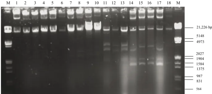
3.4. Plasmid profiles
The plasmid contents and the plasmid profiles of E.
faecalis isolates are shown in Table 2 and Figures 1 and
2, respectively. All the E. faecalis strains carried a certain number of plasmids with different molecular sizes. The number of plasmids varied between 2 and 5 with a molecular size of 21,226–1584 bp or larger.
3.5. Detection of virulence genes
The presence of virulence genes among the tested strains is shown in Table 2. All the E. faecalis isolates (n = 23) tested in this study, including 2 control strains, contained some sex pheromone determinants (cpd, ccf, and cad), some cytolysin determinants (cylM and cylA), the metalloendopeptidase gene (gelE), and the adhesion-encoding efaAfs gene. Certain E. faecalis isolates did not contain some virulence genes such as agg2 (22%), cob
(4%), cylB (17%), espfs (17%), and espfm (4%). None of the tested E. faecalis isolates contained the adhesion-encoding
efaAfm gene.
Furthermore, the tested E. faecalis isolates did not show phenotypic beta-hemolytic activity on blood agar with sheep blood.
4. Discussion
Enterococcus faecalis strains were predominantly isolated
from the breast milk and colostrum samples in this study (Table 2). Jimenez et al. (2008) reported similar results for colostrum samples. They also noted that skin
contamination was almost unavoidable during sampling of breast milk and colostrum for microbiological analysis. Therefore, they stated that there was no certainty as to the original location (internal mammary gland or skin) of the isolated bacteria. In this study, the samples were also collected by manual expression using sanitized hands and so there was no certainty about the original location of the isolated bacteria. As indicated by Jimenez et al. (2008), advanced studies will be required to explain the origin of the bacterial flora in breast milk and colostrum.
Antibiotic resistance is an important characteristic of enterococcal strains. Most of the tested E. faecalis strains are sensitive to ampicillin, penicillin G, chloramphenicol, vancomycin, gentamycin, and tetracycline. Similar to the results of the present study, penicillin, ampicillin, and vancomycin susceptibility and the absence of vanA and vanB genes were stated by Jimenez et al. (2008) in E.
faecalis strains isolated from colostrum. In this study, 9
strains showed multiple antibiotic resistance.
Although glycopeptide antibiotics like vancomycin are frequently a last resort for treatment of nosocomial infections with multidrug-resistant pathogens, resistance to these antibiotics is a source of concern. It has also been indicated that enterococci are opportunistic pathogens. However, enterococci that are not involved in infections are generally sensitive to clinically relevant antibiotics, including vancomycin (Franz et al., 2001; Jimenez et al., 2008).The enterococci strains isolated from the breast milk and colostrum samples in this study were sensitive to the relevant antibiotics, as was the case among the enterococci of the study by Jimenez et al. (2008).
M 1 2 3 4 5 6 7 8 9 10 11 12 13 14 15 16 17 18 M 21,226 bp 5148 4973 2027 1904 1584 1375 987 831 564
Figure 1. Plasmid profiles of Enterococcus faecalis isolates from human milk (1. A17–1, 2. A17–2, 3. A17–3, 4. A17–4, 5. A17–5, 6. A17–6, 7. A17–7, 8. A17–8, 9. A17–9, 10. A17–10, 11. A21–1, 12. A21–2, 13. A21–3 14. A40–1, 15. A40–2, 16. A40–3, 17. A40–4, 18. A40–5, M: Lambda DNA
Multiple-antibiotic–resistant E. faecalis isolates contained between 2 and 5 plasmids, usually with a molecular size of 21,226–1584 bp or larger. Coleri et al. (2004) determined that clinical enterococci isolates carried between 1 and 11 plasmids, ranging in size from 2.08 to 56.15 kb. They also reported plasmid-mediated antibiotic resistance in enterococci. Abriouel et al. (2006) indicated that virulence determinants and antibiotic resistance traits of enterococci may be plasmid-borne; therefore, their potential risk in food applications needed to be carefully evaluated. Fortunately, most of the multiply resistant strains were intermediate-level resistant to the relevant antibiotics in this study.
The genes coding for enterococcal surface protein and cell wall adhesin (espfs, espfm, and efaAfs) and sex pheromone determinants (cpd, cob, ccf, and cad) were identified in a large number of the E. faecalis strains in this study. Jimenez et al. (2008) reported that all the tested
E. faecalis strains isolated from colostrum contained the efaAfs gene and the sex pheromone determinants, with
the exception of a single isolate in which ccf could not be detected. An important property that is desirable in probiotic bacteria is the adhesion of the probiotic cells onto the surface of intestinal mucosa(Ouwehand et al., 1999). The presence of surface protein and cell wall adhesin genes in the enterococci strains may also reflect, at least partially, that the enterococci strains in the breast milk or colostrum samples have this important probiotic property.
Although beta-hemolytic activity was not present in any of the tested isolates, some isolates carried hemolysin-related genes (cylM, cylB, cylA). It was thought that the cytolysin determinants (cylM, cylB, cylA) behaved as silent genes in most nonhemolytic isolates (Eaton and Gasson, 2001; Semedo et al., 2003).
It was stated that the incidence of virulence determinants, antibiotic resistance pattern, or gene transfer potential appears to be strain-specific. Therefore, the safety of any enterococcal strain of clinical or industrial interest should be carefully and individually evaluated (Eaton and Gasson, 2001; Jimenez et al., 2008). It was found that there were similar structures among some of the tested
E. faecalis strains in terms of virulence genes, antibiotic
resistance, and plasmid profiles. Therefore, molecular-based studies such as protein profile studies and RAPD-PCR studies continue to be performed with the aim of finding out whether the E. faecalis strains isolated from the same human milk and colostrum samples have the same phylogenetic structure or not.
In conclusion, this study indicated that Enterococcus
faecalis is the predominant enterococcal species in breast
milk and colostrum. Although all the E. faecalis isolates carried a certain number of plasmids with different molecular sizes, the major strains were satisfactorily susceptible to the antibiotics, including vancomycin. The
vanA and vanB genes were not detected in any isolate
or in the control strains. None of the E. faecalis isolates contained the efaAfm gene, and some of the tested strains were found to be free from certain virulence determinants. Although a few E. faecalis strains as well as the positive control strain have some virulence factors, beta-hemolytic activity, a tested phenotypic characteristic, was not detected in any of the strains. Therefore, it is thought that further investigations are needed for the determination of virulence gene expressions in the phenotype of these strains. In addition, the enterococcal isolates from human milk may have potential as a functional or probiotic culture for the food industry.
Acknowledgments
The authors would like to thank the staff at Ankara University’s Biotechnology Institute Genomics Unit for 16S rDNA sequencing analysis of the isolates. We are also grateful to the staff at the Institute of Child Health of Hacettepe University and the Department of Obstetrics and Gynecology of Hacettepe University for supplying the human milk and colostrum samples.
1 2 3 4 5 6 M 21,226 bp 5148 4973 2027 1904 1584 1375 987 831 564
Figure 2. Plasmid profiles of Enterococcus faecalis isolates from human colostrum (1. K19–1, 2. K19–2,3. K19–3,4. K19–4,5. K19–5, 6. E. faecalis ATCC 29212 reference strain, M: Lambda DNA
References
Abriouel H, Ben Omar N, Lucas R, Martinez-Canamero M, Galvez A (2006). Bacteriocin production, plasmid content and plasmid location of enterocin P structural gene in enterococci isolated from food sources. Lett Appl Microbiol 42: 331–337.
Albesharat R, Ehrmann MA, Korakli M, Yazaji S, Vogel RF (2011). Phenotypic and genotypic analyses of lactic acid bacteria in local fermented food, breast milk and faeces of mothers and their babies. Syst Appl Microbiol 34: 148–155.
Anderson DG, McKay LL (1983). Simple and rapid method for isolating large plasmid DNA from lactic Streptococci. Appl Environ Microb 46: 549–552.
Brede DA, Snipen LG, Ussery DW, Nederbragt AJ, Nes IF (2011). Complete genome sequence of the commensal Enterococcus
faecalis 62, isolated from a healthy Norwegian infant. J
Bacteriol 193: 2377–2378.
Canzek Majhenic A, Rogelj I, Perko B (2005). Enterococci from Tolminc cheese: population structure, antibiotic susceptibility and incidence of virulence determinants. Int J Food Microbiol 102: 239–244.
Charteris WP, Kelly PM, Morelli L, Collins JK (1998). Antibiotic susceptibility of potentially probiotic Lactobacillus species. J Food Protect 61: 1636–1643.
CLSI (2006). Clinical and Laboratory Standards Institute. Performance Standards for Antimicrobial Disk Susceptibility Tests: Approved Standard, 9th ed. M2-A9, 26. Wayne, PA, USA: CLSI.
Coleri A, Cokmus C, Ozcan B, Akcelik M, Tukel C (2004). Determination of antibiotic resistance and resistance plasmids of clinical Enterococcus species. J Gen Appl Microbiol 50: 213– 219.
Çıtak S, Yücel N, Orhan S (2004). Antibiotic resistance and incidence of Enterococcus species in Turkish white cheese. Int J Dairy Technol 57: 27–31.
De Vuyst L, Foulquie Moreno MR, Revets H (2003). Screening for enterocins and detection of hemolysin and vancomycin resistance in enterococci of different origins. Int J Food Microbiol 84: 299–318.
Dutka-Malen S, Evers S, Courvalin P (1995). Detection of glycopeptide resistance genotypes and identification to the species level of clinically relevant enterococci by PCR. J Clin Microbiol 33: 24–27.
Eaton TJ, Gasson MJ (2001). Molecular screening of Enterococcus virulence determinants and potential for genetic exchange between food and medical isolates. Appl Environ Microb 67: 1628–1635.
Franz CMAP, Holzapfel WH, Stiles ME (1999). Enterococci at the crossroads of food safety? Review. Int J Food Microbiol 47: 1–24.
Franz CMAP, Muscholl-Silberhorn AB, Yousif NMK, Vancanneyt M, Swings J, Holzapfel WH (2001). Incidence of virulence factors and antibiotic resistance among enterococci isolated from food. Appl Environ Microb 67: 4385–4389.
Franz CMAP, Stiles ME, Schleifer KH, Holzapfel WH (2003). Enterococci in foods - a conundrum for food safety. Review article. Int J Food Microbiol 88: 105–122.
Foulquie Moreno MR, Sarantinopoulos P, Tsakalidou E, De Vuyst L (2006). The role and application of enterococci in food and health. Review. Int J Food Microbiol 106: 1–24.
Garcia MC, Rodriguez MJ, Bernardo A, Tornadijo ME, Carballo J (2002). Study of enterococci and micrococci isolated throughout manufacture and ripening of San Simon cheese. Food Microbiol 19: 23–33.
Giraffa G, Olivari AM, Neviani E (2000). Isolation of vancomycin-resistant Enterococcus faecium from Italian cheeses. Food Microbiol 17: 671–677.
Hugas M, Garriga M, Aymerich MT (2003). Functionality of enterococci in meat products. Int J Food Microbiol 88: 223– 233.
Jeurink PV, van Bergenhenegouwen J, Jiménez E, Knippels LMJ, Fernández L, Garssen J, Knol J, Rodríguez JM, Martín R (2013). Human milk: a source of more life than we imagine. Benef Microbes 4: 17–30.
Jimenez E, Delgado S, Fernandez L, Garcia N, Albujar M, Gomez A, Rodriguez JM (2008). Assessment of the bacterial diversity of human colostrum and screening of staphylococcal and enterococcal populations for potential virulence factors. Res Microbiol 159: 595–601.
Jurkovic D, Krizkova L, Dusinsky R, Belicova A, Sojka M, Krajcovic J, Ebringer L (2006). Identification and characterization of enterococci from bryndza cheese. Lett Appl Microbiol 42: 553–559.
Klein G (2003). Taxonomy, ecology and antibiotic resistance of enterococci from food and gastro-intestinal tract. Review. Int J Food Microbiol 88: 123–131.
Lara-Villoslada F, Olivares M, Sierra S, Rodriguez JM, Boza J, Xaus J (2007). Beneficial effects of probiotic bacteria isolated from breast milk. Brit J Nutr 98: 96–100.
Lopez-Alarcon M, Villalpando S, Fajardo A (1997). Breast-feeding lowers the frequency and duration of acute respiratory infection and diarrhea in infants under six months of age. J Nutr 127: 436–443.
Mannu L, Paba A, Daga E, Comunian R, Zanetti S, Dupre I, Sechib LA (2003). Comparison of the incidence of virulence determinants and antibiotic resistance between Enterococcus
faecium strains of dairy, animal and clinical origin. Int J Food
Microbiol 88: 291–304.
Martin R, Langa S, Reviriego C, Jimenez, E, Marin ML, Xaus J, Fernandez L, Rodriguez JM (2003). Human milk is a source of lactic acid bacteria for the infant gut. J Pediatr 143: 754–758. Martin R, Langa S, Reviriego C, Jimenez E, Marin ML, Xaus J,
Fernandez L, Rodriguez JM (2004). The commensal microflora of human milk: new perspectives for food bacteriotherapy and probiotics, Trends Food Sci Tech 15: 121–127.
Martin R, Olivares M, Marin ML, Fernandez L, Xaus J, Rodriguez JM (2005). Probiotic potential of 3 lactobacilli strains isolated from breast milk. J Hum Lact 21: 8–17.
Miteva VI, Abadjieva AN, Grigorova RT (1991). Differentiation among strains and serotypes of Bacillus thuringiensis by M13 DNA fingerprinting. J Gen Microbiol 137: 593–600.
Oryaşın E, Bıyık HH, Başbülbül G, Bozdoğan B (2013). Antimicrobial susceptibility patterns of environmental and hospital isolations of enterococci in Aydın. Turk J Biol 37: 514–519.
Ouwehand AC, Kirjavainen PV, Grönlund MM, Isolauri E, Salminen SJ (1999). Adhesion of probiotic micro-organisms to intestinal mucus. Int Dairy Journal 9: 623–630.
Özden Tuncer B, Ay Z, Tuncer Y (2013). Occurrence of enterocin genes, virulence factors, and antibiotic resistance in 3 bacteriocin-producer Enterococcus faecium strains isolated from Turkish tulum cheese. Turk J Biol 37: 443–449.
Peters J, Mac K, Wishmann-Shauer H, Klein G, Ellerbroek L (2003). Species distribution and antibiotic resistance patterns of enterococci isolated from food of animal origin in Germany. Int J Food Microbiol 88: 311–314.
Poeta P, Costa D, Rodrigues J, Torres C (2006). Antimicrobial resistance and the mechanisms implicated in faecal enterococci from healthy humans, poultry and pets in Portugal. Int J Antimicrob Ag 27: 131–137.
Reviriego C, Eaton T, Martín R, Jiménez E, Fernández L, Gasson MJ, Rodríguez JM (2005). Screening of virulence determinants in
Enterococcus faecium strains isolated from breast milk. J Hum
Lact 21: 131–138.
Sánchez Valenzuela A, Ben Omar N, Abriouel H, López RL, Veljovic K, Cañamero MM, Milan Topisirovic KL, Gálvez A (2009). Virulence factors, antibiotic resistance, and bacteriocins in enterococci from artisan foods of animal origin. Food Control 20: 381–385.
Semedo T, Santos MA, Lopes MFS, Figueiredo Marques JJ, Barreto Crespo MT, Tenreiro R (2003). Virulence factors in food, clinical and reference enterococci: a common trait in the genus? Syst Appl Microbiol 26: 13–22.
Wright AL, Bauer M, Naylor A, Sutcliffe E, Clark L (1998). Increasing breastfeeding rates to reduce infant illness at the community level. Pediatrics 101: 837–844.