An unpredictable complication during
percuta-neous endoscopic tube placement: esophageal
perforation
A percutaneous endoscopic gastrostomy (PEG) was performed to provide an enter-al feeding route. Major and minor compli-cations related to this procedure include, aspiration, wound infections, necrotizing fasciitis, peritonitis, abscess, peristomal leakage, pneumoperitoneum, ileus, porto-mesenteric venous gas, colon injury, gastrocolocutaneous fistulae, abdominal wall bleeding, intra-abdominal bleeding, gastric outlet obstruction, inadvertent gastrostomy tube removal, spleen trauma, clogged PEG tube, tumor implantation at PEG site, and buried bumper syndrome [1, 2].
An 88-year-old woman with Alzheimer’s disease required PEG tube placement as an alternative feeding route. The proce-dure was performed using the
pull-through technique (Flexiflo kit; Abbott, Sligo, Ireland). On endoscopy, tertiary contractions occurred in the esophagus. The procedure was uneventful until moments before the insertion of the tube into the esophagus through the mouth. A moderate resistance was felt while pulling the tube wire. Inspection of the tube after removal revealed deformation of its tip (
●
" Fig. 1). A new tube was inserted andthen the upper esophagus was checked endoscopically. Severe lacerations and minimal bleeding were observed in the upper part of the esophagus (
●
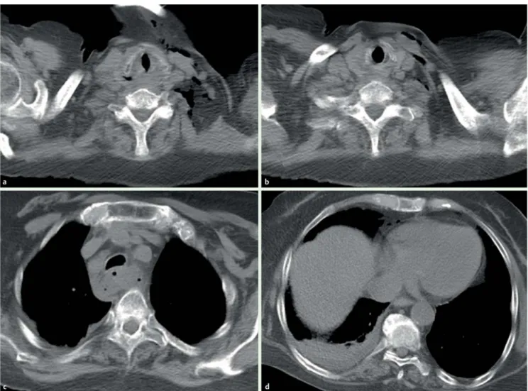
" Fig. 2).Shortly thereafter, the patient presented dyspnea, followed by edema and crepita-tion in the neck. Thoracic computed tomography indicated the presence of air in the mediastinum (
●
" Fig. 3). The patientFig. 1 The deformation of the tube tip.
Fig. 2 Minimal bleeding and laceration in the esophagus.
Fig. 3 Axial noncontrast-enhanced computed tomography images after the procedure showed free air bubbles in the left cervical region (a, b), and the peri-esophageal (c) and right paracardiac (d) spaces.
Cases and Techniques Library (CTL)
E413
Agan Ahmet Faruk. Esophageal perforation during PEG tube placement… Endoscopy 2014; 46: E413–E414
presented mild peripheral cyanosis, tachycardia, tachypnea, and a subfebrile rise in body temperature. She was cared for in the intensive care facility.
A few hours later, she reported feeling better. Vital signs returned to normal. Feeding via the PEG tube was initiated 2 days later, and the tube functioned prop-erly. The patient was hospitalized for 32 days after the procedure.
This report describes the occurrence of an esophageal perforation during the inser-tion of a PEG tube. As this complicainser-tion was detected early, conservative treat-ment was sufficient for its successful reso-lution, and the patient healed satisfac-torily.
Endoscopy_UCTN_Code_CPL_1AH_2AI Competing interests: None
Ahmet Faruk Agan
Department of Gastroenterology, Medical School, Medipol University, Istanbul, Turkey
References
1 Jarnagin WR, Duh QY, Mulvihill SJ et al. The efficacy and limitations of percutaneous en-doscopic gastrostomy. Arch Surg 1992; 127: 261–264
2 Rimon E. The safety and feasibility of percu-taneous endoscopic gastrostomy placement by a single physician. Endoscopy 2001; 33: 241–244
Bibliography DOI http://dx.doi.org/ 10.1055/s-0034-1377403 Endoscopy 2014; 46: E413–E414 © Georg Thieme Verlag KG Stuttgart · New York ISSN 0013-726X
Corresponding author Ahmet Faruk Agan, MD Department of Gastroenterology Medical School Medipol University Haydrapasa-Harem Yolu Kadikoy 34718 Istanbul Turkey Fax: +90-216-5454511 [email protected]
Agan Ahmet Faruk. Esophageal perforation during PEG tube placement… Endoscopy 2014; 46: E413–E414