22q13.3 Deletion Syndrome: An Underdiagnosed Cause of
Mental Retardation
22q13.3 Delesyon Sendromu: Mental Retardasyonun Az Tan
ınan Bir Nedeni
İlknur Erol1
, Özge Sürmeli Onay2, Zerrin Yılmaz3, Özge Özer3, Füsun Alehan4, Feride İffet Şahin5
1Baskent University Faculty of Medicine, Department of Pediatrics, Neurology Division, ADANA
2Baskent University Faculty of Medicine, Department of Pediatrics , 3Department of Genetics, 3Department of
Medical Genetics, 5Department of Pediatrics, Neurology Division, ANKARA
Cukurova Medical Journal 2015;40(1):169-173.
ABSTRACT
Phelan-McDermid syndrome, also known as 22q13.3 deletion syndrome, is characterized by global developmental delay, absent or delayed speech, generalized hypotonia, and minor physical anomalies. The deletion typically involves the terminal band 22q13.3 and has been associated with both familial and de-novo translocations. We report the case of an 11-year-old Turkish girl with 22q13.3 deletion syndrome presenting with repeated seizures during the course of a rubella infection. We also review the clinical features of 22q13.3 deletion syndrome and emphasize the importance of considering a rare microdeletion syndrome for idiopathic mental retardation when results of a routine karyotype analysis are normal. To the best of our knowledge, this is the first reported case of a Turkish patient with isolated 22q13.3 deletion syndrome.
Key words: 22q13.3 deletion syndrome, Aphasia, Epilepsy, Mental retardation
ÖZET
Phelan-McDermid sendromu olarak da bilinen 22q13.3 delesyon sendromu, global gelişme geriliği, gecikmiş ve gelişmemiş konuşma, jenaralize hipotoni ve minör fiziksel anomaliler ile karekterizedir. Delesyon tipik olarak 22q13.3'ün terminal bandını içermekte ve ailesel veya denova translokasyon ile ilişkilidir. Bu yazıda 11 yaşında, Rubella enfeksiyonu esnasında tekrarlayan nöbetlerle başvurduğu sırada 22q13.3 delesyon sendromu tanısı konulan bir türk kız hasta sunulmuştur. Ayrıca 22q13.3 delesyon sendromun klinik bulgularıda derlenerek, karyotip analizi normal olan idyopatik mental retardasyonu olan olgularda microdelesyon sendromlarının önemine dikkat çekilmiştir. Bilgilerimiz dahilinde, bu olgu ilk izole 22q13.3 delesyon sendromu olan Türk hastadır.
Anahtar kelimeler: 22q13.3 delesyon sendromu, afazi, epilepsi, mental retardasyon
INTRODUCTION
Recent advances in genetic testing can help provide a specific diagnosis for children born with congenital anomalies and who show subsequent developmental delay. The introduction of subtelomeric fluorescence in-situ hybridization (FISH) analysis has revealed 22q13.3 deletion as
a relatively widespread and underdiagnosed cause of mental retardation1,2. However, the true prevalence of 22q13.3 deletion syndrome remains unknown.
The 22q13.3 deletion syndrome is
characterized by neonatal hypotonia, global developmental delay, absent to severely delayed
speech, autistic behavior, normal to accelerated growth, and minor physical anomalies including dolichocephaly, abnormal ears, ptosis, puffy eyelids, wide nasal bridge, puffy cheeks, pointed chin, dysplastic toe nails, and relatively large hands1,2. Most individuals have severe to profound mental retardation, while a minority are in the mild to moderate range. The deletion typically involves the terminal band of the 22q13.3 region and is usually the result of de novo translocation of the paternal chromosome, although familial inheritance has been described1-3. In general, the severity of the neurological deficit is not well correlated with the deletion span but rather on the deletion of the autism-associated SH3 and multiple ankyrin repeat domains 3 protein (Shank3)4.
CASE REPORT
An 11-year-old girl with mental and motor retardation was admitted to our hospital with fever, rash, and seizures. She had been febrile for four days and had maculopapular lesions for the previous three days. On the day of admission, she had an afebrile generalized, tonic-clonic seizure that lasted nearly 10 minutes.
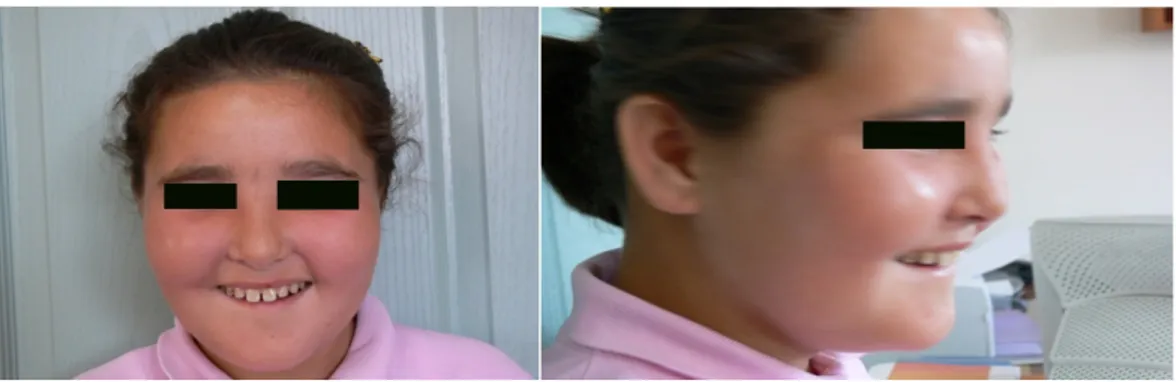
The patient was the first child of nonconsanguineous parents; her mother was 30 years of age and her father was 38. She had been born at term by vaginal delivery. Her birth weight, length, and head circumference were not recorded. The pregnancy was uncomplicated, and there was no history of maternal teratogenic agent exposure. Her neuromotor development was delayed and she had marked mental retardation. She was unable to talk, except for a few words. The patient had been diagnosed at another center with idiopathic mental retardation associated with severely compromised language development and dysmorphic features. On admission, her weight was 42 kg (within the 50th–75th percentile for her age), her length was 150 cm (within the 75th–90th percentile for her age), and her head circumference was 52 cm. Dysmorphic features included an elongated face with thick eyebrows, a
smooth philtrum, a thin upper lip, narrow palpebral fissures, broad nasal root, and mild micrognathia (Figures 1A-B). On neurologic examination, generalized tonic-clonic seizure activity with a loss of consciousness was observed. The results of an ophthalmologic examination were unremarkable. There was no focal neurologic deficit or meningeal irritation. Laboratory analyses determined that serum C-reactive protein (3 mg/L), erythrocyte sedimentation (15 mm/h), complete blood count, thyroid functioning, and standard blood chemistry values (including lactate, pyruvate, ammonia, and amino acids) were within normal ranges. Neither computed tomography nor magnetic resonance imaging revealed any gross structural abnormalities in the brain. An electroencephalogram showed diffuse slowing without an epileptic abnormality. The results of a cerebrospinal fluid examination were normal. Tandem mass spectrometry of the blood was negative for organic acid and acylcarnitine abnormalities. Results of cold agglutinin, Gruber-Widal agglutination, and brucella agglutination tests were all negative. Acyclovir and ceftriaxone were given empirically. Serologic tests were negative for cytomegalovirus, herpes virus, and parvovirus B19, but positive for Rubella IgM and IgG on the third day of admission. Cerebrospinal fluid serologic titers for Rubella Ig M and G were negative.
Phenytoin was given at a bolus dose of 20 mg/kg and a maintenance dosage of 6 mg/kg/day after the seizure; however, the seizure reoccurred several times during the next two days. Valproic acid was added to the regimen, and seizure control was achieved. The patient began to improve on the fourth day of hospitalization. The antibiotic and antiviral treatment was completed in 10 days. Results of the patient’s hearing tests and echocardiography were normal. Her global IQ score was 45 as assessed by the Wechsler Intelligence Scale for Children-Revised.
Cytogenetic analyses performed on a peripheral blood lymphocyte sample by the
Giemsa-trypsin banding method revealed a normal karyotype.5 Dysmorphic findings of the patient led us to perform a FISH analysis to test for DiGeorge/velocardiofacial syndrome (VCFS). The probe, an LSI TUPLE1 (HIRA) DiGeorge region probe with an LSI ARSA control probe (Vysis Inc.,
Downers Grove, IL, USA) revealed 22q11.2 (HIRA×2), del(22)(q13.3)(ARSA-). We therefore diagnosed 22q13.3 deletion syndrome as the cause of the patient’s delayed speech, mental and motor retardation, and dysmorphic features.
Figure 1 A-B. Frontal and lateral view of the patient at 11 years of age showing the elongated face with thick eyebrows, smooth philtrum, thin upper lip, narrow palpebral fissures, broad nasal root, and mild micrognathia.
DISCUSSION
The 22q13.3 deletion syndrome is characterized by global developmental delay, generalized hypotonia, absent or delayed speech, normal to advanced growth, and minor anomalies of the face, head, ears, hands, and feet.1 Seizures have been described in 27% of children with the 22q13.3 deletion.2 We report the case of an 11-year-old child with 22q13.3 deletion syndrome presenting with repeated seizures during the course of a rubella infection.
The first case of pure 22q13.3 deletion was described in 1985 by Watt and associates.3 in a 14-year-old boy exhibiting profound mental retardation, absent speech, and minor dysmorphic features. Phelan and associates6 were the first to describe generalized hypotonia in association with the terminal deletion of 22q13.3 in a newborn boy. They later identified the breakpoint at 22q13.31. At 26 months, this boy had normal growth but with developmental delay and diffuse hypotonia. Prior to 1994, only 8 patients had been diagnosed with this disorder. In 1994, Nesslinger and associates suggested that the 22q13.3 deletion was a recognizable clinical syndrome characterized by
global developmental delay, severe delay in expressive speech, normal or accelerated growth, and generalized hypotonia with minor facial anomalies.7 The present case shared many of these features but exhibited normal growth and head circumference.
The largest study to date was reported by Phelan and associates in 20011. They defined 37 individuals with 22q13.3 deletion and compared them with 24 previously published cases. The major findings of global developmental delay, generalized hypotonia, absent or delayed speech, and normal to advanced growth were confirmed by this study. The authors also identified common minor anomalies of the face, head, ears, hands, and feet. Only 32% of the individuals in this sample had prior genetic evaluations and normal standard chromosome analyses, suggesting that 22q13.3 deletion syndrome remains underdiagnosed. Similarly, the present patient had a normal standard chromosome analysis but was never tested for microdeletions.
Seizures have been described in 27% of children with 22q13.3 deletion2 but there are no characteristic electroencephalographic features that distinguish 22q13.3 deletion syndrome. High 171
voltage polymorphic delta activity and parietotemporal spikes and waves have been reported in patients with the 22q13.3 deletion syndrome but these are not distinguishing features8,9. Similarly, our patient had unremarkable changes in her electroencephalogram, and her seizures responded well to phenytoin and valproic acid. The patient had been examined previously at another hospital for mental retardation, severely compromised language development, and dysmorphic features, but 22q13.3 deletion syndrome was not considered until she was admitted to our institute with repeated seizures during rubella infection.
Diagnosis of the 22q13.3 deletion syndrome is established only by demonstrating a deletion or disruption of 22q13.3 by routine chromosome analysis or FISH testing1,2. Although our patient fulfilled the diagnostic criteria for 22q13.3 deletion syndrome (severely compromised language development, dysmorphic features, mental retardation, and seizures), she was diagnosed serendipitously after the telomeric control probe signal at 22q13.3 was absent on a FISH test for DiGeorge/VCFS (22q11.2 deletion syndrome). Similarities between VCFS 22q11.2 deletion syndrome and 22q13.3 deletion syndrome include hypotonia, epicanthal folds, narrow palpebral fissures, broad nasal root, speech delay, renal abnormalities, and developmental delay2,10-12. The neurologic problems observed in VCFS are not as severe as those in the 22q13.3 deletion syndrome13. Hypotonia, epicanthal folds, narrow palpebral fissures, broad nasal root, and speech delay were the most significant symptomes in our patient, so we performed a FISH analysis for the 22q11.2 deletion. Velocardiofacial syndrome is frequently associated with cardiac and/or palatal defects, immune deficiency, and hypocalcemia, however, and these symptoms are absent in 22q13.3 deletion syndrome including the case described here. The 22q11.2 deletion syndrome is diagnosed by FISH using the N25 or Tuple probe set with ARSA as the control probe because ARSA
maps to 22q13. The N25 or Tuple probe set has detected the deletion of 22q13, resulting in the diagnosis of the 22q13.3 deletion syndrome in several individuals tested for VCFS1,2,4,9,10. Precht and associates serendipitously also detected 22q13.3 deletion in one child by a FISH analysis for 22q11.2 DiGeorge/ VCFS10.
In 1995, Flint and associates developed a strategy to simultaneously screen 28 subtelomeric regions of the human chromosomes, including 22q11. They studied 99 patients with idiopathic mental retardation of varying degrees and detected submicroscopic deletion of 22q13.3 in two patients. The authors emphasized the importance of analyzing variations involving the ends of chromosomes, and in particular, testing for the 22q13.3 deletion in individuals with idiopathic mental retardation.
Determining the etiology of idiopathic mental retardation is a frequent and challenging problem in pediatric neurology. Clinicians should consider screening for 22q13.3 deletion syndrome in any child with hypotonia, persistent lack of speech, and global developmental delay, particularly if the routine karyotype is normal. There is no specific therapy for 22q13.3 deletion syndrome, but early rehabilitative and educational interventions improve the prognosis and considerable progress has been made in the social adjustment of 22q13.3 deletion syndrome patients.
Koç A et al. reported a patient with ring chromosome 22 associated with 22q13.3 deletion. Their patient had the major features of ring chromosome 22, including mental retardation, hypotonia, motor delay, microcephaly, dysplastic large ears, lack of speech, and hyperactivity disorder14. Since our patient had normal karyotype analysis, she is the first reported case of a Turkish patient with isolated 22q13.3 deletion syndrome.
In conclusion, although the dysmorphic features observed in this condition are nonspecific, they can aid in the differential diagnosis of children with developmental delay, hypotonia, marked speech and language disability, autistic features,
and normal growth and head circumference. We recommend high resolution chromosome analysis, FISH studies or molecular analyses to exclude this diagnosis in patients with idiopathic mental retardation, especially when associated with severely compromised language development.
REFERENCES
1. Phelan MC, Rogers RC, Saul RA, et al. 22q13 deletion syndrome. Am J Med Genet. 2001;101: 91-9.
2. Havens JM, Visootsak J, Phelan MC, Graham JM Jr. 22q13 deletion syndrome: an update and review for the primary pediatrician. Clin Pediatr. 2004;43:43-53.
3. Watt JL, Olson IA, Johnston AW, Ross HS, Couzin DA, Stephen GS. A familial pericentric inversion of chromosome 22 with a recombinant subject illustrating a 'pure' partial monosomy syndrome. J Med Genet. 1985;22:283-7.
4. Wilson HL, Wong AC, Shaw SR, et al. "Molecular characterisation of the 22q13 deletion syndrome supports the role of haploinsufficiency of SHANK3/PROSAP2 in the major neurological symptoms". J. Med. Genet. 2003;40:575–84.
5. Verma RS, Babu A. Human chromosomes. In: Verma and Babu A (eds). Tissue Culture Techniques and Chromosome Preparation. McGraw-Hill, New York, 1995;6-71.
6. Phelan MC, Rogers RC, Stevenson RE. A de novo terminal deletion of 22q. Am J Hum Genet. 1988;43:A118.
7. Nesslinger NJ, Gorski JL, Kurczynski TW, et al. Clinical, cytogenetic, and molecular characterization of seven patients with deletions of chromosome 22q13.3. Am J Hum Genet. 1994;54:464-72.
8. Anderlid BM, Schoumans J, Annerén G, et al. FISH-mapping of a 100-kb terminal 22q13 deletion. Hum Genet. 2002;110:439-43.
9. Herman GE, Greenberg F, Ledbetter DH. Multiple congenital anomaly/mental retardation (MCA/MR) syndrome with Goldenhar complex due to a terminal del(22q). Am J Med Genet. 1988;29:909-15.
10. Precht KS, Lese CM, Spiro RP, et al. Two 22q telomere deletions serendipitously detected by FISH. J Med Genet. 1998;35:939-42.
11. Flint J, Wilkie AO, Buckle VJ, Winter RM, Holland AJ, McDermid HE. The detection of subtelomeric chromosomal rearrangements in idiopathic mental retardation. Nat Genet. 1995;9:132-40.
12. Ghariani S, Dahan K, Saint-Martin C, Kadhim H, Morsomme F, Moniotte S et al. Polymicrogyria in chromosome 22q11 deletion syndrome. Eur J Paediatr Neurol. 2002;6:73-7.
13. Hay BN. Deletion 22q11: spectrum of associated disorders. Semin Pediatr Neurol. 2007;14:136-9.
14. Koç A, Karaer K, Ergün MA, et al. A case with a ring chromosome 22. Turk J Pediatr. 2008;50:193-6
Yazışma Adresi / Address for Correspondence:
Dr.Ilknur Erol
Baskent University Faculty of Medicine Division of Pediatric Neurology
Adana Teaching and Medical Research Center, ADANA
E-mail: [email protected] Geliş tarihi/Received on : 04.06.2014 Kabul tarihi/Accepted on: 01.08.2014