ABSTRACT
Background: The incidence of sternal dehiscence follow-ing cardiothoracic surgery via sternotomy is rare. It causes serious patient dissatisfaction and leads to higher hospital costs. For years, each clinic has made efforts to reduce this complication. Here, we aimed to summarize our techniques to prevent dehiscence.
Material: This retrospective study included two groups operated via median sternotomy from March 2009 to May 2019. The first g roup i ncluded 1 ,105 c onsecutive p atients who only received sternum wire for sternum closure from March 2009 to October 2013. The second group included 1,559 consecutive patients operated from January 2014 to May 2019; preventive closure techniques were performed for predefined high-risk patients in this group. These closure techniques included polyglyconate (Maxon) or simple longi-tudinal reinforced sutures, sternal cable or sternoband, ster-nal plate, and Robiscek technique.
Results: All patients in Group 1, and 63.8% (995/1559) patients in Group 2 received sternal wire only (P < .001). In Group 2, we applied preventive closure techniques to 564 (36.2%) patients. There was no sternal dehiscence in Group 2, whereas 29 (2.6%) patients postoperatively suffered ster-nal dehiscence in Group 1; this was statistically significant (P = .001, OR:85.5, 95%CI:5.22-1400.4). The overall inci-dence of mediastinitis was 0.94%. The inciinci-dence significantly was lower in Group 2 (P = .004, OR:3.6, 95%CI:1.52-8.82). Sternum-related mortality in Group 2 also was lower (0.54% versus 0.06%, P = .048, OR:8.5, 95% CI: 1.02-70.75).
Conclusion: Sternal dehiscence can be avoided by care-ful perioperative risk assessment and enhanced closure tech-niques. The same special consideration may significantly reduce mediastinitis and sternal-related mortality.
INTRODUCTION
Sternal dehiscence (SD) is a rare complication of median sternotomy and can be seen in almost all cardiac and tho-racic surgery tertiary centers. Chest pain and discomfort caused by SD are the leading cause of severe postoperative patient dissatisfaction and readmission [Olbrecht 2006].
Since the sternum repair procedure is more troublesome than the first closure and requires general anesthesia, it poses a risk in patients with poor cardiac and pulmonary function. Furthermore, the extended hospital stay may burden health providers and increase loss of labor for patients. Thus, pre-venting the development of median sternotomy complica-tions is rewarding to curb costs incurred by both patients and health care systems [Mehaffey 2018].
The incidence of SD was reported from 0.25% to 5% in the different studies [Eklund 2006; Gårdlund 2002; Kamiya 2012]. However, the exact incidence still is unknown because of patient heterogeneity and vast differences between the centers. SD commonly occurs in the first month after the operation; but, it also can be seen after the first month [Royse 2020]. In delayed dehiscence, the sternal repair is more complicated, due to the adhesions of underlying structures. Therefore, it is of significant importance to take the necessary measures to prevent these scenarios [Olbrecht 2006].
The most effective way to eliminate SD is to identify risk factors well and apply the most appropriate preven-tive measures. SD can be aseptic or caused by infection [Robicsek 2000]. Infection-related SD largely can be
Sternum Dehiscence: A Preventable Complication of Median Sternotomy
Emin Can Ata, MD, Metin Onur Beyaz, MD
Department of Cardiovascular Surgery, Medipol Mega University Hospital, Istanbul, Turkey
Received May 31, 2020; accepted June 15, 2020.
Correspondence: Emin Can Ata, Medipol Mega University Hospital, Department of Cardiovascular Surgery, Istanbul, Turkey; 90-506-3893476; fax: 90-212-460-7070 (e-mail: [email protected]).
Figure 1. Chest X-ray images of different sternum closure techniques. A, standard eight wire closure; B, sternoband; C, bilateral longitudinal single wire reinforcement
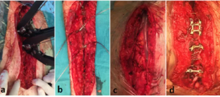
Figure 2. Operative view of the different sternum closure. A and B, sternal cable placement; C and D, fractured sternum and plate fixation
prevented by strict follow up of sterilization rules, antibiotic treatment guidelines, and DM management [Engelman 2007; Lador 2012]. However, there is far more risk factor, which leads to aseptic SD. Most of the time, these risks are patient- or technical-related. Patient-related risks include DM, obesity, COPD, osteoporosis, advanced age, and renal insufficiency [Abu-Omar 2017; Fu 2016; Schimmer 2008]. Technical-related factors include off-midline sternotomy, bilateral IMA use, insufficient wiring, and the excessive opening of the thoracic retractor. However, in rare cases, unpredictable scenarios, such as wire breakage or fragmentation of the sternum due to saw effect of wire, may cause SD [Schimmer 2008] (Table 1).
Although several specific techniques can reduce sternal complications, there is no study to show altogether how to avoid it. In this study, we aimed to summarize several differ-ent techniques we used to eliminate SD in line with the risk stratification we have determined in our clinic.
MATERIALS AND METHODS
Ethical consideration: This retrospective study was car-ried out after the approval of the Istanbul Medipol University Ethics committee (Number: 10840098-604.01.01-E.65343). The hospital authority accepted the study results.
Study concepts – study design: This retrospective mul-ticenter study was performed on all patients undergoing median sternotomy for cardiothoracic surgery at three car-diac centers from March 2009 to May 2019. A total of 2,664 eligible patients were identified out of 3,514 patients operated by the same surgical team. Eligible patients were investigated under two groups by the date of the operation.
Group 1 consisted of 1,105 consecutive patients, who only received a sternum wire from March 2009 to October 2013 for sternum closure. At the end of 2013, we created a serial of risk stratification (Table 1) and took preventive measures with the hope of reducing sternal complications. Since then, the ster-num closure process was performed using additional materi-als in compliance with this risk stratification. Group 2 included 1,559 consecutive patients in this context operated from January 2014 to May 2019. In this study, we mainly focused on this group. X-ray images, used wires, and other materials of all patients were reviewed. The two groups were analyzed in terms of aseptic SD, mediastinitis, and sternal-related mortality.
Inclusion: All patients who underwent on-pump or off-pump cardiac operations, cardiac reoperations, non-cardiac operations, emergency cardiac trauma operations via median sternotomy through March 2009 to May 2019 were included.
Exclusion: Patients operated through thoracotomy, mini-mally invasive and robotic surgery, and those who required more than two reentries were not included in this study. Patients who did not come for a regular check-up after sur-gery, and patients resulting in mortality within the first six months also were excluded from the study.
Identification of SD: The majority of SD within the first month after surgery were diagnosed by simple palpation. In delayed suspicious patients, the thoracic CT scan was per-formed to confirm the diagnosis.
Figure 3. Maxon suture reinforcement. A, Maxon sutures placement; B, after sutures tied
Figure 4. Dehicence and mediastinitis rates of the two groups
Table 1. Risk stratification for sternal dehiscence of our clinic
Patient related Technical related Unpredictable risks
Advanced age Off-midline sternotomy Wire break Osteoporosis Insufficient wire Wire cut sternum COPD Retractor opening Postoperative delirium Chronic cough Prolonged operation Postoperative reentry Improper movement Bilateral IMA harvest Prolonged immobilization Uncontrolled DM* Inadequate wound
handling Morbid obesity
Reoperation
Identification of mediastinitis: The diagnosis was con-firmed by the open wound or closed subcutaneous abscess with or without dehiscence and positive wound culture results.
Sternum closure techniques – standard wire closure: This technique includes two single wire at manubrium, one inverted figure-of-eight wire through 1st and 2nd intercostal, and another four single wire through the rest of each inter-costal space. This technique was applied to all of the patients in Group 1 (Figure 1A).
Non-standard preventive closure: Preventive closure tech-niques, materials, and indications, which differ from standard closures, are summarized below:
Sternobant and 4-6 single wire through the manubrium and intercostals (Figure 1B). This technique is preferred in patients with an overly rigid sternum and BSA> 30.
Standard wire besides additional bilateral longitudinal reinforced single wire tied upper and below the sternum. (Figure 1C) This technique especially is preferred in patients with osteoporosis and the elderly, drug abusers, and patients under antipsychotic medication.
Two figure-of-eights sternal cable and 4-6 single wire through the manubrium and intercostals (Figures 2A and 2B). This technique is preferred in patients with morbid obesity and rigid sternum.
Sternal plates are preferred in redo patients with frag-mented sternum fractures beside the adequate number of wires (Figures 2C and 2D).
Additional 3-6 polyglyconate (Covidien, Maxon 0, 75cm, absorbable monofilament) sutures besides the intercostal stan-dard single wires. This technique is applied for most of the severe osteoporotic elderly and obesity (Figure 3A and 3B).
Robiscek or modified Robiscek technique in bilateral ster-nal fractures and prolonged redo operation.
More than eight wire in patients of BMI>30kg/m2, and BSA > 2m2.
Above is our general preference, but we have no strict rules in the choice of any technique; in particular cases, it was determined by the current condition of the patient and equip-ment availability.
Postoperative following: Broad-spectrum antibiotics were given for two days after the operation. Patients provided respiratory physiotherapy and early mobilization in ICU. Patients were educated on sternum protection prior to dis-charge. Patients were followed for a minimum of six months and then directed to the primary cardiologist. Symptomatic patients (chest pain and dyspnea) with SD were immediately treated by minor debridement and rewiring. SD patients without any complaints were followed up for 2-3 weeks and ordered a sternum vest. If they did not have any sign of ster-num stabilization, they also were treated in the same way. Those with mediastinitis were applied with vacuum-assisted closure (VAC) in addition to anti biotherapy, according to the wound microgram culture result [Norman 2020]. Necrotic tissue was debrided, and the sternum was rewired after the infection was healed, and the sternum was covered with major pectus muscle [Elassal 2020].
Data collection: The patients’ medical records were col-lected in a predefined standard form and transferred to the computer. Baseline characteristics, operative variables and major complications, such as postoperative wound compli-cation, reexploration, sternal dehiscence, mediastinitis, and mortality, were obtained from the national online programs
Table 2. Preoperative patient characteristics
Group 1 N = 1105 (%) Group 2 N = 1559 (%) P Mean age 66.5±7.5 67.4±6.4 .239 Octogenarian 109 (9.9) 202 (13) .015 Female 431 (39) 685 (44) .011 BMI >30 171 (15.5) 216 (13.9) .243 DM 223 (20.2) 354 (22.7) .119 Hypertension 281 (25.4) 401 (25.7) .865 Smoker 308 (27.9) 516 (33.1) .004 COPD 77 (7.0) 138 (8.9) .079
Renal insufficiency (on dialysis) 52 (4.7) 64 (4.1) .455
Alcohol and substance user 23 (2.1) 29 (1.9) .930
Peripheral vascular disease 39 (3.5) 73 (4.7) .145
EF (%)
<30 111 (10) 203 (13) .019
30-50 260 (23.5) 375 (24.1) .754
>50 734 (66.5) 981 (62.9) .063
of Pusula and Nucleus. The sternum closure techniques were analyzed by the combination of the surgery records, list of materials used, and X-ray films. Figure-of-eight wire counted as two wires.
Statistical analysis: Statistical analysis was performed with the SPSS version 24.0 program (SPSS Inc. Chicago, IL, USA). The normal distribution of the variables was exam-ined by histogram graphs and the Kolmogorov-Smirnov test. Mean ± standard deviation values were used to present descriptive analyses. Pearson Chi-Square and Fishers Exact tests were compared with 2x2 tables. While normally distrib-uted (parametric) variables were evaluated among the groups, Student T-test was used. Mann Whitney U test was used to evaluate nonparametric variables. Logistic regression tests were performed to find the odds ratio. P-values below 0.05 were evaluated as statistically significant results.
RESULTS
The mean age of the patients was 66.8 ± 6.9; female was 41.9% (1116/2664). Group 2 had more female, elderly, and smokers in comparison with Group 1 (P < .05) (Table 2).
During the last five years, cardiac reoperation and emer-gency surgery were significantly increased (P < .001 and
P = .009). Non-cardiac surgery and off-pump cardiac surgery were decreased gradually in our practice (Table 3).
All of the patients in Group 1 and 63.8% (995) patients in Group 2 received only sternal wire for sternum closure (P < .001). In Group 2, more than one-third (36.2%) of the patients had preventive closure techniques applied by lowing our risk stratification; during the six months of fol-lowing, no SD occurred in Group 2. Meanwhile, 29 (2.6%) patients suffered SD in Group 1 (Figure 4); this was statisti-cally significant (P = .001, OR: 85.5, 95% CI: 5.22-1400.4). Preventive measures were effective in avoiding aseptic SD (Table 4). The overall incidence of mediastinitis was found to be 0.94% in this study. When comparing the groups, the incidence of mediastinitis significantly was lower in Group 2 (1.6% versus 0.45%, P = .004, OR: 3.6, 95% CI: 1.52-8.82). Preventive closure techniques remarkably reduced mediasti-nitis, but did not completely prevent it (Figure 4).
Polyglyconate and simple longitudinal reinforced sutures mostly were used in preventive closure (10.7% and 10%). The Robiscek technique was applied to only 5.3% of patients. A total of 25 (0.94%) mediastinitis patients required rewire in both groups. In Group 1, 24 (82.8%) out of 29 SD patients also were rewired due to unhealed SD; the remaining five patients’ (17.2%) early SD were treated with a custom-made thoracic vest. The postoperative rewire rates also were statistically
Table 3. Surgical types
Group 1 N = 1105 (%) Group 2 N = 1559 (%) P
On-pump elective cardiac surgery 879 (79.5) 1304 (83.6) .007
Isolated CABG 598 (68.0) 781 (59.9) <.001
Isolated valvular 111 (12.6) 158 (12.1) .649
CABG+valvular 39 (4.4) 113 (8.7) <.001
Aortic procedure 67 (7.6) 163 (12.5) <.001
Septal defect repair 37 (4.2) 48 (3.7) .532
Cardiac tumor 12 (1.4) 16 (1.2) .779
Ventricular assist device - 12 (0.9) .049
Other 15 (1.4) 13 (1.0) .164
On-pump emergency surgery 64 (5.8) 132 (8.5) .009
Isolated CABG 42 (65.6) 81 (61.4) .563
CABG+valvular 3 (4.7) 8 (6.1) .691
Aortic dissection and aneurysm 11 (17.2) 15 (11.4) .263
Cardiac tamponade (coronary perforation) 3 (4.7) 4 (3.0) .651
AVR (due to failed TAVI) - 11 (8.3) .085
Other 5 (7.8) 13 (9.8) .644
Off-pump cardiac surgery 158 (14.3) 177 (11.4) .024
Cardiac trauma 17 (1.5) 33 (2.1) .277
Non-cardiac surgery 51 (4.6) 45 (2.9) .020
Cardiac reoperation 58 (5.2) 187 (12) <.001
significant in favor of Group 2 (P < .001, OR: 9.67, 95% CI: 4.33-21.61). Postoperative bleeding complications occurred more frequently in Group 2 (1.3% versus 2.5%, P = .027); this mainly was because more emergency and redo cardiac surger-ies were performed in this group (Table 4).
In our study, the overall sternal-related mortality was 7 (0.26%), which was considerably low mortality. When com-paring the two groups, mortality in Group 2 was significantly lower than Group 1 (0.54% versus 0.06%, P = .048, OR: 8.5, 95% CI: 1.02-70.75). Preventive sternum closure reduced mortality related to sternal complications. The overall mor-tality rate from mediastinitis was 28% (7/25), which was still high. Nevertheless, mortality rates of mediastinitis decreased from 33.3% to 14.3% during the last five years; in spite of this, it was not statistically significant (P = .356) (Table 4).
DISCUSSION
The sternal wire is fundamental and sufficient for most of the low-risk sternotomy patients for sternal closure unless performing uneventful sternotomy. It was proposed that using more than 8 wire in high-risk patients reduces sternal compli-cations [Kamiya 2012; Shaikhrezai 2012]. However, there is still a group of patients who were considered to be at high-risk of SD if only wire was used. This concern and experience lead to different sternum closure materials in commerce. Nenna et al and Raman et al revealed that rigid plate fixation was the best choice in high-risk patients to reduce sternal compli-cations [Nenna 2019; Raman 2012]. We consider this tech-nique not applicable to severe osteoporotic elderly because the sternum structure must be rigid enough for the screws to
hold well. The sternalock plating system showed excellent sternum fixation for post-sternotomy elderly patients, but it requires the correct measurement of the sizing and acquain-tance of the system [Nishimura 2014]. As we know, most of the sternum closure studies performed mainly underline the superiority of a single method or form a comparison of two different methods. Nevertheless, a comprehensive compari-son of multiple techniques is still rare in the literature.
It is well known that sternal instability triggers mediasti-nitis and vice versa [Robicsek 2000]. So the main goal should be to reduce or eliminate both factors as much as possible. In the first half of our last 10 years of practice, we only used the wire closure technique. In this period, there was no evident decrease in aseptic SD although the decrease of mediastinitis is owed to the improved sterilization and strict following of the updated guideline of antibiotic proflaxy [Engelman 2007; Lador 2012]. Aseptic SD depends on multiple factors, and dos and don'ts should be clarified to prevent it. First of all, the patient-related causes for SD are established as advanced age, osteoporosis, COPD, renal insufficiency, and morbid obesity [Abu-Omar 2017].
These patients require special consideration for sternal closure. In these patients, we use one of the closure techniques mentioned above under non-standard preventive closures. Sternal cable and polyglyconate sutures are durable materi-als for sternum closure. They do not break in the postopera-tive period thanks to their slight flexibility; they’re especially useful with preventing SD as a result of wire breakage due to morbid obesity or an extremely rigid sternum. Maxon sutures have the unique advantage of not requiring special tools to tie them. These sutures also are low cost and fully available. They also can be used in any number for osteoporotic elderly
Table 4. Preventive sternal closures, complications and mortalities
Preventive sternal closures Group 1 N = 1105 (%) Group 2 N = 1559 (%) P
Standard eight wire only 1105 (100) 995 (63.8) <.001
Eight wire+Maxon reinforced suture - 167 (10.7)
Six wire+ Two figure-of-eight sternal cable - 56 (3.6)
Wire+sternal plate - 62 (4.0)
Wire+ Sternobant - 42 (2.7)
Simple longitudinal reinforcement+ Eight wire - 155 (10.0%)
Robiscek+ Eight wire - 82 (5.3)
Complications and mortalities
Dehiscence (aseptic) 29 (2.6) 0 .002
Mediastinitis 18 (1.6) 7 (0.45) .004
Superficial wound infection 32 (2.9) 28 (1.8) .063
Rewire due to
Sternal complication 42 (3.8) 7 (0.45) <.001
Postoperative bleeding 14 (1.3) 39 (2.5) .027
Mortality (sternal complication related) 6 (0.54) 1 (0.06) .048
[Ata 2015]. Related techniques have been described in detail in our previous study. Maxon sutures may be an alternative to wire for patients allergic to steel, a rarely reported condition in the literature [Ata 2018].
Secondly, technical-related sternum involvement, such as off midline sternotomy, wide opening of the IMA and thoracic retractor, use of bilateral IMA in diabetes, and excessive use of electrocautery and non-biodegradable material for hemo-stasis, should be avoided as much as possible. In our practice, bilateral IMA is not used in DM patients [Gaudino 2018]. In mitral valve procedures, if the left atrium is far posterior, we perform a transseptal approach to prevent the wide opening of the chest retractor. Performing contralateral single lung ventilation while IMA harvesting in patients with COPD and emphysematous lung with a small thoracic cavity is the method we prefer to prevent wide opening of the IMA retrac-tor. Preoperative thorax CT evaluation and carlens-type intu-bation are beneficial for this purpose. The most fundamental in sternotomy is to identify the right midline of the sternum and split then [Shafir 1988]. In the case of slightly off-midline sternotomy, strengthening the thinner edge of the sternum with longitudinal double wire is necessary. There is no need to routinely apply the Robiscek technique to all of these patients. Robiscek is useful, especially in redo cases with ster-num fragmentation [Robicsek 2000]. Studies have shown it decreases SD and mediastinitis. We prefer rigid plate fixation in addition to the wire in redo cases where the edges of the sternum are partially fractured. The plates cover the sternal defect and prevent the delay of sternum healing [Nenna 2019; Raman 2012] (Fıgure 2D).
Finally, careful questioning of patient substance use and checking up on antipsychotic medications before surgery give clues to possible postoperative complications. In our country, marijuana and opioids are the top of the list. Drug addicts and alcoholics may present extreme agitation, due to abstinence in the early postoperative period. This could lead to disruption or loosening of the sternum wire if the body is improperly positioned while getting into bed.
Postoperative delirium or psychotic disorders in comorbid patients after a prolonged operation also is a risk for early SD [Evans 2016; Giltay 2006; van Harten 2012]. We perform sternoband or preventive Robiscek technique in these high-risk patients to prevent early SD.
In the present study, although the measures taken vented isolated SD, it has not been able to completely pre-vent mediastinitis. The reported incidence varied from 0.25% to 5% after cardiac surgery in the literature. In our series, the overall incidence of mediastinitis was 0.94%, which was within the lower range of that literature [Eklund 2006; Gårdlund 2002; Abu-Omar 2017].
Fortunately, the risk of mediastinitis was decreased to 0.45% in correlation with the decrease of SD, due to the pre-ventive measures.
In conclusion, SD can be avoided with preoperative and operative careful risk stratification and preventive closure techniques. Mediastinitis and sternal-related mortality cannot be wholly preventable, but can be significantly reduced by the same special consideration.
Limitation of the study: Our risk stratification may have shortcomings, and new risk factors may emerge in the future. It is controversial whether the whole sternum we closed with preventive measures was entirely necessary. However, since it is not possible to predict all of the dehiscences, it can only be overcome by helping to prevent more patients. Finally, sternum-related mortality may not purely be related to medi-astinitis; preoperative poor ventricular and pulmonary func-tion and other comorbidities also may be attributable to this unfortunate scenario.
ACKNOWLEDGEMENT
We wish to thank Erkan Yıldız, M.D, and Korhan Erkanlı, M.D., for their enormous contribution to data arrangement and analysis.
REFERENCES
Abu-Omar Y, Kocher GJ, Bosco P, et al. 2017. European Association for Cardio-Thoracic Surgery expert consensus statement on the prevention and management of mediastinitis. Eur J Cardiothorac Surg 51:10-29. Ata EC, Boylu BB. 2018. Sternal Steel Wire Induced Persistant Intermit-tan Mediastinal Wound Drainage After CABG. Turkiye Klinikleri J Case Rep 26:98-101.
Ata EC, Dereli Y. 2015. Effects of Polyglyconate (Maxon) Suture Rein-forced Sternum Closure Technique on Aseptic Sternal Dehiscence in High Risk Patients. Kosuyolu Heart J 18:15-18.
Eklund AM, Lyytikäinen O, Klemets P, et al. 2006. Mediastinitis After More Than 10,000 Cardiac Surgical Procedures. Ann Thorac Surg 82:1784-9.
Elassal, AA, Al-Ebrahim KE, Al-Radi, OO, Jabbad HH, Eldib OS. 2020. Sternal Wound Complications: Objective Reclassification and Surgical Reconsideration. The Heart Surgery Forum 23:E076-E080.
Engelman R, Shahian D, Shemin R, Guy TS, et al. 2007. Antibiotic Pro-phylaxis in Cardiac Surgery, Part II: Antibiotic Choice. Ann Thorac Surg 83:1569-76.
Evans AS, Weiner MM, Arora RC, et al. 2016. Current approach to diag-nosis and treatment of delirium after cardiac surgery. Ann Card Anaesth 19:328-37.
Fu RH, Weinstein AL, Chang MM, et al. 2016. Risk factors of infected sternal wounds versus sterile wound dehiscence. J Surg Res 200:400-7. Gårdlund B, Bitkover CY, Vaage J. 2002. Postoperative mediastinitis in cardiac surgery – microbiology and pathogenesis. Eur J Cardiothorac Surg 21:825-30.
Gaudino M, Glieca F, Luciani N, et al. 2018. Systematic bilateral internal mammary artery grafting: lessons learned from the CATHolic University EXtensive BIMA Grafting Study. Cardiothorac Surg 54:702-7.
Giltay EJ, Huijskes RVHP, Kho KH, et al. 2006. Psychotic symptoms in patients undergoing coronary artery bypass grafting and heart valve operation. Eur J Cardiothorac Surg 30:140–7.
Kamiya H, Al-maisary SS, Akhyari P, et al. 2012. The number of wires for sternal closure has a significant influence on sternal complications in high-risk patients. Interact Cardiovasc Thorac Surg 15:665-70.
Lador A, Nasir H, Mansur N, et al. 2012. Antibiotic prophylaxis in car-diac surgery: systematic review and meta-analysis. J Antimicrob Che-mother 67:541-50.
Mehaffey JH, Hawkins RB, Byler M, et al. 2018. Cost of individual com-plications following coronary artery bypass grafting. J Thorac Cardiovasc Surg:155:875-882.e1.
Nenna A, Nappi F, Dougal J, et al. 2019. Sternal wound closure in the current era: the need of a tailored approach. Gen Thorac Cardiovasc Surg 67:907-16.
Nishimura T, Kurihara C, Sakano Y, et al. 2014. Sternalock plating system for elderly post-sternotomy patients. J Artif Organs 17:288-90. Norman G, Goh EL, Dumville JC, et al. 2020. Negative pressure wound therapy for surgical wounds healing by primary closure. Cochrane Data-base Syst Rev 5:CD009261.
Olbrecht VA, Barreiro CJ, Bonde PN, et al. 2006. Clinical outcomes of noninfectious sternal dehiscence after median sternotomy. Ann Thorac Surg 82:902-7.
Raman J, Lehmann S, Zehr K, et al. 2012. Sternal closure with rigid plate fixation versus wire closure: a randomized controlled multicenter trial. Ann Thorac Surg 94:1854-61.
Robicsek F, Fokin A, Cook J, Bhatia D. 2000. Sternal instability after midline sternotomy. Thorac Cardiovasc Surg 48:1-8.
Royse AG, El-Ansary D, Hoang W, et al. 2020. A randomized trial com-paring the effects of sternal band and plate fixation of the sternum with that of figure-of-8 wires on sternal edge motion and quality of recovery after cardiac surgery, Interactive CardioVascular and Thoracic Surgery 30:863-70.
Schimmer C, Sommer SP, Bensch M, Bohrer T, Aleksic I, Leyh R. 2008. Sternal closure techniques and postoperative sternal wound complica-tions in elderly patients. Eur J Cardiothorac Surg 34:132-8.
Shafir R, Weiss J, Herman O, Cohen N, Stern D, Igra Y. 1988. Faulty sternotomy and complications after median sternotomy. J Thorac Car-diovasc Surg 96:310-3.
Shaikhrezai K, Robertson FL, Anderson SE, Slight RD, Brackenbury ET. 2012. Does the number of wires used to close a sternotomy have an impact on deep sternal wound infection? Interact Cardiovasc Thorac Surg 15:219-22.
van Harten AE, Scheeren TW, Absalom AR. 2012. A review of post-operative cognitive dysfunction and neuroinflammation associated with cardiac surgery and anaesthesia. Anaesthesia 67:280-93.