J Exp Clin Med 2021; 38(3): 361-366
doi: 10.52142/omujecm.38.3.29
1. Introduction
Polycystic ovary syndrome (PCOS) is a multifactorial metabolic and endocrine disease and has a high incidence in women (Divyashree et al., 2019). Stein and Leventhal wanted to provide information about the morphology and clinical findings of the ovary and for the first time they used the term ‘polycystic ovary syndrome’ in 1935 (Stein, 1935). In PCOS disease, the growth of early antral follicles is mostly arresting at the early stage, and numerous follicular structures ranging in diameter from 2 to 8 mm appears (Franks et al., 2008). In addition to this, the main complications are abnormal folliculogenesis and decrease in oocyte number (Huang et al., 2013). PCOS affects approximately 5-20% of women of reproductive age, as well as those in the premenopausal period (Yildiz et al., 2012). It is also seen as one of the causes of infertility or failed births in recent years (Corbould 2008, Teede et al., 2010). Based on the diagnostic criteria, the prevalence of PCOS was determined to be approximately 4% -6.6% according to the NIH 1990 criteria and about 4% -21% according to the Rotterdam 2003 criteria (Lizneva et al., 2016). Treatment method in females with PCOS is determined depending on symptoms such as infertility, menstrual disorders, androgen-induced ovulation dysfunction (Badawy and Elnashar, 2011).
Today, chemical agents are used in almost all PCOS treatments. The most commonly used chemicals in treatment are; clomiphene citrate (Legro et al., 2007), tamoxifen
(Messinis and Nillius, 1982; Boostanfar et al., 2001), metformin (Sam and Dunaif, 2003) and various gonadotropin agonists (Artini et al., 1996). Although the therapeutic properties of these chemicals have been demonstrated clinically and experimentally, it has been noted that each of them produces different results in their single or combined use and thus have various advantages and disadvantages (Parsanezhad et al., 2002; Nardo, 2004; Hwu et al., 2005; Elnashar et al., 2006; Legro et al., 2007). In addition, there are also studies that use alternative treatment methods such as acupuncture, but acupuncture treatment has been found to be unsuccessful in women with PCOS cases with high testosterone and insulin levels (Stener-Victorin et al., 2000).
Mesenchymal stem cells (MSCs) were first found in the bone marrow and have been used to support bone healing for nearly 20 years (Hernigou et al., 1997) and later MSCs were detected in adipose tissue (Lin et al., 2011). According to its origins, bone marrow MSCs are called bone marrow stem cells (BMSCs), adipose tissue derived MSCs are called adipose stem cells (ASCs), and perivascular stem cells are called PSCs. In recent years, the effectiveness of MSCs continues to be investigated both experimentally and clinically. Minimum criteria have been set for the identification of mesenchymal stem cells. According to these criteria, MSCs should be cells that can attach plastic surfaces and differentiate into osteoblasts, chondroblasts and adipocytes. They should Journal of Experimental and Clinical Medicine
https://dergipark.org.tr/omujecm
Review Article
Mesenchymal stem cell applications in polycystic ovary syndrome treatment
Muhammet Volkan BULBUL1,2 , Berna YILDIRIM1,2 , Bircan KOLBASI1,2 , İlknur KESKİN1,2,*1Department of Histology and Embryology, Faculty of Medicine, İstanbul Medipol University, İstanbul, Turkey 2Regenerative and Restorative Medical Research Center (REMER), İstanbul Medipol University, İstanbul, Turkey
Received: 14.11.2020 • Accepted/Published Online: 19.02.2021 • Final Version: 23.04.2021 Abstract
Mesenchymal stem cells (MSCs) are highly capable of self-renewal and differentiation. They can be isolated from a variety of sources such as adipose tissue, bone marrow, umbilical cord, tooth pulp and can be cultured under in vitro conditions. MSCs have anti-inflammatory, anti-apoptotic, angiogenic, immunomodulatory and many more therapeutic effects because of paracrine factors they secrete. Today, mesenchymal stem cells are used for treatment in more than twenty diseases, from spinal cord injuries to diabetes. However, there is little mention in the literature of the use of these cells in female reproductive system diseases. In this review, a limited number of clinical and experimental studies on the use of mesenchymal stem cells in the treatment of polycystic ovary syndrome, which is quite common in women, were examined and analyzed. Keywords: BM-MSC, HUC-MSC, AMSC, PCOS
Bülbül et al. / J Exp Clin Med
express CD73, CD90 and CD105 surface biomarkers at high levels. However, CD14, CD11b, CD34, CD45, CD19 or CD79a and human leukocyte antigen-D related surface molecules should be expressed at low levels or not at all (Dominici et al., 2006). The areas of use of mesenchymal stem cells obtained from different sources in allogenic or autogenic in vitro and in vivo treatments are increasing day by day. That is; Arthritis-foot fusion, bone fracture, bone tumors, cartilage defects, menisectomy, osteodysplasia, osteogenesis imperfecta, osteonecrosis, periodontitis, spine fusion, cardiomyopathy, heart failure, ischemic heart disease, myocardial infarction, extremity ischemia, kidney disease transplant, lupus nephritis, cirrhosis, familial hypercholesterolemia, chronic obstructive pulmonary disease, multiple system atrophy, neuroblastoma, spinal cord injury, multiple sclerosis, Parkinson disease, ALS, stroke, type 1 diabetes, type 2 diabetes, diabetic wounds, systemic sclerosis, epidermolysis bullosa and many other diseases, the effectiveness of mesenchymal stem cells obtained from various sources was investigated with different injection methods (Ankrum and Karp, 2010). Although it is less in number compared to all these diseases, there are ongoing studies on the use of mesenchymal stem cells in the treatment of reproductive system disorders. The effects of BMSCs, ASCs and stem cells isolated directly from endometrial tissue or menstrual blood on the female reproductive system are investigated (Ding et al., 2011; Mutlu et al., 2015; He et al., 2018). However, when the literature review examined, we see that the studies mostly focus on premature ovarian diseases, endometriosis, endometrial tissue damage, and there are quite a limited number of studies in the use of mesenchymal stem cells directly in PCOS treatment. In this review, current studies investigating the effects of mesenchymal stem cells in PCOS treatment were evaluated.
1.1. Treatment by injecting bone marrow-derived mesenchymal stem cells from the tail vein
In 2018, a study was published showing the therapeutic effect of bone marrow derived stem cells (BM-MSCs) in the PCOS mouse model. In this study, the PCOS model was established using testosterone enanthate in mice. Mice were divided into three groups as control, PCOS and PCOS + MSCs. BM-MSCs are marked with the nucleus dye Hoechst33342. Following the formation of the PCOS model, injections of stem cells from the tail vein were performed on the 1st and 14th days.
The animals were sacrificed two weeks after the last injection. As a result, a significant increase in total antral follicle number, oocyte volume and shingles pellucida thickness and a significant decrease in primary and preantral follicle groups were seen in the PCOS + BM-MSC group compared to the PCOS group (Table 1, 2, 3). In addition, there was a significant increase in FSH and TAC serum levels in the PCOS + BM-MSCs group compared to the PCOS group, while there was a significant decrease in testosterone, LH, MDA serum level and TUNEL positive apoptotic cell count. It has been suggested
mice with PCOS and may be an operative treatment for PCOS through its anti-inflammatory, antioxidant, antiapoptotic properties (Kalhori et al., 2018).
Table 1. Comparison of the mean total volume of ovary, cortex, and medulla (mm3) in different Groups of mice post PCOS induction and
treatment with BM-MSCs. (Values are means ±SD, One-way analysis of variance and Tukey test; P<0.05, (Kalhori et al., 2018)).
Groups Volume of ovary Volume of cortex Volume of medulla
Control 2.07 ± 0.09 1.88± 0.10 0.19± 0.01
PCOS 1.58± 0.12 1.42± 0.10 0.16± 0.02
PCOS +
BM-MSCs 1.85± 0.11 1.68± 0.11 0.17± 0.01 Table 2. Comparison of the mean Number of follicles in different stages of growth in different Groups of mice post PCOS induction and treatment with BM-MSCs. (Values are means ±SD, One-way analysis of variance and Tukey test; P<0.05, (Kalhori et al., 2018)).
Groups Primordial follicles Primary follicles Preantral follicles Antral Follicles Control 1761.65± 74.57 524.23± 23.41 339.46± 22.78 130.44± 12.70 PCOS 1813.81± 75.08 658.70± 30.8 510.01± 32.57 75.49± 10.12 PCOS +BM-MSCs 1795.32± 50.43 584.91± 28.53 413.06± 21.79 103.61± 6.86 Table 3. Comparison of the mean thickness of zona pellucida (µm) in pre-antral and Antral follicles, in different Groups of mice post PCOS induction and treatment with BM-MSCs. (Values are means ±SD, One-way analysis of variance and Tukey test; P < 0.05, (Kalhori et al., 2018)). Groups Preantral follicles Antral follicles Control 12.14± 0.42 17.38± 0.60 PCOS 10.88± 0.32 15.75± 0.51 PCOS + BM-MSCs 11.94± 0.38 17.01± 0.42 1.2. Treatment by injection of mesenchymal stem cells of
umbilical cord origin from the tail vein
Chronic inflammation is considered one of the causes of ovarian dysfunction. Increasing evidence in animal studies and preclinical studies have shown that MSCs have immunomodulatory effects by interacting with immune cells (Augello et al., 2005). In the study conducted by Xie et al. In 2019, in the PCOS model induced with dehydroepiandrosterone (DHEA) in mice, the human umbilical cord-derived mesenchymal stem cells (hUC MSC's) were applied by injection from the tail vein. And this practice has been shown to effectively improve pathological changes, including ovarian histopathology and function. As a result of their experiments, hUC reported that MSCs significantly reduce the expression of proinflammatory factors (TNF-a, IL-1β and IFN-y) (Fig. 1). Fibrosis-dependent genes (CTGF) in the ovarian and uterine tissues and affect the systemic inflammatory response (Fig. 2). In the spleen, neutrophils showed that the percentage of M1 macrophages, IFN γ + CD19 + B cell, IFN-γ + CD4 + T cells (Th1) and IL-17 + CD4 + T cells (Th17) decreased significantly in hUC-MSC-treated
mice. With these results, they suggested that hUC-MSC therapy can alleviate ovarian dysfunction by inhibiting local and systemic inflammatory responses (Xie et al., 2019).
Fig. 1. hUC-MSC treatment alleviates ovarian and uterine local inflammatory response and tissue fibrosis in PCOS mice. Quantitative RT-PCR analysis of the expression of proinflammatory factors
(TFN-α, IL-1β, and IFN-γ) and anti-inflammatory factor (IL-10) in the
ovaries and uterus (Values are expressed as the means ± SEM. n = 8 per group. ∗P < 0.05 and ∗∗P < 0. 01, (Xi et al., 2019)).
Fig. 2. Quantitative RT-PCR analysis of the expression of connective tissue growth factor (CTGF) in the ovaries and uterus. (Values are expressed as the means ± SEM. n = 8 per group, ∗P < 0.05 and ∗∗P < 0.01, (Xie et al., 2019)).
1.3. The use of mesenchymal stem cells in enrichment of IVM culture medium
Women with PCOS are at high risk of ovarian hyperstimulation syndrome (OHSS) when they receive infertility treatment (Shalom-Paz et al. 2012). Therefore, immature oocytes from small antral follicles are collected to prevent OHSS, and their in vitro maturation (IVM) is then achieved (Lim et al. 2013). However, since the maturation and fertilization rate of oocytes maturing in vitro is not sufficient, the clinical applications of IVM face limitations. To eliminate this, studies examining the effects of cultural media containing different additions are performed (Jee et al., 2008; Demyda and Genero, 2011; Ishizuka et al., 2013; Ellenbogen et al., 2014; Sánchez et al., 2015). MSCs secrete various cytokines and growth factors such as, insulin-like growth factor - 1 (IGF - 1), VEGF, EGF, fibroblast growth factor (FGF), interleukin - 6, leukemia inhibitory factor (LIF), TGF – β (Yoon et al., 2010). EGF and IGF - 1 are thought to play important roles in
improving meiotic maturation directly or through cumulus cells (Ling et al., 2008). In addition, female bone marrow MSCs have been shown to differentiate to steroidogenic cells in a culture medium supplemented with high glucose, thereby increasing the potential for estrogen secretion (Li et al., 2015). Thus, in 2018, Jafarzadeh et al. used the medium (hBM - MSC - CM) in which human bone marrow-derived stem cells were grown to improve the IVM culture medium in vitro maturation of oocytes collected from polycystic ovary syndrome mice. In this study, oocytes at germinal vesicle (GV) and metaphase II (MII) stages were collected from dehydroepiandrosterone-induced PCOS mice. GVs were randomly divided into four groups. Classical IVM media (TCM199) used as control group. The dose groups are 25%, 50% and 75% supplemented with TCM199 ((PCOS-CM25E, PCOS-CM50, CM75-PCOS) IVM mediums used, and 24 hours incubated. The results suggest that supplementing the IVM medium with 50% hBM - MSC - CM increases cytoplasmic and nuclear maturation of GVs (P <0.001), as well as fertilization, two cell stage (P <0.001) and blastocyst (P<0.001) formation rate (Fig. 3). In general, considering the result in the PCOS - CM50 group with higher oocyte maturation and fertilization, it has been suggested that enrichment of the IVM medium with hBM - MSC - CM can be considered as a promising approach in improving the IVM of PCOS oocytes (Jafarzadeh et al., 2018).
Fig. 3. Fertilization and early embryo development of resultant matured oocyte, supplemented by MSC‐CM after IVF. Representative phase‐contrast micrographs (A, C, E) and quantitative data for each stage of preimplantation embryo development (B, D, F) are presented. The graphs show that supplementation of IVM medium by 50% of MSC‐CM significantly improves the fertilization outcome of PCOS oocytes. These improvements in the PCOS‐CM50 group have made the fertilization, two‐cell, and blastulation rates to be significantly superior to those of the PCOS‐CM0 and PCOS‐IVO groups. Data are presented as mean ± SEM. *, Ф, and # are statistically significant compared with the PCOS‐CM0, normal‐CM0, and PCOS‐IVO groups, respectively (* and # = P < 0.05, **, ФФ, and ## = P<0.01, and *** = P<0.001). CM, conditioned media; IVF, in vitro fertilization; MSC, mesenchymal stromal cell; PCOS, polycystic ovary syndrome; SEM, standard error of mean. (Jafarzadeh et al., 2018).
Bülbül et al. / J Exp Clin Med
1.4. The use of exosomes of stem cells originating from adipose in treatment
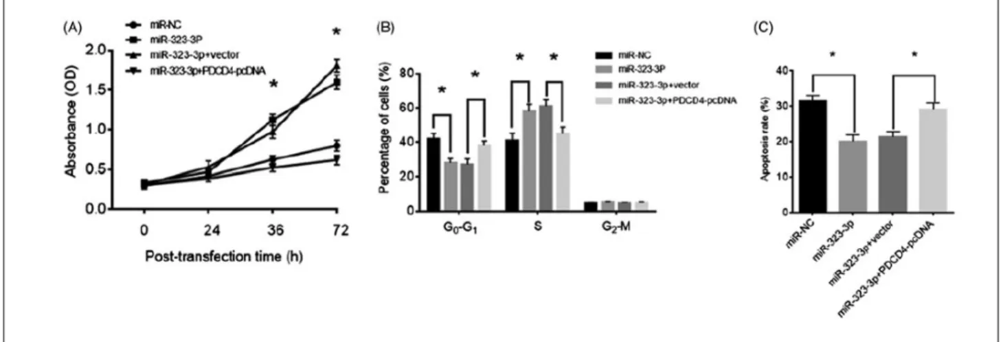
Exosomes are special nano-sized endocytic vesicles secreted by many cell types (Zomer et al., 2010). Exosomes are important mediators in intercellular communication that enable the transfer of functional miRNAs and proteins (Zhang et al., 2015). Mesenchymal stem cells (AMSCs) derived from adipose tissue can produce many exosomes. This suggests that AMSCs can be used as an agent to transfer miRNAs in exosome-mediated cell interactions (Yeo et al., 2013). AMSCs can communicate with brain parenchymal cells and provide miR-133b transfer via exosomes to regulate neurite growth (Xin et al., 2012). Exosomes produced by AMSCs modified with MiR-181-5P suppress hepatic fibrosis in hepatic stellate cells (Qu et al., 2017). In addition, exosomes derived from AMSCs designed with miR-122 have been reported to cause greater sensitivity to chemotherapeutic treatment in hepatocellular carcinoma cells (Lou et al., 2015). However, the therapeutic approach through exosomes has rarely been reported in PCOS. Recently, miR-323-3p has been reported to play a role in the regulation of steroidogenesis and apoptosis in the cumulus cells (CC) of women with PCOS (Wang et al., 2019). In 2019, Zhao et al conducted a study to determine the effects of exosomal miR-323-3p on cumulus cells (CC) of PCOS patients in the letrazol-induced mouse PCOS model. Exosomal miR-323-3p was isolated from modified AMSCs and collected. Real-time PCR, western blot, MTT, flow cytometry and luciferase analyze were performed to identify exosomal miR-323-3p mechanisms in CCs in PCOS mice. The results showed that miR-323-3p expression was upregulated in AMSCs, exosomes and CCs. The upregulated miR-323-3p promotes cell proliferation in CCs and suppresses apoptosis, while the miR-323-3p inhibitor plays opposite roles in exosome-treated CCs (Fig. 4). They reported that the upregulation of miR-323-3p suppresses the apoptosis of CCs and alleviates PCOS by targeting PDCD4 (Programmed cell death protein 4) (Fig. 5) (Zhao et al., 2019).
With this study, it has been revealed that mesenchymal stem cell exosomes may contribute to the development of new therapeutic strategies as well as it can be understood that their paracrine effects can be benefited by direct injection of mesenchymal stem cells in the treatment of PCOS.
Fig. 4. Upregulation of miR-323-3p promoted cell growth and inhibited apoptosis in cumulus cells (CCs). Detection of miR-323-3p expression in (A) human adipose tissue-derived mesenchymal stem cells (AMSCs), (B) AMSC exosome and (C) CCs. (D) Cell proliferation. (E) Cell cycle distribution. (F) Bax Bcl-2, and caspase-3 protein expression in CCs transfected miR-caspase-32caspase-3-caspase-3p-modified exosomes. (G) Apoptosis level in CCs transfected miR-323-3p-modified exosomes. (*) Denotes differences from the control group (p<.05). Values are means ± SEM. For each experiment, at least four samples were available for the analysis. (Zhao et al., 2019).
Fig. 5. MiR-323-3p promoted cell growth and inhibited apoptosis in cumulus cells (CCs) by targeting PDCD4. CCs were transfected with miR-323-3p and miR- miR-323-3p+PDCD4-pcDNA. (A) Cell proliferation. (B) Cell cycle distribution. (C) Apoptosis level. (*) Denotes differences from the control group (p<.05). Values are means ± SEM. For each experiment, at least 4 samples were available for the analysis. (Zhao et al., 2019).
2. Discussion
PCOS is a multi-factor reproductive and endocrine system disorder. However, positive results can be obtained in the long term with correct diagnosis and treatment guidelines. In recent years, mesenchymal stem cells are the focus of restorative and regenerative medicine. In many different disease types, the use of mesenchymal stem cells isolated from different sources has positive results. However, a limited number of studies have been conducted in female reproductive system disorders, especially in the treatment of PCOS. This creates a reliability problem in the use of mesenchymal stem cells in the treatment of PCOS. For this reason, experimental and clinical studies on the use of MSCs in the treatment of PCOS should be increased. Thus, according to changing diagnostic criteria, the use of MSCs with the most accurate injection technique and doses has the potential to be a promising new approach in the treatment of PCOS. Conflict of interest None to declare. Acknowledgments None to declare. References
1. Ankrum, J., Karp, J.M., 2010. Mesenchymal stem cell therapy: Two steps forward, one step back. Trends. Mol. Med. 16, 203-209.
2. Artini, P. G., de Micheroux, A. A., D'Ambrogio, G., 1996. Growth hormone cotreatment with gonadotropins in ovulation induction. J. Endocrinol. Invest. 19, 763-779.
3. Augello, A., Tasso, R., Negrini, S. M., Amateis, A., Indiveri, F., Cancedda, R., & Pennesi, G., 2005. Bone marrow mesenchymal progenitor cells inhibit lymphocyte proliferation by activation of the programmed death 1 pathway. Eur. J. Immunol. 35,
1482-1490.
4. Badawy, A., Elnashar, A., 2011. Treatment options for polycystic ovary syndrome. Int. J. Women's Health. 3, 25.
5. Boostanfar, R., Jain, J. K., Mishell, D. R., Jr, Paulson, R. J., 2001. A prospective randomized trial comparing clomiphene citrate with tamoxifen citrate for ovulation induction. Fertil. Steril. 75,
1024-1026.
6. Corbould, A., 2008. Insulin resistance in skeletal muscle and adipose tissue in polycystic ovary syndrome: Are the molecular mechanisms distinct from type 2 diabetes? Panminerva Med. 50,
279-294.
7. Demyda, S., Genero, E., 2011. Developmental competence of in vivo and in vitro matured oocytes: A review. Biotechnol. Mol. Biol. 6, 155-165.
8. Ding, D. C., Shyu, W. C., Lin, S. Z., 2011. Mesenchymal stem cells. Cell Transplant. 20, 5-14.
9. Divyashree S, Janhavi P, Ravindra PV, Muthukumar SP., 2019. Experimental models of polycystic ovary syndrome: An update. Life Sci. 237, 116911.
10. Dominici M, Le Blanc K, Mueller I, Slaper-Cortenbach I, Marini F, Krause D, Deans R, Keating A, Prockop Dj, Horwitz E., 2006. Minimal criteria for defining multipotent mesenchymal stromal cells. The international society for cellular therapy position statement. Cytotherapy. 8, 315-317.
11. Ellenbogen A, Shavit T, Shalom-Paz E., 2014. Ivm results are
comparable and may have advantages over standard ivf. Facts Views Vis. Obgyn. 6, 77.
12. Elnashar A, Abdelmageed E, Fayed M, Sharaf M., 2006. Clomiphene citrate and dexamethazone in treatment of clomiphene citrate-resistant polycystic ovary syndrome: A prospective placebo-controlled study. Hum. Reprod. 21,
1805-1808.
13. Franks, S., Stark, J., Hardy, K., 2008. Follicle dynamics and anovulation in polycystic ovary syndrome. Hum. Reprod. Update.
14, 367-378.
14. He Y, Chen D, Yang L, Hou Q, Ma H, Xu X., 2018. The therapeutic potential of bone marrow mesenchymal stem cells in premature ovarian failure. Stem Cell Res. Ther. 9, 263.
15. Hernigou P, Bernaudin F, Reinert P, Kuentz M, Vernant JP., 1997. Bone-marrow transplantation in sickle-cell disease. Effect on osteonecrosis: A case report with a four-year follow-up. JBJS. 79,
1726-1730.
16. Huang X, Hao C, Shen X, Liu X, Shan Y, Zhang Y, Chen L., 2013. Differences in the transcriptional profiles of human cumulus cells isolated from mi and mii oocytes of patients with polycystic ovary syndrome. Reprod. (Cambridge, England). 145, 597-608.
17. Hwu, Y. M., Lin, S. Y., Huang, W. Y., Lin, M. H., Lee, R. K., 2005. Ultra-short metformin pretreatment for clomiphene citrate-resistant polycystic ovary syndrome. Int. J. Gynecol. Obstet. 90,
39-43.
18. Ishizuka, Y., Nishimura, M., Matsumoto, K., Miyashita, M., Takeo, T., Nakagata, N., Hosoi, Y., Anzai, M., 2013. The influence of reduced glutathione in fertilization medium on the fertility of in vitro–matured c57bl/6 mouse oocytes. Theriogenology. 80, 421-426.
19. Jafarzadeh, H., Nazarian, H., Ghaffari Novin, M., Shams Mofarahe, Z., Eini, F., Piryaei, A., 2018. Improvement of oocyte in vitro maturation from mice with polycystic ovary syndrome by human mesenchymal stromal cell–conditioned media. J. Cell. Biochem. 119, 10365-10375.
20. Jee, B. C., Han, S. H., Moon, J. H., Suh, C. S., Kim, S. H., Seoul National University College of Medicine Assisted Reproductive Technology (SMART) Study Group., 2008. Influence of well-defined protein source on in vitro maturation of human oocyte: Human follicular fluid versus human serum albumin. Fertil. Steril.
89, 348-352.
21. Kalhori, Z., Azadbakht, M., Soleimani Mehranjani, M., Shariatzadeh, M. A., 2018. Improvement of the folliculogenesis by transplantation of bone marrow mesenchymal stromal cells in mice with induced polycystic ovary syndrome. Cytotherapy. 20,
1445-1458.
22. Legro, R. S., Barnhart, H. X., Schlaff, W. D., Carr, B. R., Diamond, M. P., Carson, S. A., Steinkampf, M. P., Coutifaris, C., McGovern, P. G., Cataldo, N. A., Gosman, G. G., Nestler, J. E., Giudice, L. C., Leppert, P. C., Myers, E. R., Cooperative Multicenter Reproductive Medicine Network., 2007. Clomiphene, metformin, or both for infertility in the polycystic ovary syndrome. N. Engl. J. Med. 356, 551-566.
23. Li, J., Peng, X., Zeng, X., Liu, B., Hao, Q., Yu, X., Zhu, L., & Hu, Q., 2015. Estrogen secreted by mesenchymal stem cells necessarily determines their feasibility of therapeutical application. Sci. Rep. 5, 15286.
24. Lim, K. S., Chae, S. J., Choo, C. W., Ku, Y. H., Lee, H. J., Hur, C. Y., Lim, J. H., Lee, W. D., 2013. In vitro maturation: Clinical applications. Clin. Exp. Reprod. Med. 40, 143.
Bülbül et al. / J Exp Clin Med
C., Chang, Y. H., Hu, Y. C., 2011. The role of adipose-derived stem cells engineered with the persistently expressing hybrid baculovirus in the healing of massive bone defects. Biomaterials.
32, 6505-6514.
26. Ling, B., Feng, D. Q., Zhou, Y., Gao, T., Wei, H. M., Tian, Z. G., 2008. Effect of conditioned medium of mesenchymal stem cells on the in vitro maturation and subsequent development of mouse oocyte. Braz. J. Med. Biol. Res. 41, 978-985.
27. Lizneva, D., Suturina, L., Walker, W., Brakta, S., Gavrilova-Jordan, L., Azziz, R., 2016. Criteria, prevalence, and phenotypes of polycystic ovary syndrome. Fertil. Steril. 106, 6-15.
28. Lou, G., Song, X., Yang, F., Wu, S., Wang, J., Chen, Z., Liu, Y., 2015. Exosomes derived from mir-122-modified adipose tissue-derived mscs increase chemosensitivity of hepatocellular carcinoma. J. Hematol. Oncol. 8, 1-11.
29. Messinis, I., Nillius, S., 1982. Comparison between tamoxifen and clomiphene for induction of ovulation. Acta Obstet. Gynecol. Scand. 61, 377.
30. Mutlu, L., Hufnagel, D., Taylor, H. S., 2015. The endometrium as a source of mesenchymal stem cells for regenerative medicine. Biol. Reprod. 92.
31. Nardo, L., 2004. Management of anovulatory infertility associated with polycystic ovary syndrome: Tamoxifen citrate an effective alternative compound to clomiphene citrate. Gynecol. Endocrinol.
19, 235-238.
32. Parsanezhad, M. E., Alborzi, S., Motazedian, S., Omrani, G., 2002. Use of dexamethasone and clomiphene citrate in the treatment of clomiphene citrate-resistant patients with polycystic ovary syndrome and normal dehydroepiandrosterone sulfate levels: A prospective, double-blind, placebo-controlled trial. Fertil. Steril. 78, 1001-1004.
33. Qu, Y., Zhang, Q., Cai, X., Li, F., Ma, Z., Xu, M., & Lu, L., 2017. Exosomes derived from mir‐181‐5p‐modified adipose‐derived mesenchymal stem cells prevent liver fibrosis via autophagy activation. J. Cell. Mol. Med. 21, 2491-2502.
34. Sam, S., Dunaif, A., 2003. Polycystic ovary syndrome: Syndrome. Trends Endrocrinol. Metab. 14, 365-370.
35. Sánchez, F., Romero, S., De Vos, M., Verheyen, G., Smitz, J., 2015. Human cumulus-enclosed germinal vesicle oocytes from early antral follicles reveal heterogeneous cellular and molecular features associated with in vitro maturation capacity. Hum. Reprod. 30, 1396-1409.
36. Shalom-Paz, E., Holzer, H., Son, W., Levin, I., Tan, S. L., Almog, B., 2012. Pcos patients can benefit from in vitro maturation (ivm) of oocytes. Eur. J. Obstet. Gynecol. Reprod. Biol. 165, 53-56.
37. Stein, I.F., 1935. Amenorrhea associated with bilateral polycystic
ovaries. Am. J. Obstet. Gynecol. 29, 181-191.
38. Stener-Victorin, E., Waldenström, U., Tägnfors, U., Lundeberg, T., Lindstedt, G., Janson, P. O., 2000. Effects of electro-acupuncture on anovulation in women with polycystic ovary syndrome. Acta Obstet. Gynecol. Scand. 79, 180-188.
39. Teede, H., Deeks, A., Moran, L., 2010. Polycystic ovary syndrome: A complex condition with psychological, reproductive and metabolic manifestations that impacts on health across the lifespan. BMC Med. 8, 41.
40. Wang, T., Liu, Y., Lv, M., Xing, Q., Zhang, Z., He, X., Xu, Y., Wei, Z., Cao, Y., 2019. Mir-323-3p regulates the steroidogenesis and cell apoptosis in polycystic ovary syndrome (PCOS) by targeting igf-1. Gene. 683, 87-100.
41. Xie, Q., Xiong, X., Xiao, N., He, K., Chen, M., Peng, J., Su, X., Mei, H., Dai, Y., Wei, D., Lin, G., Cheng, L., 2019. Mesenchymal stem cells alleviate dhea-induced polycystic ovary syndrome (PCOS) by inhibiting inflammation in mice. Stem Cells Int.
2019(6), 1-12.
42. Xin, H., Li, Y., Buller, B., Katakowski, M., Zhang, Y., Wang, X., Shang, X., Zhang, Z. G., Chopp, M., 2012. Exosome‐mediated transfer of mir‐133b from multipotent mesenchymal stromal cells to neural cells contributes to neurite outgrowth. Stem Cells. 30,
1556-1564.
43. Yeo, R. W., Lai, R. C., Zhang, B., Tan, S. S., Yin, Y., Teh, B. J., & Lim, S. K., 2013. Mesenchymal stem cell: An efficient mass producer of exosomes for drug delivery. Adv. Drug Deliv. Rev. 65, 336-341.
44. Yildiz, B. O., Bozdag, G., Yapici, Z., Esinler, I., Yarali, H., 2012. Prevalence, phenotype and cardiometabolic risk of polycystic ovary syndrome under different diagnostic criteria. Hum. Reprod.
27, 3067-3073.
45. Yoon, B. S., Moon, J. H., Jun, E. K., Kim, J., Maeng, I., Kim, J. S., Lee, J. H., Baik, C. S., Kim, A., Cho, K. S., Lee, J. H., Lee, H. H., Whang, K. Y., You, S., 2010. Secretory profiles and wound healing effects of human amniotic fluid-derived mesenchymal stem cells. Stem Cells Dev. 19, 887-902.
46. Zhang, X., Yuan, X., Shi, H., Wu, L., Qian, H., Xu, W., 2015. Exosomes in cancer: Small particle, big player. J. Hematol. Oncol. 8, 83.
47. Zhao, Y., Tao, M., Wei, M., Du, S., Wang, H., Wang, X., 2019. Mesenchymal stem cells derived exosomal mir-323-3p promotes proliferation and inhibits apoptosis of cumulus cells in polycystic ovary syndrome (PCOS). Artif. Cells Nanomed. Biotechnol. 47,
3804-3813.
48. Zomer, A., Vendrig, T., Hopmans, E. S., van Eijndhoven, M., Middeldorp, J. M., Pegtel, D. M., 2010. Exosomes: Fit to deliver small RNA. Commun. Integr. Biol. 3, 447-450.