ISOLATION AND CHARACTERIZATION OF AN ADHESIN PROTEIN FROM THE SURFACE OF A RESPIRATORY PATHOGEN MORAXELLA
CATARRHALIS
A THESIS SUBMITTED TO
THE DEPARTMENT OF MOLECULAR BIOLOGY AND GENETICS AND THE INSTITUTE OF ENGINEERING AND SCIENCE OF
BILKENT UNIVERSITY
IN PARTIAL FULFILLMENT OF THE REQUIREMENTS FOR THE DEGREE OF MASTER OF SCIENCE
BY
TOLGA TURAN AUGUST 2002
I certify that I read this thesis and that in my opinion it is fully adequate, in scope and in quality, as a thesis for the degree of Master of Science.
_____________________________
Prof. Dr. Gülay Özcengiz
I certify that I read this thesis and that in my opinion it is fully adequate, in scope and in quality, as a thesis for the degree of Master of Science.
_____________________________
Assoc. Prof. Kamruddin Ahmed
I certify that I read this thesis and that in my opinion it is fully adequate, in scope and in quality, as a thesis for the degree of Master of Science.
_____________________________
Assist. Prof. Uygar Tazebay
Approved for Institute of Engineering and Science
__________________________ Director of Institute of Engineering and Science
ABSTRACT
ISOLATION AND CHARACTERIZATION OF AN ADHESIN PROTEIN FROM THE SURFACE OF A RESPIRATORY PATHOGEN MORAXELLA
CATARRHALIS
TOLGA TURAN
M.S in Molecular Biology and Genetics Supervisor: Assoc. Prof. Kamruddin Ahmed
August 2002, 59 pages
Moraxella catarrhalis is a member of the normal flora of upper respiratory
tract. Starting in the early 1980s it gained importance as an important cause of otitis media in children and lower respiratory tract infections in adults with chronic obstructive pulmonary disease. β-lactamase producing strains of M. catarrhalis has been increasing at a very fast rate. In some locations, 100% of the strains are β-lactamase producer. The pathogenesis of infection by this bacterium is not clearly understood which hindered the development of a vaccine. In this study, a surface protein of about 55 kDa was isolated from M. catarrhalis by celite chromatography. It is a heat stable protein and is not affected by 2-mercaptoethanol or dithiothreitol treatment. The immunogenic property of the protein has been determined by
immunizing rabbits with M. catarrhalis and detecting the antibody response in serum against 55 kDa protein by Western blotting. In addition, the protein is immunogenic in humans as antibody against 55 kDa protein can be detected in the sputum of patients with M. catarrhalis infection. Moreover, we determined upto 40 amino acids at the N-terminal and also two fragments of the protein. To determine the function of the protein, attachment inhibition assays were performed and it was found that 55 kDa protein competetively inhibits attachment of M. catarrhalis to human
pharyngeal epithelial cells (HPEC). Similarly, monoclonal antibody against 55 kDa (mAb) blocks the protein and inhibits the attachment of M. catarrhalis to HPEC. These two lines of evidence show that 55 kDa protein is an adhesin of M. catarrhalis which mediate attachment to HPEC. In addition, immunoflourescence experiments further verified that 55 kDa protein binds to HPEC. To sequence the gene encoding 55 kDa protein, PCR was done using degenerate primers constructed from the N-terminal amino acid sequence. PCR amplification of the possible gene of 55 kDa protein resulted in a 500 bp fragment, but no homology can be obtained with N-terminal amino acid sequence. In addition, a genomic library of M. catarrhalis is prepared and screened with mAb and with a radiolabelled oligonucleotide probe. We isolated several positive clones; therefore in future it might be possible to sequence the gene encoding 55 kDa protein from these clones.
ÖZET
MORAXELLA CATARRHALIS BAKTERİSİNİN YÜZEYİNDEN BİR ADHESİN
PROTEİNİNİN İZOLE EDİLMESİ VE KARAKTERİZASYONU
TOLGA TURAN
Moleküler Biyoloji ve Genetik Yüksek Lisans Tez Yöneticisi: Doç. Dr. Kamruddin Ahmed
Ağustos 2002, 59 sayfa
İnsan boğaz florasının bir üyesi olan Moraxella catarrhalis, 80’li yılların başından itibaren bir patojen olarak önem kazanmaya başlamıştır. M. catarrhalis çocuklarda orta kulak iltahabı ve yetişkinlerde (özellikle kronik bronşit hastalarında) alt solunum yolu enfeksiyonunda en çok karşılaşılan üçüncü bakteridir. Antibiyotik dirençli suşların son zamanlarda çok artması (bir çok yerde 100%) ve bu bakteriye karşı başarılı bir aşı geliştirilememiş olması, M. catarrhalis üzerine yapılan
çalışmaları arttırmıstır. Bu çalışmada molekül ağırlığı 55 kDa olan bir protein M.
catarrhalis’in yüzeyinden celite kromatografisi metoduyla izole edilmiştir. Bu
proteinin ısı dirençli olduğu ve 2-merkaptoethanol ve DTT tarafından etkilenmediği bulunmuştur. Proteinin immunojenik olduğu tavşan ve insanda yapılan deneyler sonucu ispatlanmıştır. Buna ek olarak proteinin amino kısmından 40 amino asitlik bölümü Edman metoduyla belirlenmiştir. Proteinin bakteride adhsin olarak görev yaptığı bağlanma inhibisyonu deneyleriyle gösterilmiştir. Bu deneylerde 55 kDa proteininin insan boğaz epitel hücreleriyle inkübe edilmesi M. catarrhalis’in boğaz hücrelere bağlanmasını azaltmıştır. Benzer mantıkla bu proteine karşı üretilmiş monoklonal antikorun M. catarrhalis ile inkübe edilmesi bakterinin hücrelere bağlanmasını azaltmıştır. Bu proteinin boğaz epitel hücrelerine bağlandığı immunofloresans metodu ile de gösterilmiştir. 55 kDa proteini kodlayan genin klonlanması için protein sekansından elde edilen dejenere primerlerle PCR
yapılmıştır. Sonuç olarak 500 bazlık bir fragman elde edilmiştir ama bu fragmanın amino asit zinciriyle homolog olmadığı görülmüştür. Buna ek olarak M. catarrhalis genomundan hazırlanan gen kütüphanesi monoklonal antikor ve radyoaktif işaretli bir oligonukleotid ile taranmıştır. Çıkan pozitif kolonilerin sekanslanması sonucu elde edilen bilgiler amino asit zinciriyle karşılaştırılmış fakat homoloji
bulunamamıştır. Elde edilen pozitif klonlar sonuna kadar henüz sekanslanmamıştır. Anahtar Kelimeler: Moraxella catarrhalis, bağlanma, bağlanma inhibisyon deneyi, adhesin.
ACKNOWLEDGEMENTS
Thank you…
…Dr. Kamruddin Ahmed, my sensei, for all that I have learned from you, for your warmth and patience.
…Dr. Uygar Tazebay, for your help in molecular biology techniques.
…My family, for your support and help in my hardest times. I cannot pay your right even if I’d work for all my life.
…Hani, Tolga and Emre, for your help as my elder brothers.
…Tuba and Ozgur, for your companionship.
…Memed, you came in the toughest time helped me and went back as if you were here only to help me. Good luck for your future career in science.
…Pinar, for your help in attachment assays.
…Fusun, for your help in making orders easier.
…Our support units, Abdullah Bey, Sevim Hanim and Tulay for the efforts you have spent to make things run smoothly.
This project was partially supported by a grant provided from TUBITAK (TBAG-2403 101T044) and by a grant provided by Bilkent University (MBG-01-04)
Antibody detection in patients’ sputum, polyclonal and monoclonal antibody production were done in collaboration with Prof. Tsuyoshi Nagatake, Department of Internal Medicine, Institute of Tropical Medicine, Nagasaki University.
N-terminal amino acid sequencing was done in collaboration with Dr.
TABLE OF CONTENTS
SIGNATURE PAGE ………iii
ABSTRACT………iv ÖZET………v ACKNOWLEDGEMENT………...…vi TABLE OF CONTENTS………...…vii LIST OF FIGURES……….xi LIST OF TABLES………..xii ABBREVIATIONS………...xiii CHAPTER 1: INTRODUCTION………...1 1.1 MORAXELLA CATARRHALIS…...1 1.1.1 History of Classification...1
1.1.1.1 Identification and Bacteriological Properties...1
1.1.2 Importance...2
1.1.2.1 M. catarrhalis Infections...2
1.1.3 Level of Fight with M. catarrhalis...3
1.1.4 Surface Exposed Antigens...5
1.1.5 Typing and Epidemiology...7
1.1.5.1 Colonization...7
1.2 ADHERENCE...8
1.2.1 Definition and Importance...8
1.2.2 Mechanism of Adherence...9
1.2.3 Mucus and Muciciliary Action...10
1.2.4 Adhesins and Receptors for M. catarrhalis...10
1.2.5 How to Exploit Adherence...11
1.2.6 Aim of the Project...12
CHAPTER 2: MATERIALS AND METHODS...13
2.1 MATERIALS...13
2.1.2.1 E. coli Strains...13 2.1.2.2 M. catarrhalis Strains...13 2.1.3 Enzymes...14 2.1.4 Oligonucleotides...14 2.1.5 Cloning Vectors...15 2.1.6 Antibodies...16
2.1.6.1 Generation of Monoclonal Antibody against 55 kDa Protein...16
2.1.6.2 Generation of Polyclonal Antibody against M. catarrhalis...16
2.1.6.3 Secondary Antibodies...16
2.1.7 Commercially Available Kits...17
2.1.8 Apparatus and Equipment...17
2.1.9 Materials for Autoradiography...18
2.1.10 DNA and Protein Size Markers...18
2.1.11 Membranes...18
2.1.12 Computer Programs...18
2.2 SOLUTIONS AND MEDIA...19
2.2.1 Agarose Gel Electrophoresis Solutions...19
2.2.2 Microbiological Media and Antibiotics...19
2.2.3 Polyacrylamide Gel Electrophoresis Solutions ...20
2.2.4 Colony Lift Solutions...20
2.2.5 Western Blot Solutions...21
2.2.6 Colony Hybridization Solutions...21
2.3 METHODS...22
2.3.1 Growth and Maintanence of Bacteria...22
2.3.1.1 M. catarrhalis...22
2.3.1.2 E. coli...22
2.3.2 DNA Experiments...22
2.3.2.1 Preparation of Competent Cells and Transformation of E.coli...22
2.3.2.1.1 Simple and Efficient Method(SEM) for preparation of competent cells..22
2.3.2.1.2 Transformation of E. coli ...23
2.3.2.2 Plasmid DNA Isolation...23
2.3.2.3 DNA Ligation Reactions...23
2.3.2.4 Restriction Enzyme Digestion...23
2.3.2.6 Polymerase Chain Reaction...24
2.3.2.7 Genomic DNA Isolation from M. catarrhalis...24
2.3.2.8 Phenol-Chloroform Extraction...25
2.3.2.9 Electroelution...26
2.3.2.10 Butanol Concentration……….26
2.3.2.11 Agarose Gel Electrophoresis………...26
2.3.2.12 Purification of Linear Plasmid DNA from Agarose Gels………26
2.3.2.13 DNA Sequence Analysis……….27
2.3.3 Protein Experiments...27
2.3.3.1 Isolation of 55 kDa Protein...27
2.3.3.2 N-Terminal Amino Acid Sequencing……….28
2.3.3.3 Glycan Detection………....28
2.3.3.4 SDS Polyacrylamide Gel Electrophoresis and Western Blot...28
2.3.3.5 Colony Lift...29
2.3.3.6 Dot Blot...29
2.3.3.7 Attachment Inhibition Assay...29
2.3.3.7.1 Attachment Inhibition Assay with 55 kDa protein...30
2.3.3.7.2 Attachment Inhibition Assay with Antibody against 55 kDa protein...31
2.3.3.8 Quantitative Culture...31
2.3.3.9 Radial Immunodiffusion (RID)...31
2.3.3.10 Colony Hybridization...32
2.3.3.11 Immunofuorescence...33
2.3.3.12 Isolation of Sol Phase of Sputum...33
2.3.3.13 Detection of Antibody in Patients’ Sputum……….33
2.3.3.14 Detection of Antibody in Rabbit Serum………..33
CHAPTER 3: RESULTS...34
3.1 INTRODUCTION...34
3.2 ISOLATION AND CHARACTERIZATION OF 55 kDa PROTEIN ...34
3.2.1 Isolation of 55 kDa Protein...34
3.2.2 Biochemical Properties...35
3.2.3 Immunogenic Properties...37
3.2.4 N-Terminal Amino acid Sequence...38
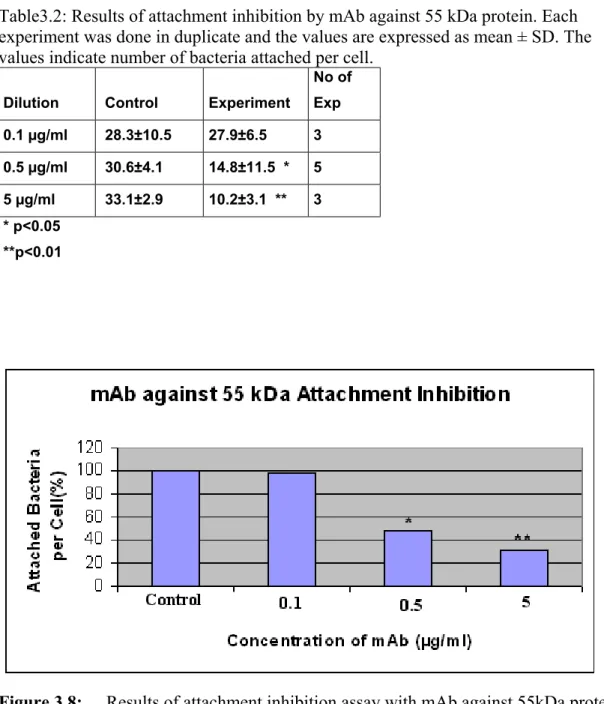
3.3.2 Attachment Inhibition Assay with mAb Against 55 kDa Protein...40
3.4 PCR AMPLIFICATION OF GENE ENCODING 55 kDA PROTEIN...42
3.5 IMMUNOFLOURESCENCE...43
3.6 MOLECULAR CLONING EXPERIMENTS...43
3.6.1 Genomic Library in pQE-31 Expression Vector...43
3.6.2 Genomic Library in pBluescript SK Vector...46
CHAPTER 4: DISCUSSION...48
4.1 CONCLUSION...51
4.2 FUTURE PERSPECTIVES...52
LIST OF FIGURES
Figure 2.1 The map of pQE expression vector...………...15
Figure 2.2 The map of pBluescript cloning vector...………...15
Figure 3.1 Elution profile of celite chromatography...………..34
Figure 3.2 SDS-PAGE analysis of elution profile of celite chromatography…35 Figure 3.3 Glycosylation determined by DIG glycan detection kit...36
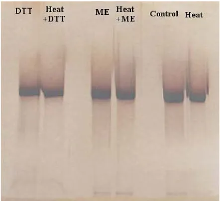
Figure 3.4 SDS-PAGE analysis of DTT, 2-ME and heat treatment of 55 kDa protein…………...36
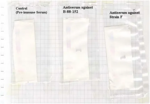
Figure 3.5 Immunogenic properties of 55 kDa protein in rabbits...37
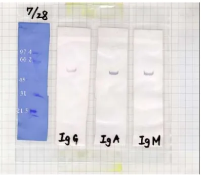
Figure 3.6 Immunogenic properties of 55 kDa protein in humans...38
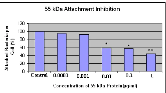
Figure 3.7 Results of attachment inhibition assay with 55 kDa protein.…...40
Figure 3.8 Results of attachment inhibition assay with mAb against 55kDa protein………...41
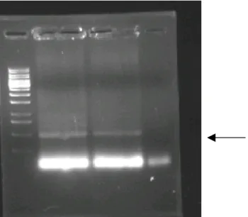
Figure 3.9 Agarose gel electrophoresis of PCR product...42
Figure 3.10 Dot blot of M. catarrhalis and E. coli detected with monoclonal antibody against 55 kDa protein...…...43
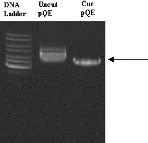
Figure 3.11 Agarose gel electrophoresis of digested and undigested pQE...44
Figure 3.12 HindIII digested M. catarrhalis genomic DNA...45
Figure 3.13 Agarose gel electrophoresis showing the insert released after restriciton enzyme digestion...45
Figure 3.14 M. catarrhalis genomic DNA was digested by different restriction enzymes...46
Figure 3.15 M. catarrhalis genomic DNA and pBluescript (pBSK) plasmid DNA were digested with EcoRV...46
Figure 3.16 Screening of HindIII-pBluescript genomic library of M. catarrhalis with radiolabelled probe...47
LIST OF TABLES
Table 3.1 Results of attachment inhibition assay with 55 kDa protein...39 Table 3.2 Results of attachment inhibition by mAb against 55 kDa protein...41 Table 3.3 Results of attachment inhibition of other strains by mAb...42
ABBREVIATIONS
RTI Respiratory tract infections
CTAB Cetyltrimethylammonium Bromide TBS Tris Buffered Saline
PBS Phosphate Buffered Saline IgA Immunoglobulin A BHI Brain Heart Infusion
COPD Chronic Obstructive Pulmonary Disease OMP Outer Membrane Protein
HPEC Human Pharyngeal Epithelial Cell mAb Monoclonal Antibody
PCR Polymerase Chain Reaction
SCID Severe Combined Immunodeficient Usp Ubiquitous Surface Protein
RFLP Restriction Fragment Length Polymorphism PFGE Pulsed Field Gel Electrophoresis
SDS Sodium Dodecyl Sulphate
SDS-PAGE SDS-Polyacrylamide Gel Electrohoresis HRP Horse Radish Peroxidase
OD Optical Density PB Phosphate Buffer BSA Bovine Serum Albumin APS Ammonium per Sulphate
TEMED N, N, N, N-tetramethyl-1,2 diaminoethane MH Mueller Hinton
CHAPTER 1: INTRODUCTION
1.1 MORAXELLA CATARRHALIS
Moraxella (Branhamella) catarrhalis, formerly called Neisseria catarrhalis
or Micrococcus catarrhalis is a gram-negative aerobic diplococcus frequently found as a normal inhabitant of human respiratory tract. Over the last 20 years, this bacterium has emerged as a pathogen (Johnson 1981) and is now considered as an important cause of respiratory tract infection and otitis media together with
Heamophilus influenzae and Streptococcus pneumoniae.
1.1.1 History of Classification
Although M. catarrhalis is now accepted as a pathogen, its classification has generated controversy since Pfeiffer’s designation as Mikrococcus catarrhalis in 1896 (Frosch 1896). Subsequent studies reported that M. catarrhalis was a common inhabitant of the oropharynx of healthy adults. It was classified as
Neisseria catarrhalis based on phenotypic characteristics and the colony
morphology (Enright 1997). Unfortunately, this latter study failed to differentiate
M. catarrhalis from the commensal bacterium Neisseria cinera. Thus M. catarrhalis was not considered as a pathogen for a significant portion of this
century and most clinical laboratories neglected to test for it in biological fluids collected from patients. In 1970, DNA hybridization studies showed that little homology existed between M. catarrhalis and Neiseriaceae species, influencing its reclassification as Branhamella catarrhalis (Karalus 2000). However, Bovre proposed a division of genus Moraxella into two subgenera, Moraxella and
Branhamella (Bovre 1979). To date the controversy over the nomenclature of
this bacterium remains unresolved, although most investigators now use
Moraxella catarrhalis.
1.1.1.1 Identification and Bacteriological Properties
Laboratory identification of M. catarrhalis is not difficult: It is a gram-negative diplococcus. It is positive for cytochrome oxidase, DNase and tributyrin, 4-methylumbelliferyl butyrate as well as indoxyl acetate esterases. It fails to produce acid from glucose, maltose, sucrose, lactose and fructose; it can grow at 22 °C on nutrient agar; it fails to grow on modified Thayer-Martin medium and it
can reduce nitrate and nitrite (Doern 1990). In addition, modern DNA technology has opened new avenues for the detection of M. catarrhalis in clinical materials. Particularly, PCR is the most convenient method of modern molecular biology to identify microorganisms (Verduin, 2002). Recently, multiplex PCR has been designed for detection of major pathogens in middle ear infections (H.
influenzae, M. catarrhalis and S. pneumoniae) (Hendolin, 2000).
1.1.2 Importance
M. catarrhalis had been known as a harmless commensal for a significant
portion of this century. How or why did it suddenly emerge as a pathogen is not yet known. However, the following two factors might be responsible:
1. The bacterium may have changed such that it is now more virulent than in past decades.
2. Since M. catarrhalis has long been regarded as a nonpathogenic commensal, infections by the organism may have been occurring but have been overlooked for years.
M. catarrhalis is an important pathogen to explore because the infections it
causes results in a high level of morbidity and mortality and a substantial
financial burden. For example, in USA, it is responsible for about 15-20 % of all otitis media cases in children (Enright 1997) and this represents a substantial financial burden ($2 billion/year in US) on the health care system (Murphy 1996).
Although, M. catarrhalis is now an established pathogen all over the world,
its virulence factors remain to be discovered. Moreover the pathogenesis of this bacterium is also not yet clear. In addition, substantial increase in β-lactamase producing M. catarrhalis makes it difficult to treat with commonly used
antibiotics (Martinez, 1998). Therefore new preventive and therapeutic strategies are urgently needed to combat infections by this bacterium. All these factors motivated us to study M. catarrhalis.
1.1.2.1 M. catarrhalis Infections
In children, M. catarrhalis is a major cause of otitis media together with H.
Norway, Finland, Japan and Spain used tympanocentesis to demonstrate that 15 to 20 % of the otitis media cases were caused by M catarrhalis (Del Beccaro 1992). This infection rate is significant as it is estimated that 70 to 80% of all children will have had at least a single episode of middle ear disease by the age of three years. Recent data from various centers throughout the US show that M.
catarrhalis is responsible for over 3 million episodes of otitis media annually and
repeated episodes of otitis media may result in hearing loss and are associated with developmental and learning problems as children reach school age (Murphy 1996).
M. catarrhalis is one of the major cause of respiratory tract infection (RTI) in
adults especially in adults having an underlying disease such as chronic
obstructive pulmonary disease (COPD). M. catarrhalis is one of the most three common causes of exacerbation in COPD patients, together with H. influenzae and S. pneumoniae. The reason why M. catarrhalis infection is low in healthy adults is thought to be due to the strong immune system in these individuals. Therefore M. catarrhalis cause more infection in children whose immune system is immature and in old people whose immune system is weak (Verduin 2002).
Occasionally, the bacterium causes systemic infections, e.g., meningitis and sepsis (Catlin 1990). In addition, M. catarrhalis may cause sinusitis, tracheitis, bronchitis, pneumonia and less commonly, ocular infections in children (Verduin 2002). Although rare, M. catarrhalis can cause sinusitis (Brorson 1976), septic arthritis (Craig 1983), bacteremia (Cimolai 1989), cellulitis (Rotta 1994), osteomyelitis (Prallet 1991), endocarditis (Douer 1977), and pericarditis. (Kostiala 1989). It is also important to note that nosocomial infections could be caused by M. catarrhalis (Omori, 1999).
1.1.3 Level of Fight with M .catarrhalis
Because very short time has passed since this organism stepped on stage as a pathogen; the level of knowledge on it is scarce. Both the virulence factors and the pathological properties of M. catarrhalis remain largely unknown. The virulence factors should be explored further to understand the pathogenesis of this bacterium and to develop protective means against the organism.
One additional reason for the low level of knowledge on M catarrhalis is the lack of a good animal model. M. catarrhalis is a strict human pathogen and it
doesn’t cause infection in mouse or in other small laboratory animals. The best models available are the Murine Pulmonary Clearance Model (Unhanand 1992) and the SCID (Severe Combined Immunodeficient) mouse model (Harkness 1993). In the pulmonary clearance model, mice are challenged by introducing bacteria into their lungs and the rate of clearance is followed as a measure of the immune response. In SCID mouse model, bacteria are introduced by multiple routes including intranasal and intravenous, but the symptoms produced in these animals do not mimic those seen in human infection.
It has been difficult to combat with M. catarrhalis as more and more β-lactamase positive strains emerge. The first β-β-lactamase-positive clinical strain was isolated in 1977 (Hoi-Dang 1978). Subsequently, resistance to β-lactams has increased at very fast rate. Presently, in North America and Europe almost 100% of the strains are β-lactamase producer (Clavo-Sanchez 1997). However it varies from country to country such as in Japan it is 94% (Martinez 1998), in Kuwait it’s 57% (Ahmed 1999) and in Uganda it’s 100% (Yoshimine 2001). Although the reason is not known, it is thought that increased use of antibiotics has
contributed for this dramatic increase of β-lactamase producing strains. However, such increase has not been demonstrated in other bacteria under the same
selective pressure (Felmingham 1999).
The lactamase enzymes of M. catarrhalis have been designated as BRO β-lactamase. BRO is different from other β-lactamases and has only been identified in two other Moraxella species (Wallace 1989). The suspected location of the
bro genes on the chromosome and the high efficiency of transfer tend to support
theory of a conjugal transposon as a method for spread of resistance (Wallace 1989). This different means of spread of resistance might be the reason of the steep increase of antibiotic resistance in M. catarrhalis.
The emergence of β-lactamase-positive strains motivated scientists to find a vaccine against M. catarrhalis. But vaccine development for M. catarrhalis is only in the antigen identification stage.
Surface exposed antigens of M. catarrhalis are mainly the outer membrane
proteins, other surface proteins and the lipooligosaccharide. Studying surface exposed antigens will not only be a progress in vaccine development but also help us to understand the virulence properties of M. catarrhalis since the surface
exposed antigens are the sites where the bacterium interacts with the host. In this respect our work on the characterization of a surface exposed protein of M.
catarrhalis will be an important contribution to the understanding of
pathogenesis and prevention of infection caused by this bacterium.
1.1.4 Surface Exposed Antigens
The outer membrane proteins (OMPs) of M. catarrhalis are similar to other gram-negative bacteria in that 10 to 20 OMPs are present with 6 to 8 of these proteins predominating (Murphy 1990). The outer membrane of M. catarrhalis contains 8 major OMPs that are labeled OMP A through OMP H. The molecular weight of these proteins ranges from 98 to 21 kDa. An interesting finding was the striking homogeneity of OMPs among 50 strains. This means that an immune response against one strain will recognize a subsequent strain also (Bartos 1988).
Following is the list of the major OMPs of M. catarrhalis:
CopB:
CopB was one of the first OMP studied. It exhibits moderate degree of antigenic conservation among strains tested (Karalus 2000). There are 3 lines of evidence to say that an antigen is conserved:
• Antibody reactivity
• Restriction Fragment Length Polymorphism (RFLP) • Comparison of sequence of the gene
The protein is immunogenic as the antibody against it could be detected in convalescent sera of patients (Sethi 1995). Moreover, passive immunization of mice with the antibody against CopB resulted in enhanced clearance of M.
catarrhalis from the lungs of these animals (Karalus 2000). Mutation and
expression analysis show that CopB is involved in iron acquisition. Iron is an important element for many bacteria to live on their mammalian hosts. Therefore bacteria developed strategies to transfer low concentration of iron from their environment.
Sequence analysis and RFLP studies on OMP CD show high degree of conservation among different strains of M. catarrhalis. It is a heat-modifiable protein that was initially reported as OMP C and OMP D. Then it was realized that these two bands were the two forms of a single antigen. BLAST search shows that the protein has homology with Pseudomonas aeruginosa porin protein (Murphy 1993). The antibody against OMP CD elicit complement-mediated bactericidal activity against various strains of M. catarrhalis which indicate it may stimulate a protective immune response if used as a vaccine.
UspA1 and UspA2:
They were first named as High Molecular Weight OMP (HMW-OMP) since the protein forms oligomeric complexes. Later on it was realized that the
complex was composed of two proteins and named as ubiquitous surface protein A1 and A2 (UspA1 and UspA2). They were characterized as a single protein not only because of a single band in Sodium Dodecyl Sulphate Polyacrylamide Gel Electrophoresis (SDS-PAGE ) but also because they share a common epitope. In fact they share a common 140-aminoacid region that is 93% identical (Cope 1999). In addition, passive immunization of mice with the antibody against CopB resulted in enhanced clearance from the lungs of these animals (Karalus 2000).
Fimbriae :
Fimbriae have been described on the surface of M. catarrhalis by electron microscopy (Ahmed 1994). In M. catarrhalis, fimbriae are responsible for adherence and hemagglutination, therefore thought to be an important virulence factor (Ahmed 1992a). It was also found that fimbriae are present on all fresh isolates of M. catarrhalis, and it decreases in number upon repeated in vitro passages (Ahmed 1992). Since fimbriae has not been isolated, little information available regarding the characterization of these structures.
Lipooligosaccharide(LOS)
Another prominent bacterial surface component of M. catarrhalis is the LOS.
M. catarrhalis LOS shares homology with LOS of other gram-negative bacteria.
for M. catarrhalis i.e. resistance to bactericidal activity mediated by normal human serum (Zaleski 2000). There are three major LOS serotypes in M.
catarrhalis: A, B and C. these structures encompass 95% all strains studied to
date (Rahman 1996). Recently, it has been suggested that LOS may act as an adhesin of M. catarrhalis (Hu, 2001).
1.1.5 Typing and Epidemiology
The worldwide distribution of M. catarrhalis as well as nosocomial outbreaks by this bacterium, provide an opportunity for considering epidemiological
studies. Several epidemiological typing methods have been used to characterize strains of M. catarrhalis, such as phenotypic characterization, electrophoretic mobility of esterases, OMPs, whole-cell proteins, plasmid profile, isoelectric focusing of β-lactamase enzymes, bacteriocin typing, haemagglutination, fimbriation and serotyping; however, these methods could not satisfactorily discriminate interstrain variability. The RFLP has been also used for M.
catarrhalis; however the result was not satisfactory due to many fragments,
which are difficult to compare. Pulsed field gel electrophoresis (PFGE) was successfully used to find out inter-strain variation of M. catarrhalis isolated from sputum of patients with RTI from various parts of the world (Martinez 1999). The PFGE analysis also showed that there were wide variations among the strains of M. catarrhalis isolated from the same and different geographical location. However, strains isolated from same location showed some common bands shared by the strains. In conclusion, the study showed that the strains of M.
catarrhalis may originate from ancestor strains and they have been proliferating
with slow genomic rearrangement.
1.1.5.1 Colonization
M. catarrhalis is capable of colonizing humans without causing disease,
which was one of the reasons it was characterized as a commensal bacterium. The rate of colonization is affected by age, health, socioeconomic condition, season and geographic location. M. catarrhalis is a frequent inhabitant of oro- and nasopharynx of children 0-10 years of age with a prevalence rate of 25-35%. Healthy adults are less frequently colonized (Ejlertsen 1991). In a study done on 1520 patients (Ejlertsen 1994), M .catarrhalis colonization was examined with
respect to prevalence, time of colonization and association with respiratory tract infections (RTI). In the age group of 1-47 months, children with healthy
respiratory tracts were colonized with a prevalence of 36%. However there was a significant increase in colonization rate in children with upper and lower RTI. (68 %). Children with RTI may be prone to be colonized as long as their respiratory epithelium is damaged. Subsequently, they eliminate M .catarrhalis as they recover from their RTI, their colonization rate returning to the non-RTI children.
Colonization with M .catarrhalis is not static. Molstad et al. (Molstad 1988) found a high rate of acquisition and elimination of M .catarrhalis in children. Nearly 100% of children in their study group were colonized at various times with a mean prevalence rate of 62%.
In the study of Chen et al (1999), they found that antibody levels against an OMP of M.catarrhalis (UspA1) are higher in 2 months old children when compared to 18 months old children due to the passage of maternal antibodies to the fetus during pregnancy. The low antibody levels and the low bactericidal activities seen in sera of children in 6-18 months of age are consistent with the epidemiological finding that children at this age have the highest rate of colonization and highest incidence of M. catarrhalis infection.
In a different study on nasophryngeal cultures from children of 0-2 years of age demonstrated that 66% of the infants become colonized within first year of life and increasing to 77.5% by two years of age (Faden 1994). This study also revealed that in the first 2 years of life a child acquires and clears 3 to 4 different strains of M. catarrhalis. Therefore, the rate of colonization in infants and young children is much more greater than that of adults.
1.2 ADHERENCE:
1.2.1 Definition and Importance
Bacterial adherence (also called attachment) is the term used to describe when bacteria attach to different types of surfaces present in the environment. Bacterial attachment is the initial step in the pathogenesis of infections. In M.
shown to be the initial event of infection (Mbaki 1987). Therefore studies on the attachment mechanism of M. catarrhalis is helpful for the understanding of pathogenesis as well as to find new strategies for the treatment and prevention of infection.
1.2.2 Mechanism of Adherence
Several nonspecific factors are involved in the process of adhesion such as surface electric charge, hydrophobicity and van der Waals forces etc. It is important to note that these are initial factors; they are required, but not enough for firm binding. The specific factor for attachment is the interaction of the
adhesin on the bacterium with the receptor on the host cell. Adhesin is a
molecule on the surface of bacteria that mediates adherence by interacting with a receptor on the substratum. Chemically, adhesins may be proteins,
polysaccharides, teichoic acids or lipids in nature. Many bacterial species have been found to express more than one type of adhesins and, in some cases, more than 10 have been described (London 1996). A receptor is any surface molecule of a substratum that is complementary to a bacterial adhesin. Chemically
mammalian cell receptors are composed of proteins, glycoproteins and glycolipids.
Other factors to affect adherence are the seasonal changes (high in winter and low in summer), defects in immune system, repeated sub-culturing of bacteria in
vitro, prior viral infection and underlying COPD (Rikitomi 1997, Rikitomi 1986).
Before the interaction of adhesin and the receptor, bacteria should overcome the repulsive electrostatic forces since both the bacterium and the epithelial cell are negatively charged. At this time point the filamentous structures such as fimbriae, flagella and fibrillae, act as bridging structures helping bacteria to overcome repulsion forces. The bacterium can now move closer to the surface of the epithelial cell, but the amount of water adsorbed around the surface of the epithelial cell or bacterium is the last barrier to overcome. Hydrophobic groups located on fimbriae or proteins of the cell surface, may play a major role, allowing the displacement of water molecules (Braga 2000). On the contrary, a recent study showed that bacteria with its negative charge bind with the
showed that HPEC is not entirely negatively charged rather composed of positively and negatively charged domains (Ahmed 2000).
1.2.3 Mucus and Mucociliary Action
During a normal day a person inhales about 10,000 microorganisms per 24 hours (Mims 1995). To prevent infection by this huge amount of
microorganisms, mucosal surfaces have several mechanisms: • Secretion of mucus
• Mucociliary clearance • Cellular defenses
• Immunological defenses.
The mucus in the respiratory tract is a mixture of the secretions of different types of cells present in or under the mucosa: goblet cells, epithelial serous cells, secretory cells in submucosa and ciliated cells. It is a gel like structure consists of mucus glycoproteins called mucins. It acts as a barrier for microorganisms to overcome before reaching to the epithelial cell layer. Other functions of bronchial mucosa are to entrap microorganisms and repel them out of the respiratory tract by the mucociliary action. In addition, mucosa acts as an extracelullar surface for immunoglobulin action.
Some bacteria can produce factors that disturb the mucociliary system by slowing or disorganizing the beating of cilia (Wilson 1996). Some of these factors exert their effects by increasing the mucus production, which in turn, decrease the mucociliary action. Till now no such effect has been reported in M.
catarrhalis. However, report has shown that M. catarrhalis can bind with human
nasopharyngeal mucin (Reddy 1996).
1.2.4 Adhesins and Receptors for M. catarrhalis
Adhesion is a complex process which might include more than one adhesin-receptor interaction. For M. catarhalis, fimbriae are found to be adhesins.
Fimbriae are composed of repeating subunits, the part that interacts with the host cell is on the tip of the pilus (London 1996). Other adhesins for M. catarrhalis are hemagglutinins, UspA1(Aebi 1988) and CD protein(Murphy 1993). In addition, in this study we proved that our protein of interest is an adhesin of M.
catarrhalis. The receptors for M. catarrhalis identified so far are ganglioside
GM2 (Ahmed 1996) and asialoganglioside GM1 (Ahmed 2002).
1.2.5 How to Exploit Adherence
The study on bacterial adherence to host is not only a basic science to
understand the pathogenesis and virulence factors, but also it has several practical applications. These applications center on the prevention of adhesion before or after the bacterium attaches to host. One of them is to provide the adhesin or its analogue into the respiratory tract. This might be done by spraying it to the pharynx of the patient. The adhesin or its analogue will bind to complementary receptor on host cells. This will block the receptor and will prevent the
attachment of bacteria to the epithelial cells. The same logic can also work for the receptor. There are receptor analogs, which can mimic the receptor and bind to the adhesin on the surface of bacterium. The binding of the receptor analog to the adhesin will block the adhesin-receptor interaction.
Another strategy is to use the adhesin as a vaccine. Since it is a surface protein, the immune response primed by adhesin, would be protective. The immune system will not only act by conventional means to kill the bacterium, but also prevent the adherence by the binding of antibodies to adhesin.
The scope of adhesion research can be enlarged by using anti-attachment drugs. These can be divided into two:
1. Low-dose antibiotics. 2. Non-antibiotic drugs.
Low-dose antibiotics don’t kill the bacteria but prevent the attachment by changing the surface structure of the bacterium. But the weakness of this strategy is the rapid emergence of the antibiotic resistant bacteria.
The non-antibiotic drugs have the ability to prevent attachment of M.
catarrhalis and nontypeable H. influenzae. One of them is a widely used
mucolytic drug: carboxymethyl cysteine(CMC). The mechanism how S-CMC prevent attachment is not yet clear, however, it has been shown that it can remove the carbohydrate from the surface of HPEC (Zheng 1999). The receptors for bacteria are mainly glycolipid in nature therefore it is thought that S-CMC may remove the receptor from cell surface. Another study showed that S-CMC
can decrease the positive charge of the microplicae hence decrease the attraction needed for bacteria to attach with HPEC (Ndour 2001).
1.3 Aim of the Project
The aim of this project can be categorized as the following: 1. Isolation of an adhesin from the surface of M. catarrhalis.
2. To understand the biochemical properties of the protein such as heat sensitivity, molecular weight, sensitivity to reducing agents and glycosylation.
3. To determine the immunogenic potential of the protein: a. In animal by inoculating bacteria
b. In human during M. catarrhalis infection 4. Production of monoclonal antibody
5. Attachment inhibition assay to prove the protein as an adhesin. 6. N-terminal amino acid sequence.
7. Identification of the gene of the protein
CHAPTER 2: MATERIALS AND METHODS
2.1 MATERIALS
2.1.1 Chemicals
All chemicals were of analytical grade and supplied by Sigma-Aldrich Chemie GmbH (Germany) except for:
• 4-chloro-1-naphthol was purchased from Wako Pure Chemical Industries Ltd. (Japan).
• Ampicillin and kanamycin were purchased from Roche Diagnostics GmbH (Germany).
• IPTG, X-Gal were purchased from MBI Fermentas Inc. (USA). 2.1.2. Bacterial Strains
In this study, for all purposes M. catarrhalis was used and only for cloning purposes E. coli was used.
2.1.2.1 E. coli Strains
Strain Genotype Usage Source
M15 F-,NalS,StrS,rif8,lac-,ara-,gal- Production of expression Bio-Rad
mtl-,rec+,uvr+ libraries (Hercules
USA) DH5α F-supE44 hsdR17 recA1gyrA96 Production of library Bio-Rad
endA1 thi-1 relA1 deoR lsmbda- and propagation of plasmids (Hercules ,USA)
2.1.2.2 M. catarrhalis strains
Strains of M. catarrhalis, B-87-34, B-87-69, B-87-94, B-88-152 and strain F
B-88-152 was mainly used unless otherwise stated. Except strain F, all the strains were fimbriated (Ahmed 1990).
2.1.3 Enzymes
All restriction enymes (HindIII, PstI, and EcoRV), DNA modifying enzymes (T4 ligase, calf intestine alkaline phosphatase) and Taq polymerase were
purchased from MBI Fermentas Inc. (USA). RNase A, Proteinase K and Lysozyme were purchased from Roche Diagnostics GmbH (Germany). Lysine endopeptidase was purchased from Wako Pure Chemical Industries Ltd. (Japan).
2.1.4 Oligonucleotides
All primers used in this study were custom synthesized and purchased from Iontek (Bursa, Turkey). The sequences of the primers were designed by
converting the N-terminal amino acid sequence of 55 kDa protein to DNA sequence. These primers were used to amplify possible gene of 55 kDa protein by PCR for sequencing. The sequences of the primers were as follows:
Forward: 5’-ATTACTCATCAAAATATGCTTCC-3’ 3rd base is degenerate: T, C or A 6th base is degenerate: T, C, A or G 9th base is degenerate: T or C 12th base is degenerate: A or G 15th base is degenerate T or C 19th base is degenerate: T or C 21st base is degenerate: T, C, A or G Reverse: 5’- TTAGCAGGAAACATATTTTGATG- 3’ 3rd base is degenerate: T, C, A or G 6th base is degenerate: T, C, A or G 9th base is degenerate: T, C, A or G 11th base is degenerate: A or G 15th base is degenerate: A or G 18th base is degenerate: T or C 21st base is degenerate: A or G
Apart from these degenerate primers, a 23-mer oligonucleotide has been designed according to the preferred codon usage in gram-negative bacteria. This oligonucleotide was used in colony hybridization experiment as the radiolabelled probe.
Probe: 5’- AAAATCCAYACCCCGAGCATCAC-3’ Y= C or T
We also used several primers for sequencing purposes which are not shown here for limitation of space.
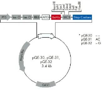
2.1.5 Cloning Vectors
pQE-31 (Qiagen Inc. USA) and pBluescript SK (Stratagene Inc. USA) were the vectors used in this study.
Figure 2.1: The map of pQE expression vector. It contains lac operon and is suitable for expression studies.
Figure 2.2: The map of pBluescript cloning vector. It contains lacZ gene and suitable for α-complementation
2.1.6 Antibodies
The primary antibodies used in this study were the monoclonal antibody against 55 kDa protein and polyclonal antibody against whole cell M. catarrhalis.
2.1.6.1 Generation of monoclonal antibody against 55 kDa protein
mAb 4H3, a murine antibody of IgM class (Hybridoma Subisotyping Kit, Calbiochem-Novabiochem Corporation, USA), was prepared by fusing spleen cells from BALB/c mouse immunized with 55 kDa protein with SP-2 mouse myeloma fusion partner (Oishi 1996) and screening the resulting hybridomas by enzyme-linked immunosorbent assay (ELISA) employing microtiter wells coated with 55 kDa protein. mAb to 55 kDa protein was measured in polystyrene
microtiter plate caoted with 1 µg of purified protein per ml suspended in coating buffer and left to stand overnight. mAb 4H3 was cloned and adapted as an ascites tumor in pristine-primed BALB/c mice. Ascitic fluid was collected, centrifuged and the supernatant was stored at -80 °C until use.
2.1.6.2 Generation of polyclonal antibody against M. catarrhalis
Whole cell antibody against strain B-88-152 was generated by injecting a rabbit with live organism as described previously (Ahmed 1992). A dose of 1 ml bacterial suspension in 1 ml Freund’s adjuvant (Difco Laboratories, USA) was injected each time in equally dividing doses into two subcutaneous and two intramuscular sites. A total of four doses was administered at 2-week intervals. Two weeks after the last injection, blood was collected and the serum was stored at -80 °C.
2.1.6.3 Secondary Antibodies
Secondary antibody for colony lift, dot blot and Western blotting was purchased from Sigma (Sigma-Aldrich Chemie GmbH Germany). It was anti-mouse IgM antibody conjugated to horse radish peroxidase (HRP) enzyme to permit the visualization of the signals. The secondary antibody for
immunofluorescence was purchased from Zymed (Zymed Laboratories, Inc., USA). It was goat anti-mouse IgM antibody conjugated to fluorscein
isothiocyanate (FITC). Secondary antibodies for sputum Western blot were anti-human IgA, IgG and IgM conjugated with HRP. The secondary antibody for
rabbit antiserum experiment was anti-Rabbit IgG conjugated with HRP and it was purchased from Zymed.
2.1.7 Commercially Available Kits
Qiaex II Gel Extraction Kit (by Qiagen Inc., USA) was used to purify DNA
from agarose gels.
MN Nucleospin Plasmid (Macherey-Nagel, GmbH& Co, Germany) was used
to purify plasmids from E. coli.
DIG Glycan Detection Kit (Boehringer Mannheim Corp. USA) was used to
determine the gycosylation status of proteins.
HexaLabel Plus DNA Labeling Kit (MBI Fermentas Inc. NY, USA) was used
to radioactively label the oligonucleotide probe for colony hybridization experiment.
Silver Stain Kit (Wako Pure Chemical Industries Ltd. Japan) was used to stain
the SDS-PAGE gels.
Mouse IgM LL Nanorid Kit (Binding Site Limited, England) was used to
determine the concentration of the primary antibody.
Hybridoma Serotyping Kit (Calbiochem–Novabiochem Corporation, USA) was used to determine the class of the antibody produced.
DYEnamic ET Terminator Cycle Sequencing Kit (Amersham Biosciences UK
Limited, England) was used for sequencing reactions.
Bio Rad Protein Assay Kit ( Bio Rad Laboratories, USA) was used to
determine concentration of proteins.
2.1.8 Apparatus and Equipment
Vertical mini gel apparatus for polyacrylamide gel electrophoresis was a product of Atto Corp. (Japan). The power supply was Power Pac 200 from Bio Rad Laboratories (USA). Horizontal mini gel apparatus used for agarose gel electrophoresis was Mupid-2 purchased from Advance Co., Ltd., Japan. Thermal cycler for PCR was a product of Perkin Elmer (USA). Slab gel dryer is from Savant Instruments Inc. (USA). Wet transfer unit for Western Blotting and GelDoc 2000Image analyzer for agarose gel electrophoresis were purchased from Bio Rad Laboratories (USA). Cytospin for attachment assays was purchased
from ThermoShandon Inc. (USA). UV cross linker, the UV Stratalinker 1800, was from Stratagene Inc (USA). Fluorescence microscope was from Zeiss (Carl Zeiss Jena GmbH, Germany)
2.1.9 Materials for Autoradiography
Radiolabelled dCTP (containing 32P) was purchased from NEN ( NEN Life Science Products, Inc. USA) The light-proof film cassette (Hypercassette) and film developing unit were products of Amersham Biosciences UK Limited (England) Medical x-ray films were purchased from Fuji (Tokyo, Japan).
2.1.10 DNA and Protein Size Markers
As DNA size marker 1 kb DNA ladder from MBI FERMENTAS Inc. (USA) was used.
SDS-PAGE protein size markers, the Rainbow Marker and Low Range Protein Size Marker were purcahsed from Bio Rad Laboratories (CA, USA).
2.1.10 Membranes
Nitrocellulose membranes for colony lift and colony hybridization were purchased from Shleicher & Schuell GmbH (Germany)
PVDF membranes for Western blots were purchased from Bio Rad Laboratories (USA)
2.1.11 Computer Programs
DNASIS Mac V3.6 and Oligo 4.0s were used to design oligonucleotide primers.
GENETYX-MAC 10.1 was used to translate the DNA sequence into amino acid sequence in three reading frames. Pairwise BLAST and protein BLAST was used for homology search.
2.2 SOLUTIONS AND MEDIA
2.2.1 Agarose Gel Electrophoresis Solutions
50X Tris-Acetic acid-EDTA (TAE) Per Liter: 242 g Tris Base
57.1 ml Glacial Acetic acid
100 ml of 0.5 M EDTA
Ethidium bromide 10 mg/ml in water (stock solution)
30 ng/ml(working solution)
2.2.2 Microbiological Media and Antibiotics
Luria-Bertani Medium(LB) Per Liter: 10 g Bacto tryptone,
5 g Yeast extract
5 g NaCl
15 g Agar(for solid media)
Blood Agar %7 Rabbit blood in BHI agar
Brain Heart Infusion (BHI) Agar Per Liter: 52 g of BHI agar powder
BHI Broth Per Liter: 37 g of BHI broth powder
Mueller Hinton (MH) Broth Per Liter:21 g of MHB powder
Ampicillin 100 mg/ml(stock solution) 100 µg/ml (working solution) in dH2O
Kanamycin 25 mg/ml (stock solution)
2.2.3 Polyacrylamide Gel Electrophoresis Solutions Separating Gel (10%) 7.8 ml dH2O 5 ml 1.5 M Tris(pH 8.8) 6.7 ml acylamide-bisacrylamide(30:0.8) 200 µl 10% SDS 200 µl 10% APS 6.7 µl TEMED Stacking Gel (5 %) 3.3 ml dH2O 1.5 ml 0.5 M Tris(pH 6.8) 1 ml acylamide-bisacrylamide(30:0.8) 60 µl 10% SDS 120 µl 10% APS 3 µl TEMED Sample Buffer 10 ml 10 % SDS 4 ml 0.25 M Tris-HCl (pH 7.6) 6 ml dH2O 2 ml Glycerol 30 mg EDTA.2Na
and per ml of the above solution:
20 µl Bromophenol Blue (Saturated Soln)/ml
75 µl 2-mercaptoethanol OR
0.13 g of DTT/ml
Electrophoresis Buffer(10X) Per 500 ml:15.14 g Tris
72 g Glycine
5 g SDS
Staining Solution 2 g Coomassie Blue
100 ml Trichloro acetic acid (100%)
400 ml Ethanol
500 ml dH2O
Destaining Solution 1950 ml dH2O
750 ml Ethanol
240 ml Acetic Acid
2.2.4 Colony Lift Solutions
10X TBS Per Liter: 80 g NaCl 2 g KCl
Blocking Solution 5% non-fat dry milk or 5% BSA
2.2.5 Western Blot Solutions
10X Tris-HCl Buffer Per Liter: 12.4 g Tris-HCl (pH 7.3)
85 g NaCl
Electrode Solution Per Liter: 6 g Tris
28.8 g Glycine
600 ml Methanol
Blocking Solution 5% non-fat dry milk
2.2.6 Colony Hybridization Solutions
Denaturation solution 0.5 NaOH
1.5M NaCl
0.1% SDS
Neutralization Solution 1.0 M Tris-HCl, pH 7.5
1.5 M NaCl
20X SSC 3M NaCl
0.3M Sodium Citrate, pH7.0
Blocking Solution 0.5M Sodium Phosphate Buffer, pH7.0 7% SDS 1mM EDTA 2X Wash Solution 2X SSC 0.1% SDS 0.1X Wash Solution 0.1X SSC 0.1% SDS
2.3 METHODS:
2.3.1 Growth and Maintenance of Bacteria
2.3.1.1 M. catarrhalis
M. catarrhalis strains were stocked in MH broth containing 5% rabbit blood
and stored at -20C until use. For stocking, the bacteria were cultured on 7% rabbit blood agar plates and the colonies were taken with a sterile cotton swab and transferred to the cryotube containing 1 ml of media. During experiments, the bacteria were cultured on BHI agar or BHI broth. The plates were incubated in a box containing 5% CO2 at 37 °C. The CO2 was generated by the CO2 Gen
Compact (Oxoid Limited, UK). All media were purchased from Merck Kga, (Germany).
2.3.1.2 E. coli
E. coli strains were stored in 50% sterile glycerol at -70 °C until use.
Overnight grown cultures to saturation were mixed with sterile glycerol in a ratio of 1:1, mixed to homogeneity and stored at -70 °C until required. Bacteria were recovered by scraping a small amount of cells from the stock with an inoculation loop and streaking onto a LB-agar plate. Experiments with E. coli were done by culturing the bacteria on LB agar or LB broth. Liquid cultures were incubated in a rotary shaking incubator at 37 °C (~200 rpm).
2.3.2 DNA EXPERIMENTS
2.3.2.1 Preparation of Competent Cells and Transformation of E. coli
2.3.2.1.1 Simple and Efficient Method (SEM) for Preparation of Competent Cells
A single colony of appropriate E. coli strain was inoculated into 15 ml of LB broth (containing the appropriate antibiotics) and grown overnight at 37 °C. The starter culture was diluted to OD600 of 0.2-0.3 in 250 ml of SOB medium (2%
with shaking at 200-250 rpm. The culture was chilled on ice for 10 minutes, centrifuged at 2500 Xg for 10 minutes at 4 °C. The pellet was then resuspended in 80 ml of ice-cold TB (10mM Pipes, 55mM MnCl2, 15mM CaCl2, 250mM
KCl, pH 6.7, sterilized by filtration through 0.45µm filter, stored at 4 °C ), and incubated on ice for 10 minutes. The mixture was pelleted as above, resuspended gently in 20 ml of TB. DMSO was added to a final concentration of 7%, mixed gently and incubated on ice for 10 minutes. Aliquots of this mixture were then immediately chilled in liquid nitrogen, and stored at -70 °C up to 3 months without loss of transformation efficiency (Inoue et al. 1990).
2.3.2.1.2. Transformation of E. coli
For transformation using supercompetent cells, an aliquot of frozen
competent cells was thawed on ice. 200 µl of the cells was mixed with plasmid DNA (less then 100ng) in an eppendorf tube and incubated on ice for 30 minutes. The cells were then exposed to a 90 seconds heat-shock at 42 °C and then chilled on ice for 2 minutes. 800 µl of SOC (SOB with 20mM glucose) was added and the cells were grown at 37 °C for 45 minutes with vigorous shaking at 250 rpm. The culture was then centrifuged and the pellet was resuspended in 100µl of SOC, and spreaded on LB agar plates containing the appropriate antibiotic.
2.3.2.2 Plasmid DNA Isolation
The plasmid DNA was isolated with MN Nucleospin Plasmid kit according to the manufacturers instructions.
2.3.2.3 DNA Ligation Reactions
DNA fragments were ligated into plasmid vectors in 15 µl reaction volumes containing 0.3-1.0 µg of linearized plasmid vector and 3-5 times molar excess of insert DNA in the presence of 1-4 Weiss units of T4 ligase and 1 X concentration of the standard ligation buffer supplied with the enzyme. The reaction mixture was incubated at 16 °C for overnight.
2.3.2.4 Restriction Enzyme Digestion
Restriction enzyme digestions were performed in 10-20 µl reaction volume for 1.5 to 4 hours and typically 1-10 µg of DNA and 5-15 units of restriction
enzymes were used. Reactions were carried out with the appropriate reaction buffer and conditions according to the manufacturers recommendations.
2.3.2.5 Alkaline Phosphatase Reaction
Calf intestine alkaline phosphatase enzyme was used in order to prevent the religation of the linearized plasmids in the ligation reaction. In 50 µl of total volume, 1 µl of enzyme and 5 µl reaction buffer was used to remove phosphates from DNA.
2.3.2.6 Polymerase Chain Reaction
Polymerase chain reaction (PCR) was performed to amplify and sequence the possible gene encoding 55 kDa protein. PCR reactions were performed in
ThermowellTM tubes (Corning Costar Corp.) using GeneAmp PCR system 9600
(Perkin Elmer).
PCR reactions were carried out in a reaction volume of 50 µl containing 100 ng of M. catarrhalis genomic DNA, 5 µl 10X PCR buffer, 4 mM MgCl2, 0.32
mM of each dNTP 20 pmol of each primer and 2.5 unit Taq DNA polymerase. The reaction mixture was preheated to 94 °C for 5 minutes and subjected to 30 cycles of denaturation (30 seconds at 94 °C), annealing (45 seconds at 55 °C) and elongation (2 minutes at 72 °C). At the end of the 30 cycles a final extension at 72 °C for 10 minutes was also applied. PCR products were assessed by agarose gel electrophoresis and EtBr staining.
2.3.2.7 Genomic DNA Isolation from M. catarrhalis
Large scale genomic DNA isolation was performed by the standard methods of Current Protocols (John & Wiley press 1998)for gram-negative bacteria.
100 ml BHI broth was inoculated with 1 or 2 colonies of M. catarrhalis. After overnight incubation the bacteria were harvested by centrifugation at 10000 rpm. The pellet was resuspended in 8.75 ml of 10X TE (100mM Tris-HCl, pH 7.5, 10 mM EDTA) by repeated pipetting and then transferred to a disposable 50 ml centritube. The cell suspension was freezed at -20 °C for 60 minutes. 1.0 ml of freshly prepared lysozyme(10 mg/ml in 0.25 Tris-HCl, pH 8.0) solution was added to the frozen cells and the cells were thawed by mixing in a
room-minutes. 0.25 ml of 20 % SDS, 50µl of 20 mg/ml proteinase K and 10 µl of 10 mg/ml DNase-free RNase was added to the cell suspension. After mixing
thoroughly the solution was incubated at 55 °C for 1 hour with occasional gentle mixing. Then 1.8 ml of 5 M NaCl was added and the tube was mixed thoroughly by inverting for 5 minutes. 1.5 ml of CTAB/NaCl solution was added and the tube was mixed thoroughly and then it was incubated at 65 C for 20 minutes with occasional shaking.
Approximately equal volume of chloroform: isoamyl alcohol (49:1) was added, the tube was mixed thoroughly by inverting the tube for 5 minutes, and it was spin down 10 min at 3000 rpm. The chloroform phase was removed and the supernatant and the interphase were homogenized with a plastic pipette. The suspension was centrifuged 10 min at 3000 rpm again and the aqueous, viscous supernatant was transferred to a fresh centritube, leaving the interface behind.
An equal volume of phenol:chloroform:isoamyl alcohol (25:24:1), was added and the solution was extracted thoroughly by inverting the tube and was
centrifuged 10 minutes at 3000 rpm. The aqueous, viscous supernatant was transferred to a fresh centritube, leaving the interface behind.
0.6 volume of isopropanol was added to precipitate the nucleic acids. The DNA pellet was transferred to a fresh tube containing 70 % ethanol by hooking it onto the end of a glass Pasteur pipette. The DNA was washed with 70 % ethanol to remove the residual CTAB and recentrifuged to pellet it. The supernatant was removed carefully and the pellet was dissolved in TE.
2.3.2.8 Phenol-Chloroform Extraction
Phenol-chloroform extraction was done to purify DNA after each enzymatic manipulation. First, an equal volume of phenol:chloroform:isoamyl alcohol (25:24:1) was added to the DNA solution containing the impurities. The optimum starting volume was about 100 µl. If the solution was less than this volume it was diluted with dH2O accordingly. After mixing 45 seconds by vortexing, the tube
was centrifuged for 5 minutes at 13000 rpm. The upper (water phase) was transferred to a new eppendorf tube . Then 1/10 volume of 3M NaOAc was added. After mixing by inverting the tube 2.5 volume of cold absolute ethanol was added. After mixing by inverting the tube was kept in -80C for 20 minutes. Then the tube was centrifuged for 10 minutes at 13000 rpm. After discarding the
supernatant, the pellet was washed with 200 µl of 70% ethanol. After drying all the ethanol the pellet was dissolved in TE or water.
2.3.2.9 Electroelution
This method was done to purify DNA from agarose gels that were too big for Qiagen Gel Extraction kit. First, the DNA was electrophoresed by using freshly prepared TAE buffer. Using the long wavelength UV the band was located and cut with a scalpel. The gel slice was put in the dialysis membrane containing some TAE. Then the membrane was put in the electrophoresis tank and run for 1 hour at 150 V. The electric field was reverted for 10 seconds. Then the buffer containing the DNA was taken out with a Pasteur pipette and the membrane was washed with 300 µl TE buffer.
2.3.2.10 Butanol Concentration
High volume DNA solution obtained after electroelution was concentrated using this method. Equal volume of butanol was added to DNA solution. After vortexing for 10 seconds the tube was centrifuged for 15 seconds. The upper butanol phase was discarded. This process was repeated until desired volume was reached.
2.3.2.11 Agarose Gel Electrophoresis
DNA fragments of less than 1 kb were generally separated on 1.0 % agarose gel , those greater than 1 kb were separated on 0.8 % gels. Required amount of agarose gels were completely dissolved in 1X TAE buffer by heating in
microwave, then ethidium bromide was added to a final concentration of 30 ng/ml. The DNA samples were mixed with one volume loading buffer and loaded onto gels. The gel was run at room temperature in 1X TAE at 100 V.
2.3.2.12 Purification of Linear Plasmid DNA from Agarose Gels This method was used to isolate the linear plasmid from the restriction enzyme digested plasmid DNA that contained both linear and circular bands. For this experiment Qiagen Gel Extraction Kit was used. The instructions of the manufacturer were pursued.
2.3.2.13 DNA Sequence Analysis
DNA sequence analysis of the positive clones was performed by the
dideoxy method. Cycle sequencing reactions were set up in 0.2 ml Greiner tubes. The 20 µl reaction volume contained 30-180 ng template miniprep plasmid product, 3.2 pmol of primer and 8 µl terminator ready reaction mix (DYEnamic ET Terminator Cycle Sequencing Kit, Amersham Biosciences UK Limited, England). The cycle sequenase reactions were performed in Perkin Elmer GeneAmp PCR 9600 system thermal cycler with the following parameters: 25 cycles of denaturation (95 °C for 20 seconds), annealing (50 °C for 15 seconds) and extension (60 °C for 1 minutes).
Electrophoresis was performed on the ABI PRISMTM 377 Genetic Analyzer (Perkin Elmer, USA) by loading the samples into the lanes of a vertical
polyacrylamide slab gel. The separated DNA fragments were exposed to a laser which in turn would excite the flourescent dyes attached to the fragments. These data were then collected and analyzed by the help of ABI Sequencing Analysis Software (version 3.0.1).
2.3.3 PROTEIN EXPERIMENTS
2.3.3.1 Isolation of 55 kDa Protein
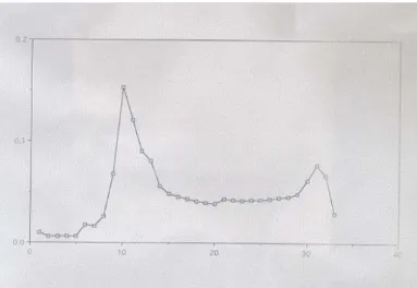
Celite chromatography was done to isolate the 55 kDa protein with the procedure described previously (Lu 1998). 1 g of activated celite was suspended in 100 mM Tris-buffered saline, pH 7.6 (TBS) and added to the column.
Surface proteins from bacteria were harvested by water extraction as described previously (Pei 1988). The water extract (WE) in 1 mM phenylmethylsulphonyl flouride at 4 °C was applied on the matrix at a flow rate of 0.5 ml/minute. The column was washed with 100 ml 100 mM TBS to remove unbound proteins. Bound protein was eluted with 10 ml stepwise gradient of 5~10 mM EDTA and finally with 1M Tris, pH 10.5.
Eluted fractions were checked by spectrophotometer (OD280) to determine the
presence of protein. Finally, the protein elution was confirmed by running the elutions on SDS-polyacrylamide gels. Protein concentration was determined by Bio Rad Protein Assay Kit according to the manufacturer’s instructions.
2.3.3.2 N-Terminal Amino Acid Sequencing
The single eluted protein was sequenced by directly applying protein through automated N-terminal Edman degradation with the High Performance Liquid Chromatography system. For determination of N-terminal amino acid sequence of 55 kDa protein fragments, the protein was dialyzed against dH2O for
overnight, then lyophilized. The protein was dissolved in 300 ml of 0.1 M NH4HCO3, then 20 ml, 1 mg/ml of lysine endopeptidase was added and
incubated at 37 °C in an incubator for 4 hours. 15 ml of the product was applied onto a C4 column and eluted with graded concentration (0-70%) of acetonitrile at a flow rate of 1 ml/min. Elutions were collected at peak. The two highest peaks were subjected to N-terminal sequencing.
2.3.3.3 Glycan Detection
DIG Glycan Detection Kit was used to determine whether our protein was glycosylated or not. Adjacent hydroxyl groups in sugars of glycoconjugates are oxidized to aldehyde groups by mild periodate treatment. The spacer linker steroid hapten digoxigenin (DIG) is then covalently attached to these aldehydes via a hydrazide group. DIG labelled glycoconjugates are subsequently detected in an enzyme immunoassay using a digoxigenin specific antibody conjugated with alkaline phosphatase.
The 55 kDa protein, transferrin and creatinase (5µg/lane) was subjected to SDS-PAGE. After transferring to PVDF membrane, the protocol was followed according to the instruction of the manufacturer.
2.3.3.4 SDS Polyacrylamide Gel Electrophoresis and Western Blot Electrophoretic separation of proteins under denaturing conditions was performed by using the vertical mini gel apparatus. For all experiments 5 % stacking gel and 10% separating gel was used and run at 40 mA. After performing SDS-PAGE, the proteins were transferred from the gel to
nitrocellulose or PVDF membrane at 100 V for 1 hour. Membranes were blocked by 5 % non-fat dry milk or BSA. For detection of the transferred proteins, 4-chloro-1-naphthol method was used (explained in section 2.3.3.5).
2.3.3.5 Colony Lift
This method was used to detect the bacteria expressing the protein of interest on their surfaces. Firstly the bacteria were streaked or spreaded on LB agar plates containing the appropriate antibiotic(s). The membrane was placed onto the plate for 1minute to lift the colonies and was incubated in petri dish containing 10 ml 5% non-fat dry milk (in TBS) for overnight at 4 °C. Then the membrane was washed 3 times with TBS each time for 3 minutes in a rotary shaker. Then the membrane was treated with primary antibody for 90 minutes at a dilution of 1:5000 (0.1 µg/ml) and was washed 3 times with TBS. After the washing step, the membrane was treated for 90 minutes with secondary antibody at a dilution of 1:5000 in TBS. Then the membrane was washed again 3 times with TBS. The detection part of the experiment was done in two different ways:
1. By using ECL reagents: The ECL reagents were mixed and spreaded over the membrane for 1 minute. The film was exposed to the membrane for 1 minute. Then the film was developed in a (name company of the
machine)
2. By using 4-chloro-1-naphthol: Solution A (15 mg 4-chloro-1-naphtol in 5 ml cold methanol) and Solution B(15 µl 30% H2O2 in 20 ml Tris-HCl)
were prepared. A and B were mixed and the solution was incubated with the membranes until the color reaction was seen.
2.3.3.6 Dot Blot
This method was used to confirm the positive signals obtained in the colony lift protocol. 2-3 colonies were picked up from the LB agar and suspended in TBS. 2-3 drops of this suspension were given on to the nitrocellulose membrane. After drying the membrane the procedure was followed as described in colony lift.
2.3.3.7 Attachment Inhibition Assay
Attachment assay is used to measure the attachment ability of bacterial cells to the human pharyngeal epithelial cells. In attachment inhibition assay, whether a molecule can prevent attachment of bacteria to human pharyngeal epithelial
cells is determined. In this study, 2 types of attachment inhibition assays were done:
1. Attachment inhibition assay with protein 2. Attachment inhibition assay with antibody.
In the first type of attachment experiment, the 55 kDa protein was incubated with the pharyngeal epithelial cells. If the protein was an adhesin as we
hypothesized, the exogenously added 55 kDa protein would bind and block the receptor on the epithelial cell and attachment of bacteria would decrease with respect to control. In the second type of attachment experiment, the mAb against 55 kDa protein was incubated with M. catarrhalis. If the protein was an adhesin as we hypothesized, the antibody would bind and block the 55 kDa protein on M.
catarrhalis and attachment of bacteria would decrease with respect to the control.
2.3.3.7.1 Attachment Inhibition Assay with 55 kDa Protein
Attachment inhibition assay was performed as described previously (Ahmed 1996). Pharyngeal epithelial cells were taken from the throat of a healthy adult individual by scraping the oropharynx with a sterile cotton swab. The cells were transferred to 1/15 mM phosphate buffer pH 7.2 (PB) and filtered to remove particles. Then in order to get rid of the normal flora that might interfere with the attachment, the cells were washed 3 times with PB each time for 10 minutes by centrifugation at 80 Xg at room temperature. The number of cells were
determined by Neubauer counting chamber and adjusted to a concentration of 2.5X 10⁴ cell/ml.
Then cells were treated with the 55 kDa protein in a shaking water bath at 37 °C for 30 minutes. Cells without 55 kDa protein treatment was taken as a control. Overnight grown M. catarrhalis was suspended in phosphate buffered saline pH 7.2 (PBS) and adjusted to a concentration of 1X 10⁸ cfu/ml by
spectrophotometer. Then the cells and the bacteria were mixed in a 1:1 ratio and centrifuged at 750 Xg for 10 minutes at room temperature and incubated for 30 minutes at 37 °C. After the incubation, 5 times washing was done with PBS to remove the unattached bacteria. The cells in the suspension, then, collected on a slide with cytospin. The slides were Gram stained and the attached bacteria were