Ankara Üniv Vet Fak Derg, 55, 145-148, 2008
Lipid peroxidation and antioxidant enzymes in rats exposed to
cigarette smoke
Funda KIRAL1, Pınar Alkım ULUTAS1, Ulvi Reha FIDANCI2
1 Department of Biochemistry, Faculty of Veterinary Medicine, Adnan Menderes University, Aydın; 2 Department of Biochemistry, Faculty of Veterinary Medicine, Ankara University, Ankara; Turkey.
Summary: Oxidative metabolism in rats exposed to long term cigarette smoking was investigated in this study. Fourty four rats (22 males and 22 females) were used in this study. 11 males and 11 female rats were assigned into cigarette smoke group and 11 male and 11 female rats were used as control group. The rats assigned into passive smoking were exposed to smoke in a unit for 120 minutes a day for five days a week during 16 weeks. At the end of this period, blood samples were collected from all rats. Plasma MDA and ascorbic acid analysis were performed immediately. Haemoglobin levels and GSH-px, SOD and CAT activities were determined in erythrocytes. The results were evaluated to student t-test. It was observed that plazma MDA levels and GSH-px, and CAT activities were high and plasma ascorbic acid levels were low compared with the control group in male and female rats. SOD activities showed no significant differences. As a result, it was concluded that high plasma lipid peroxidation and low ascorbic acid concentrations may be involved in the pathogenicity of cigarette.
Key words: Antioxidant enzymes, cigarette, lipid peroxidation, rat, vitamin C.
Sigara dumanına maruz bırakılan sıçanlarda lipid peroksidasyonu ve antioksidan enzimler Özet: Bu çalışmada uzun süre sigara dumanına maruz bırakılan sıçanlarda oksidatif metabolizma incelenmiştir. Çalışmada toplam 44 sağlıklı sıçan (22 erkek, 22 dişi) kullanılmıştır. Kontrol grubunda 11 erkek ve 11 dişi, sigara grubunda 11 erkek ve 11 dişi rat bulunmaktadır. Pasif dumana maruz bırakılan sıçanlara 16 hafta boyunca, haftada 5 gün, günde 120 dakika duman uygulanmıştır. Bu periyodun sonunda tüm hayvanlardan kan örnekleri toplanmıştır. Plazma MDA ve askorbik asit analizleri hemen yapılmıştır. Eritrositlerde hemoglobin düzeyleri ve GSH-px, SOD, CAT aktiviteleri belirlenmiştir. Sonuçlar student t-testi ile değerlendirilmiştir. Kontrol grubu ile karşılaştırıldığında erkek ve dişi sıçanların plazma MDA düzeyi ile GSH-px ve CAT aktivitelerinin yüksek, askorbik asit düzeylerinin düşük olduğu görülmüştür. SOD aktivitesi açısından ise her iki grup arasında istatistiksel farka rastlanmamıştır. Sonuç olarak, yüksek plazma lipid peroksidasyonu ve düşük askorbik asit düzeylerinin sigaranın patojenitesini arttırdığı düşünülmektedir.
Anahtar sözcükler: Antioksidan enzimler, lipid peroksidasyonu, sıçan, sigara, vitamin C.
Introduction
Cigarette smoke is a complex mixture of over 4700 chemical compounds including high concentrations of oxidant and free radicals present in gas phase and the tar phase of smoke. Those in the gas phase are both organic and inorganic including reactive oxygen species (ROS) and free radicals, aldehydes, peroxides and oxides nitrogen (23). Cigarette smoke also generates ROS indirectly from activated polymorphnuclear leucocytes or pulmonary alveolar macrophages (2). The increased production of ROS by smoke can produce a condition of oxidative stress that can result in the oxidation of lipids, induction of DNA single-strand breakage, inactivation of certain proteins, and the disruption of biological membranes (12, 24). Cigarette smoking is known to be associated with various chronic pulmoner and cardiovasculer diseases ranging from inflammation to cancer (4, 5, 7).
The damage that can be caused by free radicals is normally minimized by biological antioxidant systems
including enzymatic and non-enzymatic systems. Important antioxidant enzymes include copper (Cu) and zinc (Zn) superoxide dismutase (SOD), manganese SOD, ceruloplasmin (Cp), selenium glutathione peroxidase, glutathione reductase, and catalase. Non-enzymatic antioxidants include vitamin C, α-tocopherol, provitamin A carotenoids, and urate can also act to reduce of free radicals (10, 13, 24).
The aim of this study was to investigate the oxidative metabolism in rats exposed to cigarette smoking.
Materials and Methods
In an attempt to do experiments simultaneously, we brought 20 female and 10 male Sprague Dawley rats from Experimental Animal Unit of Veterinary Medicine School. The animals were put in cages, with two female and one male rat in each cage. They were made to mate and then the male rats were taken from the cages. The rats, born 21 days later, were kept with their mothers for
Funda Kıral - Pınar Alkım Ulutas - Ulvi Reha Fidancı 146
three weeks. Forty four healty rats (22 males and 22 females) were used in this study. 11 males and 11 females rats were assigned into passive smoking for 120 days and 11 male and 11 female rats into control group. The rats were put in the polycarbonate cages 28x28x16cm in size, with two rats in each cage. They were fed with a special feed taken from Gebze-Best Yem Factory and given ad libitum water. So that their feed should not smell smoke and the rats should not receive nicotine orally, their feed were taken away from the cages for two hours every day. After smoking was completed, the rats were fed. At weekends, they were given ad libitum feed.
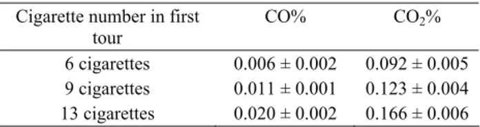
The rats assigned into passive smoking were exposed to smoke in a unit for 120 minutes a day for five days a week during 16 weeks. The unit was made of glass and nylon and 210x70x60cm in size. The smoke entered from one side of the unit and the air in the unit was circulated by an aspirator, the power of which could be adjusted, on the other side of the unit. The label of the cigarette was Birinci (85mm, Tekel, Turkey), with a high rate of nicotine. So that the experimental animals should comply with the regimen, the dose of the smoke was gradually increased. The animals were exposed to smoke of six cigarettes every round lasting for 120 minutes for two weeks, the smoke of nine cigarettes for the next two weeks and the smoke of 13 cigarettes for the following 12 weeks. Percentages of CO and CO2 were measured at
certain intervals (Sun Modular Gas Analyzer 1200, England). The results were shown in Table 1.
Table 1. Percentage of carbonmonoxide (CO%), carbondioxide (CO2%) in smoking environment.
Tablo 1. Sigara dumanı ortamında ölçülen karbonmonoksit ve karbondioksit yüzdeleri
Cigarette number in first tour
CO% CO2%
6 cigarettes 0.006 ± 0.002 0.092 ± 0.005 9 cigarettes 0.011 ± 0.001 0.123 ± 0.004 13 cigarettes 0.020 ± 0.002 0.166 ± 0.006
Blood samples were collected and animals were sacrificed by cervical dislocation under ether anesthesia. For plasma, blood was collected in a heparinized vial and separated in a centrifuge for 15 minutes at 3500 rpm.
The lipid peroxidation of plasma was measured by the TBA method as described by Yoshioka et al. (30). Malondialdehyde (MDA), formed from the breakdown of polyunsaturated fatty acids, was considered as an index for the peroxidation reaction. The absorbance of the reaction product of MDA with TBA was measured at 532 nm. Quantitation was based upon a molar extinction coefficient of 1.56 x 10⁵ M-1 cm-1. The plasma ascorbate
was measured by the phosphotungstic acid method of Kway (14).
For erythrocyte separation, heparinized blood was centrifuged at 3000 rpm for 10 min to separate the red blood cells (RBCs) from the plasma. The sediment containing the erythrocytes were washed repeatedly with an isotonic solution of 0.9% (w/v) NaCl until a colorless supernatant was observed. The cells were destroyed by adding four volumes of double distilled water. The resulting suspension was centrifuged twice to eliminate all of the cell membranes: first by 10 min in the tube centrifuge at 1300 g followed by centrifugation in an Eppendorf centrifuge at 12440 g for 5 min (28).
The hemoglobin was determined by Drabkin’s method in a 0.1/ml aliqout of hemolysate. Absorbance of the resulting cyanomethemoglobin was read 540 nm against a suitable blank in duplicate runs (27).
Glutathione peroxidase (GSH-px) activity was determined by measuring the rate of oxidation of NADPH at 340 nm (21). A unit of enzyme activity is expressed as the amount of enzyme needed to oxidize 1 nmol of NADPH oxidized/min, and the specific activity is expressed as nmol of NADPH oxidized/min/mgHb.
Superoxide dismutase activity estimation was based on the generation of superoxide radicals produced by
xanthine on xantine oxidase, which reacts with 2-(4-iodophenyl)-3-(4-nitrophenol)-5 phenyltetrazolium chloride to form a red formazon dye. The SOD activity is then by the degree of inhibition of this reaction. Erythrocyte SOD activity was expressed as U/gHb (26).
The activity of catalase (CAT) was measured by following the rate H2O2 hydrogen peroxide decomposition
at 240 nm (15). The enzyme activity is expressed as k/gHb.
Differences between the two study groups were established by means of Student’s t-test with p<0.05 as limit of significance.
Results
Results of TBA tests, serum ascorbic acid levels and GSH-px, SOD, CAT activities have been presented in Table 2 and Table 3 female and male rats.
Table 2. Parameters in control and cigarette smoke group of female rats. Data expressed as x±SD.
Tablo 2. Sigara dumanına maruz bırakılan gruptaki ve kontrol grubundaki dişi sıçanlara ait istatistiksel değerler
Parameter Controls (n=11) x ± SD Cigarette smoke group (n=11) x ± SD p Malondyaldehyde (μmol/L) 18.35 ± 1.23 27.19 ± 2.77 <0.05 Ascorbic acid (mg/dl) 0.62 ± 0.07 0.15 ± 0.07 <0.001 GSH-Px (nmol/NADPH+H+/ min/ mg-Hb) 2234 ± 152 6470 ± 1410 <0.05 SOD (U/g-Hb) 141.35 ± 13.9 229.77 ± 40.2 >0.05 CAT (k/g-Hb) 16.66 ± 1.7 24.12 ± 2.5 <0.05
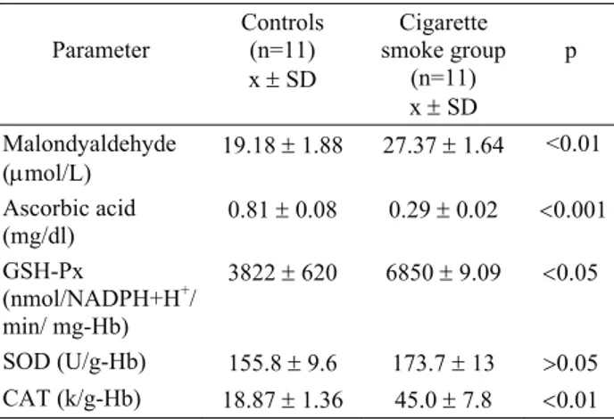
Ankara Üniv Vet Fak Derg, 55, 2008 147 Table 3. Parameters in control and cigarette smoke group of
male rats. Data expressed as x ±SD.
Tablo 3. Sigara dumanına maruz bırakılan gruptaki ve kontrol grubundaki erkek sıçanlara ait istatistiksel değerler
Parameter Controls (n=11) x ± SD Cigarette smoke group (n=11) x ± SD p Malondyaldehyde (μmol/L) 19.18 ± 1.88 27.37 ± 1.64 <0.01 Ascorbic acid (mg/dl) 0.81 ± 0.08 0.29 ± 0.02 <0.001 GSH-Px (nmol/NADPH+H+/ min/ mg-Hb) 3822 ± 620 6850 ± 9.09 <0.05 SOD (U/g-Hb) 155.8 ± 9.6 173.7 ± 13 >0.05 CAT (k/g-Hb) 18.87 ± 1.36 45.0 ± 7.8 <0.01
Plasma MDA levels were increased in all groups. Mean MDA levels were significantly high (p<0.05) to compare with control groups in female rats. Mean MDA levels in male rats were high (p<0.01) to compare with control group.
Mean plasma ascorbic acid levels in female and male experimental group rats were determined significantly low (p<0.01). However SOD activities were shown no significant differences. Erytrocyte GSH-px and CAT activities were found to be significantly high in female and male rats (p<0.01 and p< 0.01).
Discussion and Conclusion
Two major phases were identified in cigarette smoke: a tar phase and a gas phase; both phases are rich in oxygen-centered, carbon-centered and nitrogen-centered free radicals as well as non-radical oxidants (8). From the analysis of each phase, it was estimated that a single cigarette puff contains approximately, 1014 free
radicals in the tar phase, and 1015 radicals in the gas
phase (23). Free radicals in the tar phase of cigarette smoke appear to be relatively stable semiquinone and those in the gas phase include short lived carbon and oxygene centered organic radicals (11).
It is reported that alveolar macrophage numbers in cigarette smokers were increased by at least 2 - 4 times and neutrophil numbers by 10 times (29). In vitro studies have shown that alveolar leukocytes and macrophages from cigarette smokers spontaneously release increasing amounts of oxidants such as O2 and H2O2 compared with
non-smokers (23). Similarly, in pasive smokers, it was determined that peripheral blood leucocyte numbers and releasing of oxidants are increased (1). These oxidants which are released from the inflamatuar cells may cause cell damage (11).
Plasma MDA (a product of lipid peroxidation) levels can be expected to be high in smokers since the cigarette smoke is known to contain reactive peroxyl radicals and acetaldeyte which can increase lipid
peroxidation (6,9,18). It was determined that mean plasma MDA levels were significantly higher in both female and male rats than the control group (p<0.05) (p<0.001). This result might be connected to the increase in production of free oxygen radicals by polymorph nuclear leucocytes. Free oxygen radicals also caused tissue and microvasculer damage. These changes may result in disturbed membrane transport and altered membrane integrity. Smoking is an important risk factor in the course of ischemic heart disease. Atherosclerosis in coroner arters was determined in patients with ischemic heart disease. Malondialdehyde can alter LDL: This altered LDL can be taken up avidly by macrophages. The LDL in the atherosclerotic plaque has been shown to be structurally similar to LDL alterly by MDA (18, 22).
Ascorbic acid (vitamin C) is the major essential water-soluble antioxidant (19, 25). It is present extracellularly in relatively high concentrations in the blood plasma. Vitamin C can protect DNA from oxidant-mediated damage, and has been reported to neutralize phagocyte-derived oxidants protecting the α1-protease inhibitor (API) from oxidant-mediated functional inactivation (16). It was determined that plasma ascorbic acid levels in smokers were lower than in non-smoking individuals (20, 25). Similarly, plasma ascorbic acid levels were determined low in both groups.
The vitamin C concentration is lower in blood plasma of cigarette smokers compared to non-smokers, possibly due to increased turnover of the vitamin, increased oxidative stress in smokers, or other mechanisms as yet unknown. It could also be due to its increased consumption during recycling of vitamin E or β-carotene that are directly oxidized in the course of scavenging free radicals and reactive oxidant species in cigarette smoke. Nevertheless, it has also been suggested that neither of these antioxidants is an extremely efficient scavenger of cigarette smoke-induced radicals (13).
It was stated that erythrocyte enzyme activity in cigarette smokers are increased (29, 31). This is a natural result of the increase of alveolar macrophage numbers and excessive increase of H2O2 and products of H2O2
derives which is released from the neutrophiles (17). In this study, GSH-Px and CAT activities were determined high both in female and male rats but increase of SOD activities were not significantly important.
Bolzan et al. (3) have observed no differences in the antioxidant enzyme activities of smokers and nonsmokers, besides catalase activity were determined lower in females than males.
Erythrocyte antioxidants have protective effects against oxidative damage. It is known that alterations in erythrocyte antioxidants in smokers might cause emphysema and vascular damage. The development of smoking-related diseases depends on a fine balance between oxidant and antioxidant levels in the tissues. Changes in the erythrocyte antioxidants could be able to reveal this balance. In these conditions, determination of
Funda Kıral - Pınar Alkım Ulutas - Ulvi Reha Fidancı 148
the production of oxygen metabolites could play a key role in determination of tissue damage.
As a result, it was observed that, increase of free oxygen radicals which are released from inflammatory cells in cigarette smokers, causes the increase of MDA and erythrocyte antioxidants. The increase in the production of erythrocyte antioxidants may be parallel to the increase in lung and other tissue antioxidants. Because of that, this would be an effective marker of oxidant stress in lung and other tissue. Changes of erythrocyte antioxidants may be important to predict the respiratory or cardiovascular system diseases and individual sensivity.
As a result, it was concluded that high plasma lipid peroxidation and low ascorbic acid concentrations may be involved in the pathogenicity of cigarette.
References
1. Andersen R, Theron AJ, Richards GA, Myers MS, Rensberg JV (1991): Passive smoking by human sensitizes circulating neutrophils. Am Rev Respir Dis, 144, 570-574.
2. Behr J, Nowak D (2002): Tobacco smoke and respiratory disease. Eur Respir Monograph, 21, 161-179.
3. Bolzan AD, Bianchi MS, Bianchi NO (1997): Superoxide dismutase, catalase and glutathione peroxidase activities in human blood: influence of sex, age and cigarette smoking. Clin Biochem, 30, 449-454.
4. Clara JG, Coelho C, Breintenfelt L, Siqueira C (2001): Acute effects of tobacco and vascular risk modulated by genetic factors. Rev Port Cardiol, 20, 103-109.
5. Crowley-Weber CL, Dvorakova K, Crowley C, Bernstesin H, breinstein C, Garewal H, Payne CM (2003): Nicotine increases oxidative stress, activates NF-kappaB and GRP78, induces apoptosis and sensities cells to genotoxic/xenobiotic stresses by a multiple stress inducer, deoxycholate: relevance to colon carcinogenesis. Chem Biol Interact, 145, 53-66.
6. Demir S, Özkurt S, Köseoğlu M, Enli Y, Aslan D, Gümüşsu. N (2001): Sigara içenlerde plazma lipid peroksidasyonu. Solunum, 3, 57-59.
7. Doll R (1996): Cancers weakly releated to smoking. Br Med Bull, 52, 35-49.
8. Fisher EB, Debra JR, Morgan DG, Rehberg H, Rost K (1990): Smoking and smoking cessation. Am Rev Respir Dis, 142, 702-720.
9. Gültekin F, Gökduman A, Özgüner F, akdoğan M, Sütçü R, Delibaş N (2001): Sigara içenlerin antioksidan savunma sistemindeki değişmeler. Tuberk Toraks Derg, 49, 259-264.
10. Halliwell B, (1994): Free radicals, antioxidants, and human disease: curiosity, cause, or consequence? Lancet, 344, 721-724.
11. Hunninghake WG, Crystal RG (1993): Cigarette smoking and lung destruction. accumulation of neutrophis in the lungs of cigarette smoking. Am Rev Respir Dis, 128, 833-838. 12. Kalra J, Chaudhary AK, Prasad K (1991): Increased
production of oxygen free radicals in cigarette smokers. Int J Exp Path, 72, 1-7.
13. Kim HS, Lee BM (2001): Protective effects of antioxidant supplementation on plasma lipid peroxidation in smokers. J Toxicol Environ Health, 24, 583-98.
14. Kway A (1978): A simple colorimetric method for ascorbic acid determination in blood plasma. Clin Chim Acta, 86, 153-157.
15. Luck H (1965): Catalase. 855-884, In: Methods in Analysis, HU Bergmeyer (Ed), Academic Press, London. 16. Marrow JD, Frei B, Longwire AW, Gaziano JM, Lynch
SM, Shyr J (1995): Increase in circulating products of lipid peroxidation in smokers. Smoking as a cause of oxidative damage. N Eng J Med, 332, 1198-203.
17. Mccusker K, Hoidal J (1990): Selective increase of antioxidant enzyme activity in the alveolar macrophages from cigarette smokers and smoke-exposed hamsters. Am Rev Respir Dis, 141, 678-682.
18. Nadiger HA, Mathew CA, Sadasivudu B (1987): Serum malondialdehyde (TBA reactive substance) levels in cigarette smokers. Atherosclerosis, 64, 71-73.
19. Naidu KA (2003): Vitamin C in human healty and disease is still a mystery? an overwiev. Nutr J, 21, 2-7.
20. Ozan E, Çolakoğlu N, Sönmez MF, Ozan S, Yılmaz S, Taşdemir B, Ozan G (2005): Sigara inhalasyonunun trakeada oluşturduğu yapısal değişiklikler üzerine melatonin ve C vitamininin etkileri. Fırat Tıp Derg, 10, 40-44. 21. Paglia DE, Valentine WN (1967): Studies on the
qualitative and quantitative characterization of erytrocyte glutathione peroxidase. J Lab Clin Med, 70, 158-169. 22. Prasad K, Kalra J (1989): Experimental atherosclerosis
and oxygen free radicals. Angiology, 40, 835-843. 23. Rahman I, Macnee W (1996): Oxidant-antioxidant
imbalance in smokers and chronic obstructive pulmonary disease. Thorax, 51, 348-350.
24. Sohn HO, Lim HB, Lee DW, Kim YT (1993): Effect of subchronic administration of antioxidants aganist cigarette smoke exposure in rats. Arch Toxicol, 67, 667-673. 25. Steinberg FM, Chait A (1998): Antioxidant vitamin
supplementation and lipid peroxidation in smokers. Am J Clin Nutr, 68, 319-27.
26. Sun Y, Oberley LW, Li YA (1988): A simple for clinical assay of superoxide dismutase. Clin Chem, 34, 397-500. 27. Tietz WN (1987): Measurement of plasma haemoglobin.
803-813. In: Fundamental of Clinical Chemistry. Third ed, Saunders Company, Philadelphia.
28. Winterborn CC, Hawkins RE, Brain M, Carrel W (1975): The estimation of red cell superoxide dismutase activity. J Lab Clin Med, 55, 337-341.
29. Yıldız D (2004): Nicotine, its metabolism an overview of its biological effects. Toxicon, 43, 619-32.
30. Yoshioka T, Kawada K, Shimada T (1979): Lipid peroxidation in maternal and cord blood and protective mechanism against active-oxygen toxicity in the blood. Am J Obstet Gynecol 135, 372-379.
31. Yöntem M, Çevrim EE, Kaleli S (2002): Sigara içen ve içmeyen kişilerde süperoksit dismutaz, glutatyon redüktaz, ferritin ve hemoglobin düzeylerinin araştırılması. Süleyman Demirel Üniv Fen Bil Enst Derg, 6, 79-86.
Geliş tarihi: 26.02.2008 / Kabul tarihi: 04.12.2008 Yazışma adresi
Funda Kıral
Adnan Menderes Üniversitesi Veteriner Fakültesi
Biyokimya Anabilim Dalı Işıklı / Aydın