Does acoustic trauma occur in pointers due to firearm noise?
A prospective study on 50 hunting dogs
Özlem ŞENGÖZ ŞİRİN
1, Yusuf Sinan ŞİRİN
1, Ömer BEŞALTI
21Mehmet Akif Ersoy University Veterinary Faculty Surgery Department, Burdur, Turkey, 2Ankara University Veterinary Faculty Surgery Department, Ankara, Turkey.
Summary: This study investigates the possibility of acoustic trauma occurence in hunting dogs exposed to high noise with pure stimulant such as firearms. Ten puppies (Control Group-CG) were used to establish reference for Brainstem Auditory Evoked Response (BAER) and tympanometry records. BAER and tympanograms were collected from the 40 cases in active hunting group (Acoustic Trauma Group-ATG). The severities of trauma of which the cases were exposed to were determined by field study. While tympanometric pressure values of ATG cases were higher than that in CG, they were found to be in normal range (ATG left: 35,63±38,79/right: 34,00±38,25; CG left: -23,90±44,30/ right: -29,20±36,87 daPa). BAER records were saved using both click and tone burst stimuli. Records were taken at the frequencies of 0,5, 1, 2, 4, 6, 8-kHzwith tone burst stimulus. Wave I latency values obtained with click stimulus were only found to be significant in right ear at 30 dB intensity. Wave V was observed to be the last disappearing and the most observed wave among the waves obtained with high frequency stimulus. Based on this study, it was concluded that dogs experience acoustic trauma due to firearm noise, as revealed by a substantial decline in amplitude values in BAER records obtained with click stimulus. It can be said that the use of products developed for dogs and raising awareness in hunters may also be beneficial to prevent harmful effects of acoustic trauma.
Keywords: Acoustic trauma, BAER, hearing loss, hunting dog, tympanogram.
Av köpeklerinde ateşli silah gürültüsü ile akustik travma olur mu?
50 av köpeğinde prospektif bir çalışma
Özet: Bu çalışmada, ateşli silahlar gibi saf uyaranlı yüksek şiddetli gürültüye maruz kalan av köpeklerinde akustik travma olup olmadığının araştırılması amaçlandı. Referans Beyin Sapı İşitsel Uyandırılmış Yanıtları (BAER) ve timpanometri kayıtlarını oluşturmak için 10 av köpeği (Kontrol Grubu-CG) kullanıldı. 40 olgudan oluşan aktif av grubundan (Akustik Travma Grubu-ATG) BAER ve timpanogramlar alındı. Olguların maruz kaldığı travmanın şiddeti saha çalışması ile belirlendi. ATG olgularının timpanometrik basınç değerleri CG'deki değerlerden daha yüksek iken (ATG left: 35,63 ± 38,79 / right: 34,00 ± 38,25; CG left -23,90 ± 44,30 / right: -29,20 ± 36,87 DaPa) BAER kayıtları hem klik hem de tone burst uyaranları kullanılarak kaydedildi. Kayıtlar, tone burst uyarı ile 0,5, 1, 2, 4, 6, 8-kHz frekanslarında alındı. Klik uyaranı ile elde edilen Dalga I gecikme değerlerinin sağ kulakta 30 dB yoğunlukta anlamlı olduğu bulundu. Dalga V, yüksek frekans uyarısı ile elde edilen dalgalar arasında en son kaybolan ve en çok gözlenen dalga olarak gözlendi. Bu çalışma ile ateşli silah gürültüsüne bağlı akustik travmanın köpeklerde klik uyaranla elde edilen BAER kayıtlarında amplitüd değerlerindeki belirgin düşüşle ortaya konulabileceği sonucuna varıldı. Akustik travmanın zararlı etkilerinden korunmak için avcıların bilinçlendirilmesi ile köpekler için geliştirilen ürünlerin kullanımının da faydalı olabileceği söylenebilir.
Anahtar sözcükler: Akustik travma, av köpeği, BAER, işitme kaybı, timpanogram.
Introduction
With the increased consideration given to pet animal healthcare by their owners, expectations from veterinary surgeons are increasing. To meet these requests high technology medical devices are being extensively used to get a detailed diagnostic and prognostic data. Electrodiagnostic examinations have gradually becoming more widely used tests. When the diagnostic tests are interpreted in conjunction with signalement, anamnesis,
physical and neurological examinations that are fundamental in a neurological study, they provide avoidance from invasive applications. Determination of the prognosis and support or confirmation of suspected neuroanatomical diagnosis before starting treatment is possible. Deafness is the absence of sense of hearing (1). Deafness can be classified as congenital or late presented; hereditary or acquired; conductive or sensorineural (2). Outer and middle ear are responsible for conduction of
sound signal to inner ear; therefore, conductive deafness is associated with the conditions involving these sites. Sensorineural deafness refers to the disorders of cochlea involving the sensory hair cells. Excessive noise causes acquired sensorineural hearing loss (3).
Peak sound pressure level (SPL) of more than 140 dB (4) was reported in some of the studies performed with firearms and more than 155 dB in some others. These values substantially exceed the maximum permissible noise level (5). Exposure to firearms noise causes a sensorineural type hearing loss (6,7). Dogs with conductive hearing disorder can be successfully treated medically and surgically, and hearing may be regained in some cases. As the death of hair cells cannot be repaired, there is no treatment in dogs with sensorineural hearing loss (3).
History, clinical examination, impedance audiometer and BAER tests are utilized for the diagnosis of hearing loss (8). BAER is a test used in objective examination of hearing (9). This test shows brain activities induced with auditory stimulus, and reflects the electrophysiological process of sensory cells through all hearing pathways (10). After it was established at the end of 1970s, BAER took its place in quantitative analysis of hearing functions in dogs as a reliable, objective, reproducible and noninvasive test (3,11). The purpose of BAER examination is to determine whether there is a deafness (uni- or bilateral) or not, its severity and localization of the lesion (central/peripheral hearing pathways) (10). It is also used in conditions such as brain stem injury due to demyelination, brain stem tumors, vascular insufficiencies and trauma (12).
Tympanometer involves the measurement of middle ear compliance by changing the pressure in outer ear canal from +200 mmH2O to -200 mmH2O. The obtained graph is called tympanogram. Tympanometer also provides an indirect assessment of middle ear pressure via determination of the pressure in which the compliance is maximum (8). Tympanometry provides information regarding the function of the middle ear and helps to determine whether the hearing loss is conductive or sensorineural (3). At the present time, tympanometric data in clinical conditions in dogs are limited to a few reports. Normal tympanogram in an animal with hearing disorder supports a sensorineural deficit. A variation in tympanogram shape reveals the shape of abnormality in middle ear. Presence of fluid in middle ear generally causes a shift to the left in maximum compliance or a straight trace which shows minimal compliance in tympanic membrane and middle ear cavity. Another reason for shifting to the left is the dysfunction of Eustachian tube causing negative pressure in middle ear (8).
When a loud noise is applied to a healthy animal’s ear, muscles in middle ear are contracted. This activity causing a temporary change in impedance properties of the structures in middle ear is called acoustic reflex (AR). It is a noninvasive, objective and reliable method in the examination of the integrity of tympanic membrane. The results of AR test provide useful information to diagnose audiologic, otologic and neurologic disorders. AR threshold in dogs was reported to be 70-90 dB SPL for ipsilateral reflexes and 70-100 dB HL for contralateral reflexes. Biphasic responses are generally related with high stimulus severity in dogs and cats (8).
In human medicine, there are acoustic trauma studies performed in shooters (policemen, soldiers, hunters) exposed to noise with high-intensity pure stimulant such as firearm noise (20). As a result of those studies, the presence of acoustic trauma in shooters due to firearm noise has been revealed. However, no studies evaluating or investigating the effect of acoustic trauma in hunting dogs exposed to high noise during hunt and the corresponding hearing loss was found in literature. Therefore, uncertainties regarding hearing and acoustic trauma were tried to be answered with this prospective study in which a substantial uniformity was ensured (same breed hunting dog, same type and brand firearm, restricting the position of dog and hunter with 1 m during hunting).
Materials and Methods
Study material consisted of 50 German Shorthaired Pointer (GSP) dogs in total. Since no reference values were found for GSP days in literature, reference BAER test values (Synergy CareFusion 5 channel EMG-EP® device) were obtained from 10 dogs with normal neurological and video-otoscopic examination findings which have not been to hunting before. Acoustic Trauma Group (ATG) of the study consisted of 40 GSP dogs (20 females, 20 males) exposed to firearm noise. Control Group (CG) and ATG ages ranged from 12 - 18 weeks (4,2 months) and 18 - 46 months (3,8 years), respectively. As study details were shared with hunters, dogs were brought to the clinic to perform hearing tests right after hunting. The intensity of firearm noises exposed during field studies was measured by a special device (Sinus, Tango, Sound Level Meter, Sinus Messtechnik GmbH®, Germany) as Lcpeak value. After the procedure repeated 10 times, 143,1 dB Lcpeak value, which was the maximum value which the device can measure, was determined. By imitating the hunting position of the dog in the field study, microphone was placed in approximately 1 m front of the barrel, and then the noise caused by gunfire was recorded as measured. The reason for this distance was that the hunters preferred approximately 1 m between them and their dogs.
Generated values were used to determine the intensity of the noise of which the hunting dogs were exposed and whether it caused acoustic trauma or not.
It was crucial to select the dogs without any history of ear problem and thus only the dogs without any previous history of ear disorder were included in the study. After detailed neurological and video-otoscopic examination (Karl Storz Tele Pack VetX®) tympanograms
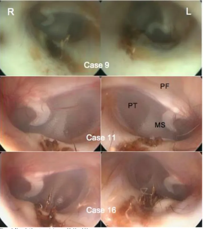
(Interacoustics AT235h®) of the patients were recorded under anesthesia with xylazine-ketamine combination, and their ears were evaluated with video-otoscopy. Vertical - horizontal canal of outer ear and parts of tympanic membrane were viewed and recorded. The exemplary normal video-otoscopic images of 9, 11 and 16 cases are shown in Figure 1.
Figure 1. Normal video-otoscopic images of 9, 11 and 16 cases. Şekil 1: Olgu 9, 11 ve 16’nın normal video-otoskopik görünümleri.
Figure 2. BAER of a CG case obtained with click stimulus at different hearing levels (70 to 20 db NHL)*.
Şekil 2: Bir CG olgusunun değişik işitme seviyelerinde klik uyaranla elde edilmiş BAER (70- 20 db NHL)*db NHL)*.
Figure 3. BAER of a ATG case obtained with click stimulus at different hearing levels (70 to 30 db NHL)*.
Şekil 3: Bir ATG olgusunun değişik işitme seviyelerinde klik uyaranla elde edilmiş BAER (70- 20 db NHL)*.
Figure 4. BAER of a CG case obtained with high frequency toneburst stimulus at different hearing levels (60 to 10 db NHL)*.
Şekil 4: Bir CG olgusunun değişik işitme seviyelerinde toneburst uyaranla elde edilmiş BAER (60-10 db NHL)*.
Figure 5. BAER of a ATG case obtained with high frequency toneburst stimulus at different hearing levels (60 to 10 db NHL)**. Şekil 5: Bir ATG olgusunun değişik işitme seviyelerinde toneburst uyaranla elde edilmiş BAER (60-10 db NHL)**.
*BAER recordings at 1 𝜇V sensitivity level. *BAER kayıtları 1 𝜇V hassasiyetinde gösterildi. ** BAER recordings at 500 nV sensitivity level. **BAER kayıtları 500 nV hassasiyetinde gösterildi.
For tympanometric examination, right and left ears of the cases were evaluated individually, and their volume, compliance and pressure values were recorded along with acoustic reflex data (Interacoustics AT235h®). After insertion of the mushroom-shaped ear tips into the ear canals in a well seated position to prevent air leakage; typanograms, ipsilateral and contralateral acoustic reflexes recorded respectively.
Obtained data was examined using Student t test (Table 1). Cases which were found to be completely normal in all of these examinations underwent BAER test. Same anesthesia protocol (Xylazine; 1-2mg/kg, IM-Alfazyne®2% injectable, Egevet and Ketamine; 2-4 mg/kg, IM-Ketasol® 10% injectable, Interhas) was applied to all animals after neurological examination. The depth of the anesthesia was monitored at 10-minute intervals by checking palpebral-corneal reflexes, jaw tonus, respiration and eye position. BAER recordings were taken using both clique and tone burst stimuli. Records were taken at 0,5 kHz-1 kHz-2 kH-–4 kH-6 kHz-8 kHz frequency with tone burst stimulus. As BAER records are affected from patient’s body temperature changes, patients body temperatures were monitored (core temperature) through all tests, and heated table was used to avoid hypothermia during anesthesia. Moreover, as hypotension may affect BAER test, mean arterial pressure was measured in a noninvasive manner at 10-minute intervals, and was ensured to remain in the normal range (> 60 mmHg). Stimulus intensity was dropped by 10 dB at a time starting from 70 dB NHL, and recording was taken up to threshold value at which trace disappeared. Same type foam-end earphones were used to provide stimuli. Teflon coated stainless steel needles were placed into vertex, and mastoid region of ipsilateral ear via subcutaneous route. Needle with same properties was placed into neck region as a ground electrode. The impedance of electrodes was ensured to be less than 5kΩ. Latency, amplitude, I-III and I-V interpeak latencies, differences within the same ear
and contralateral ear of the obtained peaks (I to VI) were evaluated by measuring the cranial diameters of the dogs (cranial diameter was measured between the broadest arcus zygomaticus locations with calipers). Latency and amplitude values of waves I to VI and I-III, III-V and I-V interpeak latencies were compared between groups by Student t test (Figure 2, 3). BAER waves obtained with tone burst stimulus at various frequencies (8, 6, 4, 2, 1 and 0,5 kHz) were evaluated morphologically (Figures 4, 5). Normal latency, amplitude and central conduction rate were utilized in determination of the compliance of traces between hearing level and both ears.
Statistical Analysis: Student t test was used for
BAER and tympanometric results. For tympanometric results, right and left ears of the cases were evaluated individually, and their volume, compliance and pressure values were recorded along with acoustic reflex data. For BAER results, latency and amplitude values of waves I to VI and I-III, III-V and I-V interpeak latencies were compared between groups by Student t test. The study datas were assessed using means and standard deviations (mean ± SD).
Results
Study material consisted of 50 GSP dogs in total, 40 of them being in acoustic trauma group (ATG) and 10 of them in control group (CG).
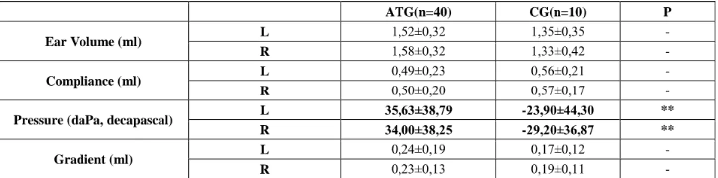
As a result of clinical, video-otoscopic and tympanometric examinations, dogs which were determined to have healthy ears were included into the study and their BAER records were collected. Tympanometric results were not significantly different according to ear volume, compliance and gradient. Statistically significant ATG and CG tympanometric pressure values were found 35,63±38,79, -23,90±44,30 daPa for left and 34,00±38,25, -29,20±36,87 daPa for right ear, respectively (Table 1).
Table 1. Comparison of various tympanometric values of left and right ears between groups.
Tablo 1: Gruplar arasında değişik timpanometrik verilerin sol ve sağ kulakta karşılaştırılması (mean±SD).
ATG(n=40) CG(n=10) P
Ear Volume (ml) L 1,52±0,32 1,35±0,35 -
R 1,58±0,32 1,33±0,42 -
Compliance (ml) L 0,49±0,23 0,56±0,21 -
R 0,50±0,20 0,57±0,17 -
Pressure (daPa, decapascal) L 35,63±38,79 -23,90±44,30 **
R 34,00±38,25 -29,20±36,87 **
Gradient (ml) L 0,24±0,19 0,17±0,12 -
R 0,23±0,13 0,19±0,11 -
*p<0.05 and **p< 0,001 was accepted as statistically significant. * p<0.05 ve **p< 0,001 istatistiksel olarak anlamlı kabul edildi. - not significant.
Table 2. Comparison of BAER wave I amplitude values (µV) of left and right ears obtained with click stimulus at various decibel levels between groups.
Tablo 2: Gruplararasında değişik desibel seviyelerinde klik uyaranla sol ve sağ kulaklardan elde edilen BAER’de I.dalga amplitüd değerlerinin (µV) karşılaştırılması.
dB NHL ATG CG n ATG/CG P 70L-I 2,05±0,78 3,57±1,18 40/10 ** 70R-I 2,20±0,83 3,72±1,57 40/10 * 60L-I 1,77±0,75 3,36±1,07 40/10 ** 60R-I 1,85±0,74 3,31±1,49 40/10 * 50L-I 1,36±0,85 2,70±1,20 40/10 * 50R-I 1,43±0,77 2,61±1,05 40/10 * 40L-I 0,59±0,49 1,50±0,96 35/9 * 40R-I 0,59±0,50 1,29±1,34 36/8 - 30L-I 0,37±0,31 0,78±0,49 14/7 - 30R-I 0,31±0,36 0,37±0,21 12/6 - 20L-I 0,31±0,12 0,38±0,12 3/6 - 20R-I - - - - 10L-I - - - - 10R-I - - - -
*p<0.05 and **p< 0,001 was accepted as statistically significant. * p<0.05 ve **p< 0,001 istatistiksel olarak anlamlı kabul edildi. - not significant.
- istatistiksel olarak anlamlı bulunmadı.
Table 3. Comparison of BAER wave II amplitude values (µV) of left and right ears obtained with click stimulus at various decibel levels between groups.
Tablo 3: Gruplararasında değişik desibel seviyelerinde klik uyaranla sol ve sağ kulaklardan elde edilen BAER’de II.dalga amplitüd değerlerinin (µV) karşılaştırılması.
dB NHL ATG KG n ATG/KG p 70L-II 1,64±0,78 3,50±1,48 40/10 ** 70R-II 1,89±0,96 3,30±2,03 40/10 - 60L-II 1,43±0,65 3,01±0,99 40/10 ** 60R-II 1,56±0,72 2,94±1,67 40/10 * 50L-II 1,16±0,65 2,39±0,96 40/10 ** 50R-II 1,34±0,68 2,40±1,25 40/10 * 40L-II 0,61±0,45 1,58±0,77 36/9 * 40R-II 0,68±0,53 1,15±0,45 37/9 * 30L-II 0,39±0,33 0,88±0,42 21/7 * 30R-II 0,33±0,41 0,50±0,38 27/7 - 20L-II 0,42±0,33 0,49±0,19 4/7 - 20R-II 0,72±0,76 0,25±0,04 3/3 - 10L-II - - - - 10R-II - - - -
*p<0.05 and **p< 0,001 was accepted as statistically significant. * p<0.05 ve **p< 0,001 istatistiksel olarak anlamlı kabul edildi. - not significant.
- istatistiksel olarak anlamlı bulunmadı.
Table 4. Comparison of BAER wave III amplitude values (µV) of left and right ears obtained with click stimulus at various decibel levels between groups.
Tablo 4: Gruplararasında değişik desibel seviyelerinde klik uyaranla sol ve sağ kulaklardan elde edilen BAER’de III.dalga amplitüd değerlerinin (µV) karşılaştırılması.
dB NHL ATG CG n ATG/CG P 70L-III 1,04±0,52 2,15±1,11 40/10 * 70R-III 1,12±0,59 2,21±1,26 40/10 * 60L-III 0,74±0,32 1,66±0,69 40/10 ** 60R-III 0,83±0,53 1,62±0,87 40/10 * 50L-III 0,54±0,37 1,02±0,61 40/10 * 50R-III 0,56±0,37 1,07±0,55 40/10 * 40L-III 0,32±0,16 0,81±0,54 31/9 * 40R-III 0,38±0,26 0,65±0,35 34/9 * 30L-III 0,27±0,12 0,68±0,28 18/7 * 30R-III 0,28±0,17 0,54±0,28 28/7 * 20L-III 0,23±0,09 0,36±0,16 5/5 - 20R-III 0,37±0,04 0,18±0,05 3/2 * 10L-III - - - - 10R-III - - - -
*p<0.05 and **p< 0,001 was accepted as statistically significant. * p<0.05 ve **p< 0,001 istatistiksel olarak anlamlı kabul edildi. - not significant.
- istatistiksel olarak anlamlı bulunmadı.
Table 5. Comparison of BAER wave V amplitude values (µV) of left and right ears obtained with click stimulus at various decibel levels between groups.
Tablo 5: Gruplararasında değişik desibel seviyelerinde klik uyaranla sol ve sağ kulaklardan elde edilen BAER’de V.dalga amplitüd değerlerinin (µV) karşılaştırılması.
dB NHL ATG CG n ATG/CG p 70L-V 1,27±0,62 2,72±0,62 40/10 ** 70R-V 1,10±0,58 2,53±0,74 40/10 ** 60L-V 1,01±0,54 2,55±0,76 40/10 ** 60R-V 1,00±0,47 2,28±0,78 40/10 ** 50L-V 0,80±0,52 1,97±0,61 40/10 ** 50R-V 0,78±0,36 1,62±0,60 40/10 ** 40L-V 0,56±0,35 1,77±0,60 37/9 ** 40R-V 0,59±0,32 1,34±0,74 39/9 * 30L-V 0,38±0,31 1,56±0,62 26/9 ** 30R-V 0,40±0,34 0,75±0,66 27/8 - 20L-V 0,43±0,34 1,28±0,56 5/6 * 20R-V 0,46±0,51 0,89±0,82 8/3 - 10L-V - - - - 10R-V - - - -
*p<0.05 and **p< 0,001 was accepted as statistically significant. * p<0.05 ve **p< 0,001 istatistiksel olarak anlamlı kabul edildi. - not significant.
BAER obtained with click stimulus with different hearing levels revealed statistically significant lower amplitudes in ATG than CG except of recorded from the right ears at 70 db NHL for V. and 30 db NHL for II. waves (Table 2-5). There was no statistically significant difference between groups only for the IV. waves at any hearing level.
Discussion and Conclusion
There are many studies indicating the effect of cranial diameter on BAER values (10,11). In order to minimize this possible effect, cranium diameters were measured to calculate latency and amplitude values accordingly. To prevent interbreed variation only GSP dogs were included in this study. Moreover, in order to minimize the age effect, dogs which completed their development in terms of BAER traces, and which do not have age-induced changes in hearing (3 months-5 years) were included in the study. In dogs, adult data for BAER is achieved at the age of 6-8 weeks (8). As differences between females and males were reported in some human studies, study material was ensured to consist of equal numbers of female and male dogs both in control and trauma groups. But it was no statistically significant differences between male and female dogs in control and trauma groups.
Though tympanometric recordings provide useful information easily about middle ear pathology, it is underused in veterinary practice. Also it is very helpful to rule out middle ear abnormalities that cannot be detected easily without the tympanometry device. Although tympanometric pressure values of ATG cases were higher than CG, they were found to be in normal range. In humans +50 to -50 daPa tympanometric pressure is considered as normal (16). In one of the few studies performed in dogs, mean tympanometric values were reported to be 53,94 ± 55,26 daPa in one group and 65,61 ± 35,13 daPa in the other (17). According to these information it was concluded that middle ears of these dogs were typmanometrically normal.
Waves I and V are used to predict the hearing threshold using click stimulus in dogs. As wave V is the least affected and the last disappearing one among the waves obtained with high frequency stimulus, it is used for the determination of threshold (18). As the waves obtained in certain frequencies with tone burst stimuli had small amplitude and as their location cannot be determined always compared to the waves obtained with click stimuli, latency and amplitude values were evaluated morphologically without measurement. Consistent with literature data, wave V was observed to be the last disappearing and the most observed wave. Generally, waves obtained with tone burst stimulus in ATG cases
were observed to be more undetermined and with much lower amplitude than CG.
Exposure to noise higher than 140 dB NHL may cause permanent hearing loss (4). Firearm noise measured to be 143,1 dB Lcpeak in field was found to be high enough to cause hearing loss due to acoustic trauma. In a detailed study in which the risk of hearing loss of a person who is 1 m away from a shooter with firearm, none of the noise levels of the several guns were found to be safe in audience without protective earphones (15). In another study performed with firearms, the individuals next to the shooter were found to be exposed to a higher impulse level than the shooter (15,19). In addition, the pointer which gets position in front of the barrel during shooting was pointed to be exposed a much higher level (19). 143,1 dB as the maximum value which could be determined by the decibel meter is an important finding regarding the noise of which the dogs were exposed to, and explains the cause of acoustic trauma with the other accompanying findings. Poncelet et al. (12) have shown that click BAER has a low clinical potential for determination of high-frequency hearing losses. Click BAER method cannot determine high-frequency losses up to 4 kHz which can be considered to be low according to the hearing frequency range of dogs. In the same study, determination of wave V threshold was found to be more consistent. In dogs’ audiogram, it was shown to be best evaluated between 2 and 8 kHz with behavioral methods (12). The frequency at which the humans are the most sensitive is 5 kHz, which was very similar to dogs. Dogs, as humans, were reported to be sensitive against high frequency (2-4 kHz) which may be low for dogs hearing range in acoustic trauma, and wave I amplitude was reported to be declined in animal studies performed in cases with acquired acoustic trauma (21). When the center frequency of click stimulus also being 2-4 kHz (3) was taken into consideration, it was concluded that dogs have a sensitivity level near humans at frequency range generated by firearm noise, and that BAER’s click stimuli may provide significant data for the determination of hearing loss due to acoustic trauma. Consistent with the literature datas, the amplitude values obtained in the study with click stimulus in ATG records were found to be statistically more significantly lower than CG. This decline was found to be statistically significant up to approximately 20 dB NHL. This decline determined in ATG compared to CG, was attributed to the fact that dog haircells were affected from acoustic trauma at a frequency at which they are known to be sensitive. As a result of the affected hair cells at certain frequencies from acoustic trauma resulted with inability to evoke this fibers. So, incompletely evoked fibers resulted in waves/traces with decreased amplitudes.
Knowles (14) reported a decline in latencies and amplitudes of BAER waves in dogs with hearing loss. In
another study, it was revealed that wave I is still smaller than the control after healing of acoustic trauma (13). In another study performed in people who were exposed to noise, wave I, III, and V latency values and III-V and I-V interpeak latencies were found to be increased compared to the control (22). In this study, the fact that there is no consistent statistical difference between groups in terms of wave latency values draws attention. However, contrary to the individual wave latencies, III-V and I-V interpeak latencies which provide information on the central conduction velocity were determined to be consistently though not significantly higher. In this study, unlike Waghmare’s (22) study, comparing the groups instead of comparing latency data from the same cases, may have precluded the individual latency differences to be revealed. As amplitude data in ATG was found to be significantly lower than CG in all waves, it was thought to be a more determinative factor than latency in the diagnosis of hearing loss due to noise.
Methodological differences between the studies and the broad variation due to breeds in ear anatomy of the dogs may be the key drivers for presentation of different data. Nonetheless, including same breed of dogs with equal number of females and males at a certain age range may have facilitated the evaluation of the results. With this study, it was concluded that acoustic trauma due to firearm noise can be revealed with a substantial decline in amplitude values in BAER records obtained with click stimulus. In order to be protected from the detrimental effects of acoustic trauma, hunters should be informed about products developed for dogs, as those products may be beneficial.
Acknowledgements
The authors wish to acknowledge the support of The Scientific and Technological Research Council of Turkey (Project Number: 112O889) in enabling the completion of this study and Prof. Dr. Safa GÜRCAN for his kindly assistance in statistical analysis.
References
1. Bapat U, Tolley N (2006): Temporary threshold shift due
to recreational firearm use. J Laryngol Otol, 121, 927-931.
2. Bodenhamer RD, Hunter JF, Luttgen PJ (1985): Brain
stem auditory evoked responses in the dog. Am J Vet Res,
46, 1787-1792.
3. Borg E, Bergkvist C, Sjöback DB (2008): Effect on
directional hearing in hunters using amplifying (level dependent) hearing protectors. Otol Neurotol, 29, 579-585.
4. Cole LK, Podell M, Kwochka KW (2000): Impedance
audiometric measurements in clinically normal dogs. Am J
Vet Res, 61, 442-445.
5. Cox C (2002): Investigation of hearing loss in dogs. In Practice, 24, 494-501.
6. Dewey CW, Ducote, JM (2008): Neurodiagnostics. In, Dewey CW (ed), A Practical Guide to Canine & Feline Neurology. Wiley-Blackwell.
7. Flamme GA, Stewart M, Meinke D, Lankford J, Rasmussen P (2011): Auditory risk to unprotected bystanders
exposed to firearm noise. J Am Acad Audiol, 22, 93-103.
8. Haar G, Venker-van Haagen AJ, van den Brom WE, van
Slujis FJ, Smoorenburg GF (2008): Effects of aging on
brainstem responses to toneburst auditory stimuli: a cross-sectional and longitudinal study in dogs. J Vet Intern Med,
22, 937-945.
9. Lauer AM, El-Sharkawy AM, Kraitchman DL, Edelstein WA (2012): MRI acoustic noise can harm
experimental and companion animals. J Magn Reson
Imaging, 36, 743-747.
10. Meij BP, Venker-van Haagen AJ, van den Brom WE (1992): Relationship between latency of brainstem
auditory-evoked potentials and head size in dogs. Vet Q, 14, 121-6.
11. Munro KJ, Shiu JN, Cox CL (1997): The effect of head
size on the auditory brainstem response for two breeds of dog. Br J Audiol, 31, 309-314.
12. Poncelet L, Deltenre P, Coppens A, Michaux C, Coussart E (2006): Brain stem auditory potentials evoked
by cliques in the presence of high-pass filtered noise in dogs. Res Vet Sci, 80, 167-174.
13. Radlinksy MC, Mason DE (2005): Diseases of the Ear. In: Ettinger SJ, Feldman EC (eds). Textbook of Veterinary Internal Medicine. Elsevier Saunders.
14. Knowles KE, Cash WC, Blauch BS (1988):
Auditory-evoked responses of dogs with different hearing abilitie.
Can J Vet Res 1988; 52: 394-397.
15. Rasmussen P, Flamme G, Stewart M, Meinke D, Lankford J. (2009): Measuring recreational firearm noise, Sound & Vibration, http://www.sandv.com/downloads/ 0908rasm.pdf. Access to date: 10.11.2013.
16. Shanks J, Shohet J (2001): Tympanometry in clinical
practice. In, Katz J Mishawaka (ed), Hand Book of Clinical
Audiology, Lippincott Williams & Wilkins.
17. Sims MH (1988): Electrodiagnostic evaluation of auditory
function. Vet Clin North Am Small Anim Pract, 18, 913-943.
18. Srenk P, Flühmann G, Muhle A, Bergamasco L, Jaggy A (2010): Electrodiagnostics. In, Jaggy A, Platt SR (eds), Small Animal Neurology, Hannover, Schlütersche, 161-163. 19. Stamper GC, Johnson TA (2015): Auditory function in
normal-hearing, noise-exposed human ears. Ear Hear, 36,
172-184.
20. Stewart M (2017): Recreational firearm noise exposure, Audiology Information Series ASHA.
http://www.asha.org/uploadedFiles/AIS-Recreational-Firearm-Noise-Exposure.pdf. Access to Date: 01.02.2013. 21. Strain GM (1999): Congenital deafness in dogs and cats.
Vet Clin North Am Small Anim Pract, 29, 895-907.
22. Waghmare S (2015): Evaluation of brainstem auditory
evoked potential in noise induced deafness. Int J Bioassays,
4, 4171-4173.
Geliş tarihi: 21.04.2017 / Kabul tarihi: 26.12.2017.
Correspondence Author:
Assist. Prof. Dr. Ozlem SENGOZ SIRIN
Mehmet Akif Ersoy University Veterinary Faculty, Surgery Department, Burdur, Turkey