The association between insulin-like growth factors I, II and bovine
papillomavirus type-1 expressions in naturally occuring bovine
fibropapilloma cases
Mehmet Eray ALÇIĞIR
1, Mehmet Özkan TİMURKAN
21Ankara University, Faculty of Veterinary Medicine, Department of Pathology, Ankara; 2Atatürk University, Faculty of Veterinary
Medicine, Department of Virology, Erzurum, Turkey.
Summary: In this study, we hypothesized that insulin-like growth factors (IGFs) may have a combine effect together with
BPV-1 in the etiopathogenesis of fibropapilloma. With this aim, macroscopic and histopathological findings of the fibropapillomatous changes were described in masses collected from the skin of neck, hindlimbs and abdominal region of 15 cattle, ranging from 14 to 23 months old. In several cases, hyperkeratosis (n=7) and/or parakeratosis (n=8), acanthosis (n=15), degenerative changes (n=15) and inflammatory cell infiltrations (n=9), and also vascularization (n=15) were accompanied to the neoplastic cells comprising paranchyme and stroma. In terms of virus entity in collected samples, L1 and L2 genes of BPV-1 were found to be positive by PCR. Immunohistochemically, BPV-1, IGF-I and IGF II expressions were found parallel. IGF-I positivities were stronger and more common in the epidermal cells and connective tissue components when compared with IGF-II positivity. In general, immunolocalization of both IGF-I and IGF-II were seen in the cytoplasm and membranes of all cell layers in the epidermis and only in the fibrocytes and fibroblasts in addition to collagen bundles. We concluded that IGF-I and IGF-II could have an effect as much as BPV-1 in pathogenesis of fibropapillomatous changes in the epidermis and dermis.
Keywords: Bovine fibropapilloma, bovine papillomavirus type-1, insulin-like growth factors.
Doğal gelişen sığır fibropapilloma olgularında insülin-benzeri büyüme faktörü I, II ve sığır
papillomavirus tip-1 ekspresyonları arasındaki ilişki
Özet: Bu çalışmada, insülin benzeri büyüme faktörlerinin (IGF'ler) fibropapillomanın etiyopatogenezinde BPV-1 ile birlikte
kombine bir etkisinin olabileceği ileri sürülmüştür. Bu amaçla, yaşları 14-23 ay arasında değişen toplam 15 sığırda boyun, arka bacaklar ve abdominal bölgede fibropapillomatöz değişikliklere ait makroskopik ve histopatolojik bulgular tanımlanmıştır. Tümörün paranşim ve stromasını oluşturan neoplazik hücrelere çeşitli olgularda hiperkeratozis (n=7), ve/veya parakeratozis (n=8), acanthosis (n=15), dejeneratif değişiklikler (n=15), yangısal hücre infiltrasyonları (n=9) ve vaskülarizasyon (n=15)'un eşlik ettiği dikkati çekmiştir. Toplanan örneklerde virusun varlığı, BPV-1'in L1 ve L2 genlerine PCR ile bakılarak pozitif olduğu bulunmuştur. İmmunohistokimyasal olarak BPV-1, IGF-I ve IGF-II ekspresyonlarının birbirine paralel olduğu belirlenmiştir. IGF-I pozitivitelerinin, IGF-II ile karşılaştırıldığında epidermal ve bağ doku hücrelerinde daha şiddetli ve yaygın olduğu gözlenmiştir. Hem IGF-I hem de IGF-II için immunpozitiflikler genelde epidermisin tüm katmanlarındaki hücrelerin sitoplazma ve membranlarında, kollajen demetlerin yanı sıra fibrosit ile fibroblastlarda gözlenmiştir. Fibropapillomalarda epidermis ve dermisteki fibropapillomatöz değişikliklerin patogenezisinde IGF-I and IGF-II'nin en az BPV-1 kadar etkili olabileceği sonucuna varılmıştır.
Anahtar sözcükler: İnsulin-benzeri büyüme faktörleri, sığır fibropapillomu, sığır papillomavirus tip-1.
Introduction
Fibropapillomas are defined as benign proliferative and neoplastic changes of the skin in cattle (15). Fibropapillomas are frequently evaluated together with papillomas under the same category. It is known that these neoplastic changes are involved in the neoplastic transformations of dermal connective tissue and proliferative epithelial cells of the epidermis (5). In the virus-induced papilloma, it is mentioned that several types of bovine papillomavirus (BPV) has a great role (10). The genotypes of BPVs are typed from 1 to 6 (BPV-1 to 6) (4).
However, the number of viral genotypes has risen to 13 (BPV-1 to 13) by adding new genotypes in recent years (3, 13, 20). In general, all BPVs are mainly classified into three genera: deltapapillomavirus (BPV-1); xipapillomavirus (BPV-3, BPV-4, BPV-6, BPV-9, and BPV-10) and epsilonpapillomavirus (BPV-5, BPV-7, and BPV-8) (3). Deltapapillomavirus replicates itself in keratinocytes localized upside of stratified squamous epithelial cells; the virus leads to transformation in subepithelial fibroblasts (3, 4, 14). BPV genotypes can affect cutaneous tissues differently owing to their stimulating effects on epidermal
and dermal cells. As a result, it is generally stated that BPV-1 frequently causes papillomas or fibropapillomas on teat and penis (3).
On the other hand, insulin-like growth factors (IGFs), which have a similar molecular structure to that of proinsulin, play important roles in cellular proliferation and differentiation (24, 26, 29). IGF-I can cause transformation in cells infected through some viral proteins (21, 23, 29). However, IGF-II is primarily associated with the regulation of embryonal and fetal growth (7, 16, 27). Particularly, IGF-I demonstrates through the IGF-I receptor (IGF-IR) that it has a positive correlation between serum levels and tissue expressions in human papillomavirus (HPV)-induced cervical intraepithelial neoplasia (12, 17, 18). It has been stated that IGF-IR is required for the transformation of embryonal mouse fibroblasts in the presence of BPV E5 oncoprotein (22).
The aim of the present study was to evaluate the possible interactions between expressions of BPV-1, IGF-I and IGF-IGF-IGF-IIGF-I in the etiopathogenesis of naturally occuring bovine fibropapilloma cases.
Materials and Methods
a) Macroscopic examination: The materials were collected from slaughterhouses in Ankara and Samsun provinces of Turkey. This study was approved and confirmed by the Local Ethical Committee at Ankara University, Ankara, Turkey (number: 53184147/56050, date: 21 October 2013). Samples of tissues suspected to be tumors were collected from neck, hindlimbs, and abdominal region of fifteen 14 to 23 month-old cattle. The samples were evaluated in terms of their weight, dimension, and general and cut section views in Department of Pathology, Ankara University, Faculty of Veterinary Medicine.
b) Histopathological examination: After macroscopic examination, tissue samples were fixed in 10% buffered formalin. The samples were processed routinely. After embedding in paraffin blocks, the sections were cut at 5-μm thickness and were stained with hematoxylin and eosin (H&E) and Mallory’s trichrome staining method.
c) Immunohistochemical examination: A labeled streptavidin-biotin horseradish peroxidase (LSAB-HRP) method was preferred to show activation of IGFs according manual instruction (DAKO LSAB + System HRP- kit, cat no: K0679). After blocking of non-specific protein activity (Dako), the tissue sections were incubated with polyclonal goat anti-IGF-I (1:40 dilution, Santa Cruz, sc-7144) and polyclonal goat anti-IGF-II, (1:40 dilution, Santa Cruz, sc-7435) and monoclonal mouse anti-BPV-1 (1:100 dilution, AA182-190-Antibodies Online) primary antibodies at 45ºC for 1 hour. Until this stage, sections were treated with PBS for 5 min two times at the end of
each step. As a chromogen, diaminobenzidine-DAB (Histostain Plus IHC kit, Invitrogen) was dropped over the sections. Gill's hematoxylin was selected for counterstaining. Control sections were evaluated under three categories. For negative controls, PBS instead of primary antibodies were dropped onto fibropapilloma-diagnosed sections, healthy cow skin section, and liver tissue section suggested for positive reaction by IGFs and BPV-1. The rest of procedures was the same in primary method. Secondly, isotype control was performed. Primary serum including immunoglobulin G obtained against goat was dilued with antibody diluent and dropped onto fibropapilloma-diagnosed section, healthy cow skin section and liver tissue section suggested for positive reaction by IGFs. The rest of procedures was the same in primary method. Finally, absorption control was performed. An antibody prepared against goat having blocking primary antibodies of IGFs was used by mixing primary antibody to blocker goat antibody in 10 to 1 ratio. The mixture was incubated at an overnight in +4ºC. Then, the mixture was dilued with antibody diluent and dropped fibropapilloma-diagnosed section, healthy cow skin section and liver tissue section suggested for positive reaction by IGFs. The rest of procedures was the same as applied to other sections. For negative controls of BPV1, only PBS was used instead of primary antibody in both fibropapilloma-diagnosed tissue and healthy cow skin tissue. The rest of process was performed like other procedures.
Histopathological and immunohistochemical findings were observed with an optic light microscope (Leica DM4000). For scoring of immunohistochemistry, positive cells in 10 randomly chosen fields at High Power Field (HPF) were counted under x400 magnification. These results were scored semiquantitatively by counting positive cells at each field. Finally, scoring was performed as negative [(-), no positive staining], mild [(+), 10–30% positivity], moderate [(++), 30–70% positivity], or strong [(+++), 70–100% positivity]. For calculation of mean (%) ± standard deviation, the mean scores were entered in one of the columns on the Excel spreadsheet (Microsoft Excel Program 2016 16.0.6741.2048). After the data have been entered, formulas (fx) were configured. Mean average and
standard deviation were calculated by selecting from statistical categories.
d) Polymerase Chain Reaction: Tissue samples from the masses were sent to the Department of Virology, Atatürk University, Faculty of Veterinary Medicine for diagnosis. Typing of viruses was performed by using PCR. For this purpose, 1 g samples were used for extraction and homogenized in PBS by a homogenizer. Homogenized tissue-PBS mixtures were centrifuged at 3000 rpm, and supernatants were separated for the extraction process. For extraction, High Pure Viral
Nucleic Acid Kit was used (Roche, Germany). Probable viral DNA extraction was processed for PCR. For this, specific complementary primers were selected to L2 and L1 region of BPV-1 [(forward: 5’-ggagcgcctgctaacta tagga-3’ and reverse: 5’-atctgttgtttgggtggtgac-3’)] as indicated by Lindsey et al. (2009). And also, the same PCR conditions were followed in the paper. With this aim, the bands after PCR were visualized with UV transilluminator (Vilber Lourmat, France) using a gel documentation system (Kodak, Gel Logic 100, USA). As negative controls, aliquots of sterile ultrapure water were included in the DNA extraction procedures.
Results
a) Macroscopic findings: The exophytic tumoral growths were occasionally in solid or multiple masses, and sometimes localized dispersely on the skin of animals. The masses, developed in skin varied in sizes from 0.5 to 4 cm, had a cauliflower-like appearance, firm consistency, and grayish-white color. Their cut sections appeared homogenous.
b) Histopathological findings: Hyperkeratosis (n=7) was nearly as widespread as parakeratosis (n=8) at stratum corneum. In addition, acanthosis was not observed in the same way in each case. The epithelial cells proliferation expanding basal lamina toward connective tissue was observed moderately (n=6) and strongly (n=7) despite of being mild acanthosis two cases. Some cells having acute cell swelling findings had more eosinophilic appearance. Especially, their nuclei were depriving of condense
chromatin. The stratum spinosum and lucidum cells contained more clear vacuoles with roughly edges and eccentric location of their nuclei. However, acute cell swellings in the cells were more evident (n=10) when compared to vacuolar degeneration (n=5). In epidermis, there were also numerous atypical koilocytes with eccentric pyknotic nuclei surrounded by wide clear halos among spinosum layer cells. Also, pinkish round shaped keratohyalin granules were more observed in granular and spinal layers of the skin in almost all cases.
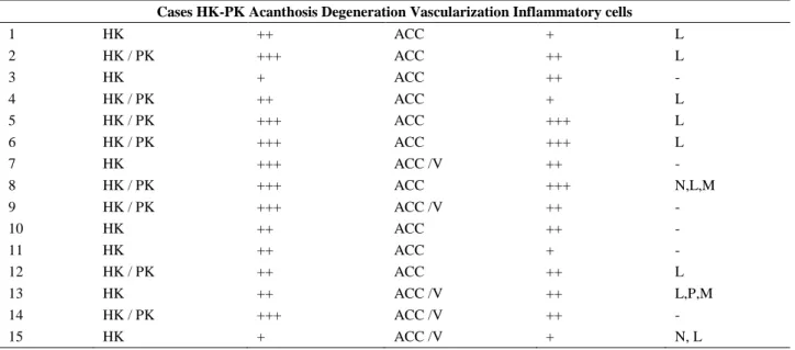
In addition to epidermal neoplastic changes, there were generally uniform fibrocytes and fibroblasts clusters arranged by whorls and interwoven in the fibrovascular stroma of the dermis (Figure 1). In some cases, collagen production was more abundant when compared with cellular elements. In general, fibropapilloma was an often encountered finding (n = 12), however, epithelial neoplastic changes were similar to those of fibroma in 3 of the cases. In those cases, acanthosis, hyperkeratosis, and epithelial proliferation were also observed, but these findings were not as conspicuous as in that of other fibropapilloma cases. In some areas, there were mononuclear cells composed of densely packed lymphocytes (n = 9). In addition, macrophages (n = 2), plasma cells (n = 1), and neutrophilic leukocytes (n = 2) were present dispersely in the fibrous stroma or at the periphery of hyperemic capillaries. Mallory’s trichrome staining method differentiated the density of fibrous tissues in all cases. The summary of all histopathological results has been shown in Table 1.
Table 1: Evaluation panel of histopathological findings. Tablo 1: Histopatolojik bulguların değerlendirme paneli.
Cases HK-PK Acanthosis Degeneration Vascularization Inflammatory cells
1 HK ++ ACC + L 2 HK / PK +++ ACC ++ L 3 HK + ACC ++ - 4 HK / PK ++ ACC + L 5 HK / PK +++ ACC +++ L 6 HK / PK +++ ACC +++ L 7 HK +++ ACC /V ++ - 8 HK / PK +++ ACC +++ N,L,M 9 HK / PK +++ ACC /V ++ - 10 HK ++ ACC ++ - 11 HK ++ ACC + - 12 HK / PK ++ ACC ++ L 13 HK ++ ACC /V ++ L,P,M 14 HK / PK +++ ACC /V ++ - 15 HK + ACC /V + N, L
Hyperkeratosis: HK, Parakeratosis: PK Acanthosis: +: mild, ++: moderate, +++strong
Degenerative changes: acut cell swelling: ACC, vacuolar degeneration: V, Vascularization: +: mild, ++: moderate, +++strong Inflammatory changes: -: no inflammation, N: neutrophile leucocyte, L: lymphocyte, P: plasma cell, M: macrophages. The inflammatory cells are put in order according to intensity in several areas.
Figure 1: Benign neoplastic proliferation of fibrocytic, fibroblastic cells in dermis (white star) and changes in epidermis (black star), x100, H&E.
Şekil 1: Dermisteki fibrosit ve fibroblastların benign neoplazik proliferasyonu (beyaz yıldız) ve epidermisteki değişiklikler (siyaz yıldız), x100, H&E.
Figure 2: Strong immunoreaction with IGF-I, particularly in cytoplasm and plasma membrane of all layer cells of epidermis (black arrows) as well as positivities in dermal fibroblasts (white arrows), and melanocytes (arrow heads), x200, immunoperoxidase staining. Şekil 2: Tüm epidermis katmanlarında özellikle sitoplazma ve plazma membranında şiddetli IGF-I ile pozitiflikler (siyah oklar), fibroblast pozitiflikler (beyaz oklar) ve melanositler (ok başları), x200, immunoperoksidaz boyama.
Figures 3: Strong positivities with IGF-I in cytoplasm and membranes of neoplastic fibrocytes and fibroblasts in dermis (arrows), x250, immunoperoxidase staining.
Şekil 3: Dermiste neoplazik fibrosit ve fibroblastların sitoplazma ve membranlarında şiddetli IGF-I pozitifliği (oklar), x250, immunoperoksidaz boyama.
Figure 4: Moderate positivities with IGF-II mainly in stratum basale, spinosum and lucidum cells of epidermis (black arrows), and melanocytes (arrow heads), x200, immunoperoxidase staining.
Şekil 4: Epidermisin bazal, spinozum ve lusidum katmanı hücrelerinde IGF-II ile orta şiddette pozitiflikler (siyah oklar) ve melanositler (ok başları), x200, immunoperoksidaz boyama.
Figure 5: Strong positivities with IGF-II in cytoplasm and membrane of fibrocytes and fibroblasts in dermis (arrows), x400, immunoperoxidase staining.
Şekil 5: Dermiste fibrosit ve fibroblastların sitoplazma ve membranlarında IGF-II ile şiddetli pozitiflikler (oklar), x400, immunoperoksidaz boyama.
Figure 6: Strong BPV-1 positivities in stratum basale and spinosum cells (black arrows), in fibrocyte and fibroblasts (white arrows), x200, immunoperoxidase staining.
Şekil 6: Stratum bazale ve spinozum hücrelerinde (siyah oklar), fibrosit ve fibroblastlarda (beyaz oklar) şiddetli BPV-1 pozitifliği, x200, immunoperoksidaz boyama.
c) Immunohistochemical findings: Expressions of IGF-I and IGF-II were localized in the cytoplasm and membranes of epidermal cells and in cytoplasms and membranes of fibrocytes and fibroblasts with collagen bundles (Figures 2-5). Taken overall, epidermal reactions had a wide distribution in the stratum corneum, lucidum, and spinosum cells. Stratum basale cells exhibited less positivity by both markers. In contrast, dermal positivities were dense in the spindle cells at the center of fibromas. Immunoreaction in spindle cells at the periphery of vessels were also attended. However, the distribution of immunostaining in connective tissue just beneath the epidermis was lesser when compared with the fibrocytes and fibroblasts found in the inner region of fibromas.
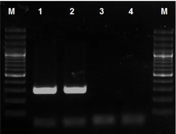
Immunoreaction to BPV-1 antibody was detected as nuclear, perinuclear and/or cytoplasmic (Figure 6). Immunohistochemical results showed that IGF-I was more evident on the epidermal and dermal components of fibropapilloma when compared with IGF-II. Moreover, the immunoreaction for IGF-I had similar distribution with that of BPV-1. The localization of all compared positive reactions was shown at Table-2 and Graph-1. In all control sections, immunopositive reaction could not be obtained in fibropapilloma cases and healthy bovine skin. d) PCR findings: The presence of BPV in tissues was indicated by amplification of a 301 base pair (bp) for BPV-1 in our samples. BPV-1 was positively detected in comparison with leader and positive controls (Figure 7).
Graph-1: Comperative scoring of IGF-I, IGF-II and BPV-1 distribution. Grafik-1: IGF-I, IGF-II ve BPV-1 dağılımının karşılaştırmalı skorlaması.
SC: Stratum corneum, SL: Stratum lucidum, SS: Stratum spinosum, SB: Stratum basale, FB: Fibrocytes, FBB: Fibroblasts Col: Collegen bundles.
Table-2: Mean scores±standard deviation (M±SD) of IGF-I, IGF-II and BPV-1 expressions according to the histological localization in fibropapilloma cases.
Tablo-2: Fibropapilloma olgularında histolojik lokalizasyona göre IGF-I, IGF-II ve BPV-1 ekspresyonlarının ortalama±standart sapması (M±SD). Location / Markers SC (n=15) SL(n=15) SS(n=15) SB(n=15) FB(n=15) FBB(n=15) Col(n=15) IGF-I 3.90±1.55 4.80±5.93 5.70±7.49 3.90±0.14 4.80±5.93 4.35±5.16 3.90±0.14 IGF-II 2.55±3.46 3.45±3.62 4.35±5.16 4.35±6.15 4.80±3.11 5.70±4.66 3.90±4.60 BPV-1 5.25±7.42 5.70±8.06 3.45±0.77 4.35±3.74 5.70±3.25 5.70±4.66 4.80±4.52 SC: Stratum corneum, SL: Stratum lucidum, SS: Stratum spinosum, SB: Stratum basale, FB: Fibrocytes, FBB: Fibroblasts Col: Collegen bundles.
Figure 7: Specific product of BPV-1 use 1F-1R (301 bp) primer pair. M: DNA ladder 100bp (Fermenthas, Lithuania), Line-1: Positive BPV-1 sample, Line-2: Positive Control of BPV-1, Line-3: Negative sample, Line-4: Negative control.
Şekil 7: BPV-1 spesifik ürünü 1F-1R (301 bp) baz çifti. M: DNA merdiveni 100bp (Fermenthas, Litvanya), Sıra-1: Pozitif BPV-1 örneği, Sıra-2: BPV-1’in pozitif kontrolü, Sıra-3: Negatif örnek, Sıra-4: Negatif kontrol.
Discussion and Conclusion
Fibropapillomas generally originate from the dermis and epidermis. In this study, the features of multiple masses were consistent with macroscopical and histopathological descriptions of the tumor characterized by numerous micropapillary extensions, parakeratosis and hyperkeratosis, acanthosis, and enlarged microscopic papilla. The stromal components of fibropapilloma have been included many whorls of or interwoven neoplastic fibrocytes and fibroblasts beneath the epidermis (15). Moreover, it is mentioned that there is also much mononuclear cell infiltration, which has a large proportion of lymphocytes in the ongoing times of virus-induced papillomas (3, 4, 10, 14, 15). In the present study, hyperkeratosis was observed in the limited areas in fibropapilloma cases and degenerative changes were common throughout the epidermis in several cases. Moreover, neoplastic fibrocytes and fibroblasts were encountered in addition to inflammatory cells at the periphery of vessels and in paranchyme. Hyperkeratosis alone was observed in seven cases, although it was found together with parakeratosis in eight cases. Acanthosis was generally mild or moderate in most of the cases. In accordance with these findings, it was interpreted that almost all cases, including fibropapillomas and fibroma-epithelial changes, occurred during the developing stage.
The previous studies reported that
deltapapillomaviruses fulfill their replications in the squamous epithelium, particularly in keratinocytes localizing in the surface of the epidermis. The virus makes its transformations in subepithelial fibroblasts (3, 4, 14). In the present study, findings related to fibromatous
changes were fairly evident and the presence of BPV-1 was found in nuclei of both epithelial and fibroblastic cells using immunohistochemistry, as also confirmed by PCR method. There are additional studies associated with other studies correlating histopathological characteristics and BPV or HPV (8, 9, 11, 28), agreeing with the results of the present study.
Moreover, there are also some suggestions in this topic about IGFs, which are endocrine hormones and they have an additional role in papillomavirus-induced tumors in the cervix of women (12, 17, 18). Specifically, IGF-I regulates cellular proliferation and differentiation and enhances transformation (30). IGF-II promotes cellular proliferation and is effective in fetal development (29). For HPVs, HPV-16 is frequently identified in cervical cancers (31). Some viral oncoproteins of HPV stimulate cell proliferation in cervical cancer and alter cellular differentiation (31). In an in vivo study with BPV, E5 (a transforming protein) transforms mouse embryonal fibroblasts in the absence of the IGF-I receptor (IGF-IR). However, it can be activated indirectly by IGF-I expression (22, 25). Insulin growth factor binding protein 3 (IGFBP-3) is a pivotal regulator of the cellular response against IGF-I and interacts directly with IGF-I for its activation (6). IGF-I-induced cell proliferation by means of IGFBP-3 may contribute to a selective growth advantage for HPV-immortalized cervical cells (2). In an in vivo study, IGF-I is synthesized and released by fibroblasts; it stimulates the growth of surrounding epithelial cells via a paracrine mechanism (1). Furthermore, in the present study, IGF-I was found like IGF-II in the epidermis and dermis, even though IGF-I activation is reported in cellular proliferation and transformation. The present study suggested that epithelial and connective tissues were heavily under the influence of IGF-I and IGF-II. Thereby, it is interpreted that IGF-I and IGF-II may enhance the capability of BPV-1 activation and transformation mainly as well as stromal elements. So, it has been thought that the expansion and growing of fibropapilloma may be related to the effect of paracrine effects of IGF(s) and BPV-1 in bovine fibropapilloma.
In conclusion, we observed some expressions in the same localizations with IGF I and IGF II and also BPV-1 markers. Polymerase chain reaction (PCR) and following immunohistochemistry confirmed the entity of BPV-1 in all sections. Expressions of IGF-I and IGF-II in those locations showed that IGF-I is clearly expressed on the epidermal and dermal components of fibropapilloma when compared with IGF-II. Moreover, IGF-I immunopositivity had similar distribution with that of BPV-1. This situation may be suggested a combined effect of BPV-1 and IGF interaction on expansion and growth of fibropapilloma. We believe that there will be a possible interaction with IGF(s) and oncogenic activity of
papillomavirus. To the best of our knowledge, the expression of the IGF complex (including IGFs, IGFBP, and IGFR) is needed to be evaluated together in progressing, developing, and regressing stages of papilloma and/or fibropapilloma cases induced with oncogenic BPV. The authors hope that the present study will be supported by further studies in this respect.
Acknowledgement
This paper has been partially derived by some additives on the basis of the project with grant number 14B0239001 from Ankara University, Commission of Scientific Research Projects.
References
1. Baege AC, Disbrow GL, Schlegel R (2004): IGFBP-3, a
marker of cellular senescence, is overexpressed in human papillomavirus-immortalized cervical cells and enhances IGF-I induced mitogenesis. J Virol, 78 (11), 5720-5727.
2. Barreca A, De Luca M, Del Monte P et al. (1992): In vitro
paracrine regulation of human keratinocyte growth by fibroblast-derived insulin-like growth factors. J Cell
Physiol, 151, 262-268.
3. Borzacchiello G, Roperto F (2008): Bovine papillomavirus, papillomas and cancer in cattle. Vet Res,
39 (5), 45.
4. Campo MS (1997): Bovine Papillomavirus and Cancer. Vet Journal, 154, 175-188.
5. Cheville NF, Olson C (1964): Epithelial and Fibroblast
Proliferation in Bovine Cutenous Papillomatosis. Epithelial and fibroblastic proliferation in bovine cutenous papillomatosis. Path Vet, 1, 248-257.
6. Clemmons DR (1997): Insulin-like growth factor binding
proteins and their role in controlling IGF actions. Cytokine
Growth Factor Rev, 8, 45-62.
7. Daughaday WH, Rotwein P (1989): Insulin-like growth
factors I and II. Peptide, messenger ribonucleic acid and gene structures, serum, and tissue concentrations. Endocr
Rev, 10, 68–91.
8. Ergunay K, Mısırlıoğlu M, Fırat P, et al. (2008):
Detection and typing of human papilloma virus by polimerase chain reaction and hybridization assay in cervical samples with cytological abnormalities. Microbiol
Bull, 42 (2), 273-282.
9. Ergunay K, Mısırlıoğlu M, Pınar F, et al. (2007): Human
papilloma virus DNA in cervical samples with cytological abnormalities and typing of the virus. Microbiol Bull, 41
(2), 219-226.
10. Goldshcmidt MH, Hendrick MJ (2002): Tumors of the
skin. Chapter 11. In: D.J. Meuten (Ed), Tumors in Domestic
Animals. Fourth Edition, Iowa State Press, USA, pp 47-50. 11. Gülbahar MY, Yüksel H, Aslan L (2003):
Angiokeratomatous papilloma associated with papillomavirus in a calf. Vet Path, 40 (5), 582-586.
12. Harris TG, Burk RD, Yu H, et al. (2008): Insulin-like
growth factor axis and oncogenic human papillomavirus natural history. Cancer Epidemiol Biomarkers Prev, 17 (1),
245-248.
13. Hatama S, Ishihara R, Ueda Y, et al. (2011): Detection of
a novel bovine papillomavirus type 11 (BPV-11) using xipapillomavirus consensus polymerase chain reaction primers. Arch Virol, 156 (7), 1281-1285.
14. Jarrett WFH (1985): The natural history of bovine
papillomavirus infections. In: G Klein (Ed): Advances in
Viral Oncology, vol 5, Raven Press, New York, pp 83-101. 15. Jelinek F, Tachezy R (2005): Cutenous Papillomatosis in
Cattle. J Comp Path, 132, 70-81.
16. Jones JI, Clemmons DR (1995): Insulin-like growth
factors and their binding proteins: biological actions.
Endocr Rev, 16, 3–34.
17. Jozefiak A, Pacholska-Bogalska J, Myga-Nowak M, et al. (2008): Serum and tissue levels of insulin-like growth
factor-I in women with dysplasia and HPV-positive cervical cancer. Mol Med Rep, 1 (2), 231-237.
18. Kuramoto H, Hongo A, Liu YX, et al. (2008):
Immunohistochemical evaluation of insulin-like growth factor I receptor status in cervical cancer specimens. Acta
Med Okayama, 62 (4), 251-259.
19. Lindsey CL, Almeida ME, Vicari CF, et al. (2009):
Bovine papillomavirus DNA in milk, blood, urine, semen, and spermatozoa of bovine papillomavirus-infected animals. Genet Mol Res, 8 (1), 310-318.
20. Lunardi M, Alfieri AA, Otonel RA, et al. (2013): Genetic
characterization of a novel bovine papillomavirus member of the Deltapapillomavirus genus. Vet Microbiol, 162 (1),
207-213.
21. Minshall C, Arkins S, Straza J, et al. (1997): IL-4 and
insulin-like growth factor-I inhibit the decline in Bcl-2 and promote the survival of IL-3-deprived myeloid progenitors.
J Immunol, 159 (3), 1225–1232.
22. Morrione A, Deangelis T, Baserga R (1995): Failure of
the bovine papillomavirus to transform mouse embryo fibroblasts with a targeted disruption of the insulin-like growth factor I receptor genes. J Virol, 69 (9), 5300-5303.
23. Parrizas M, Leroith D (1997): Insulin-like growth
factor-1 inhibition of apoptosis is associated with increased expression of the Bcl-xL gene product. Endocrinology, 138,
1355–1358.
24. Rosenfeld RG, Lamson G, Pham H, et al. (1990): Insulin
like growth factor-binding proteins. Recent Prog Horm Res,
46, 99–159.
25. Rubini M, Hongo A, DʼAmbrosio C, et al. (1997): The
IGF-I receptor in mitogenesis and transformation of mouse embryo cells: role of receptor number. Exp Cell Res, 230,
284-292.
26. Sara VR, Hall K (1990): Insulin-like growth factors and
their binding proteins. Physiol Rev, 70, 591–614.
27. Stewart CE, Rotwein P (1996): Growth, differentiation,
and survival:multiple physiological functions for insulin-like growth factors. Physiol Rev, 76, 1005–1026.
28. Tan MT, Yıldırım Y, Sözmen M, et al. (2012):
Histopathological, Immunohistochemical and Molecular Study of Cutaneous Bovine Papillomatosis. Kafkas Univ
Vet Fak Derg, 18 (5), 739-744.
29. Yu H, Rohan T (2000): Role of the insulin-like growth
factor family in cancer development and progression. J Natl
30. Zur Hausen H (1996): Papillomavirus infections—a major
cause of human cancers. Biochim Biophys Acta, 1288 (2),
55-78.
31. Zur Hausen H (2000): Papillomavirus causing cancer:
evasion from host-cell control in early events in carcinogenesis. J Natl Cancer Inst, 92 (9), 690-698.
Geliş tarihi: 26.09.2016 / Kabul tarihi: 13.02.2017
Address for correspondence:
Dr.Mehmet Eray ALÇIĞIR
Ankara University, Faculty of Veterinary Medicine, Department of Pathology, 06110, Dışkapı, Ankara, Turkey e-mail: [email protected]