Magnetic Resonans Imaging Of Intrapancreatic Collaterals in Portal
Vein Thrombosis With And Without Cirrhosis
Sirozu Olan ve Olmayan Hastalarda Portal Ven Trombozunda İntrapankreatik Kollaterallerin Manyetik
Rezonans Görüntülemesi
Nuray Haliloğlu, Esra Özkavukcu, Ayșe Erden
Ankara University Faculty of Medicine Department of Radiology
Aim: To evaluate the frequency of intrapancreatic collaterals in patients with cavernous transformation of the portal vein, in cirrhotic and non-cirrhotic groups.
Materials and Methods: We evaluated MR angiography images of 41 patients with cavernous transformation of the portal vein. Twenty-one of these patients had cirrhosis (Group I), and 20 patients had other causes of portal vein thrombosis (PVT) (Group II). MR studies were performed between September 2006-February 2010 using 1-T MRI system. MR images were reviewed by two radiologists, for the presence of intrapancreatic collaterals and the extension of the PVT.
Results: Intrapancreatic collaterals were present in 25 of 41 (61%) patients (11 patients in Group I, 14 patients in Group II). Statistically there was no significant difference between the two groups (p>0.05). Mass effect caused by the intrapancreatic collaterals was not detected in any of our patients. The PVT was extending to the superior mesenteric vein and/or splenic vein in 22 of the 25 patients (88%) with intrapancretic collaterals.
Conclusion: Intrapancreatic collaterals are common in patients with cavernous transformation of the portal vein. Cirrhosis does not seem to affect the frequency of intrapancreatic collaterals. The extension of PVT plays the major role in the formation of intrapancreatic collaterals.
Keywords: Pancreas; Portal vein; Magnetic Resonance Angiography; Cirrhosis
Amaç: Sirotik ve sirotik olmayan gruplarda portal vende kavernöz transformasyonu olan hastalarda intrapankreatik kollaterallerin sıklığını araștırmaktır.
Materyal ve Metod: Portal vende kavernöz transformasyonu olan 41 hastanın MR anjiyografi görüntüleri değerlendirildi. Bu hastaların 21’inde siroz vardı (Grup I) ve 20 hastada portal ven trombozunun (PVT) diğer nedenleri mevcuttu (Grup II). MR incelemeleri Eylül 2006- Șubat 2010 tarihleri arasında 1-T MRG cihazında yapıldı. MR görüntüleri, intrapankreatik kollaterallerin varlığı ve PVT’nin yaygınlığı açısından iki radyolog tarafından değerlendirildi.
Bulgular: İntrapankreatik kollateraller 41 hastanın 25’inde (%61) mevcuttu (Grup I’de 11 hasta, Grup II’de 14 hasta). İstatistiksel olarak iki grup arasında anlamlı bir fark yoktu (p>0,05). Hastalarımızın hiçbirinde intrapankreatik kollaterallere ikincil kitle etkisi saptanmadı. İntrapankreatik kollateralleri olan 25 hastanın 22’sinde (%88) PVT superior mezenterik vene ve/veya splenik vene uzanıyordu.
Sonuç: Portal vende kavernöz transformasyonu olan hastalarda intrapankreatik kolateraller sık görülür. Siroz intrapankreatik kollaterallerin sıklığını etkilememektedir. İntrapankreatik kollaterallerin gelișiminde ana etken PVT’nin yayılımıdır.
Anahtar Sözcükler: Pankreas, Portal ven, Manyetik Rezonans Anjiyografi,Siroz
Portal vein thrombosis (PVT) can occur in cirrhosis, or in different etiologies including hematological, inflammatory or neoplastic disorders. In such patients collateral vessels develop in order to by-pass the obstructed segment
of the portal venous system (1). Masses of collateral vessels known as cavernomas develop, and replace the occluded portal vein segments (1-3). Peripancreatic, and intrapancreatic collaterals named as pancreatic cavernoma have been
Received : May 07,2012 Accepted: March 03,2014 Corresponding Author
Doç.Dr.Nuray Haliloğlu Phone : + 90 312 595 67 81 E-mail : [email protected]
Ankara University Faculty of Medicine Cebeci Hospital Department of Radiology Research and Practice. Dörtyol /
described in the literature (4-6) but as far as we are concerned these collaterals are not mentioned in cirrhotic patients.
The aim of this study is to evaluate the frequency of intrapancreatic collaterals in patients with cavernous transformation of the portal vein, in cirrhotic, and non-cirrhotic groups. We retrospectively investigated whether the presence of cirrhosis affects the development of intrapancreatic collaterals.
Materials and Methods
Patient Population:Forty-one patients with cavernous transformation of the portal vein were enrolled. Cavernous transformation of the portal vein was shown on MR images, as multiple collateral vessels which developed in porta hepatis, and replaced the obstructed portal vein. There were 25 male, and 16 female patients with a mean age of 42 years (21-61 years).
Patients were divided into two groups; group I included 21 patients with
histopathologically proven cirrhosis. Group II consisted of 20
patients with other causes of PVT, including polycythemia vera, protein C deficiency, essential
thrombocythemia, or undetermined etiology. None of
the patients in group II had clinical, radiological or laboratory findings indicating cirrhosis. Patients with a pancreatic disease, or
an intraabdominal mass were not enrolled in the study. As this was a retrospective study we could not obtain informed consent from the patients.
MR Imaging:
MR studies were performed between September 2006-February 2010
using the same 1-T MRI system (Signa LX Horizon; General Electric Medical Systems, Milwaukee, WI) using phase array coils. Images were acquired with a breath-hold three-dimensional (3D) fast-spoiled gradient recalled (3D FSPGR) sequence in the coronal plane.
The sequence was started following a delay of 14–17 s after the manual injection of 0.2 mmol/kg of gadolinium chelate. Four consecutive sets of images were obtained at arterial, early and late portal venous, and hepatic venous phases. The imaging parameters were as follows: TR: 6 ms, TE: 1.2 ms, flip angle: 20, bandwidth: 31.2 or 62.5 kHz, image matrix: 256x160 or 256x128, field of view: 40–48 cm, and the section thickness was 4 mm. Scanning time ranged from 12 to 24 s (mean time 16 s) for each set of images. All images were reconstructed on the workstation (Advantage Windows, version 3.1; GE Healthcare, Milwaukee, Wisconsin, USA) using maximum intensity projection (MIP) technique. In order to overview the portal venous system in the axial plane postcontrast fat-suppressed fast spoiled gradient echo images (TR, 120 ms; TE, 6.3 ms; FA, 90 degrees; BW, 20.83; FOV, 32–40; slice thickness, 7 mm; spacing, 1.5 mm; matrix, 256 ×160; NEX [number of excitations], 1) of the upper abdomen were also obtained.
Image Interpretation:
Hardcopy MR images were retrospectively reviewed by two radiologists, for the presence of collateral vessels in the pancreas (intrapancreatic collaterals) and the extension of the PVT. When intrapancreatic collaterals were detected, mass effect (displacement,
encasement or invasion of neighboring structures, perilesionel edema, enlargement of the pancreas) was investigated on the postcontrast fat-suppressed gradient echo images. The craniocaudal size of the spleen was measured. The location of portosystemic collateral vessels, and the presence of pericholedochal collaterals were also noted. Final decision was made in consensus.
Pearson Chi-Square test was performed to compare the two groups. p value less than 0.05 was accepted as significant.
Results
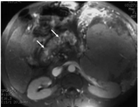
Intrapancreatic collaterals were present in 25 of 41 (61%) patients (11 of Group I patients and 14 of Group II patients). Statistically there was no significant difference between the two groups (p=0.248) (Figures 1, 2). In contrast with the previous reports, mass effect caused by the intrapancreatic collaterals was not detected in any of our patients, thus there was no diagnostic dilemma (Figure 3).
Figure 1: A-48 year old man with cirrhosis,
and PVT extending towards superior mesenteric vein, and splenic vein. Intrapancreatic collaterals are well demonstrated on axial plane postcontrast fat-suppressed fast spoiled gradient echo image (arrows).
The portal vein thrombosis was extending to the Superior Mesentenc Vein (SMV) and/or splenic vein in 21 of 25 patients (84%) with intrapancretic collaterals. Eleven of these 25 patients had extension of the thrombosis both to the splenic vein, and Superior Mesentenc Vein (SMV). Only one patient (4%) had extension of the thrombosis to the splenic vein alone. Four patients (16%) had patent SMV, and splenic vein. In three patients, intrapancreatic collaterals were not seen despite the extension of the thrombus to the SMV, and the splenic vein.
Nine patients (two in group I, seven in group II) had undergone splenectomy. The mean longitudinal size of the spleen was 19 cm in both group I (12-24 cm), and group II (11-25 cm).
The most frequently seen collateral vessels in group I were the paraoesophageal collaterals (in 12 of 21 patients). Pericholedochal collaterals were seen in five of the 21 (24%) patients in group I, and 10 of the 20 (50%) patients in group II (Figure 4).
Discussion
There is a rich venous blood flow in and around the pancreas towards the portal vein, SMV, and splenic vein (7). With the advent of high resolution scanners, thin calibered pancreatic vessels can usually be well demonstrated, and in case of a pancreatic tumor they are carefully examined to detect or exclude vascular invasion. Dilatation of the pancreatic veins, especially peripancreatic veins, can indicate the extension of a pancreatic tumor to the neighboring structures, particularly to the duodenum (8). Nevertheless the only cause of dilated pancreatic veins is not a
pancreatic tumor, and these enlarged vessels may also indicate anastomoses by-passing an obstructed vein or retroperitoneal or mesenteric portosystemic collaterals in portal hypertension. In most of the patients the drainage course of the collateral vessels has a complex, and extensive nature (9).
The causes of non-neoplastic PVT in cirrhotic patients are the reduced portal flow and the parenchymal disarchitecture. The prevalence is higher in patients with more severe liver disease. It is usually asymptomatic but can present with symptoms like abdominal pain and fever, or with life-threatening complications, such as variceal bleeding (10). The collateral veins develop gradually, and in the chronic stage, the portal vein itself may not be visualised among the network of collateral vessels, so called portal cavernoma (11). The thrombosis preferentially extends towards the SMV, and causes intestinal ischemia (10). The acute occlusion of SMV can cause severe symptoms but if the thrombus extends slow enough for the collateral vessels to develop, the symptoms would be less prominent. Even in the subacute phase of thrombosis some collaterals can be detected around the pancreas head (12).
When the splenic vein is occluded, short gastric, left gastric, omental veins, and gastroepiploic vein tend to dilate. The gastroepiploic vein terminates in the gastrocolic trunk, which drains into the SMV at the level of the pancreas head (1). Therefore pancreatic collaterals can be expected in splenic vein thrombosis, especially in, and around the pancreatic head. In patients with PVT the extent of the thrombus, and the amount, and localization of portal cavernoma should be screened, and noted on MR angiography.
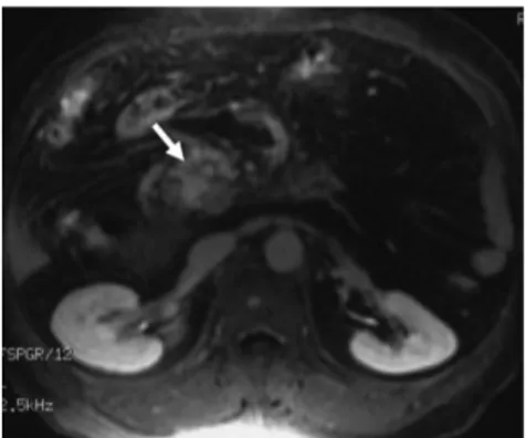
Figure 2: Mild intrapancreatic collaterals are
demonstrated on postcontrast fat-suppressed fast spoiled gradient echo image of a 21-year old non-cirrhotic man with PVT.
Figure 3: Significant amount of
intrapancreatic collaterals are seen in the pancreas head on postcontrast fat-suppressed fast spoiled gradient echo image (arrow) without any mass effect.
Figure 4: Similar amount of intrapancreatic collaterals are seen in the pancreas head (arrows in A and B) in patients with cirrhosis (A), and essential thrombocythemia (B). Note the paracholedochal collaterals in both patients (small arrow in A). Pericholecystic collaterals are also seen (small arrow in B).
An important component of intrapancreatic collaterals are the varices of the paracholedochal, and epicholedochal plexi which ensure the venous drainage of the common bile duct (CBD). The veins of the paracholedochal plexus ascend along the course of the CBD just parallel to it, and they drain into the gastric veins, the pancreaticoduodenal vein, and the the portal vein (13). The varices of these plexi can occur in cirrhosis but they are reported to be more frequent in extrahepatic portal vein occlusion (14).
Our study confirms the suggestion that intrapancreatic collaterals are common in patients with cavernous transformation of the portal vein. They are most likely located in the head of the pancreas. The extension of the PVT seems to play the major role in the formation of these collaterals (4). Peripancreatic collaterals are also
described in cavernous transformation (4). Nevertheless, as the location of many collateral veins around the splenic hilum, and in the retroperitoneal space, can also be described as around the
pancreas, we could not make a proper definition of peripancreatic collaterals. In other words, peripancreatic collaterals can have a wide definition including retroperitoneal, and perisplenic collaterals in cirrhotic patients. In this point of view we did not take collaterals around the pancreas into consideration.
The advancement of Computerized Tomography (CT) technology has enabled the vascular structures to be continuously traced. The anatomic characteristics of portosystemic collateral vessels in patients with cavernous transformation can be depicted on CT portal venography (9). However, ionising radiation exposure and use of iodinated contrast material are still considered to be major disadvantages of CT angiography. Pancreatic collateral vessels are usually well demonstrated on MR angiography which does not utilize ionising radiation, and has a multiplanar imaging capability. It has been suggested that
intrapancreatic collaterals in cavernous transformation are
associated with a heterogenous appearance, and enlargement of the pancreas on unenhanced MR images. MR angiography can provide adequate delineation of collaterals in cavernous tranformation, and no further examination would be necessary (4). One could expect that an accompanying cirrhosis might increase the tendency to develop collaterals in PVT, but cirrhosis does not seem to affect the formation of intrapancreatic collaterals.
In conclusion; intrapancreatic collaterals which are common in patients with cavernous transformation of the portal vein can be well demonstrated on MR images. Being aware of these collaterals can avoid unnecessary further examinations for the heterogenity, enlargement or the hypervascularity of the pancreas. The frequency of intrapancreatic collaterals in PVT are not influenced by an accompanying cirrhosis. The major factor in the formation of these collaterals is the extent of the PVT.
REFERENCES
1. Marn CS, Francis IR. CT of portal venous occlusion. Am J Roentgenol 1992;159:717-726.
2. Levy HM, Newhouse JH. MR imaging of portal vein thrombosis. Am J Roentgenol 1988;151:283-286.
3. Mathieu D, Vasile N, Grenier P. Portal thrombosis: Dynamic CT features and course. Radiology 1985; 154: 737-741. 4. Vilgrain V, Condat B, O’Toole D, et
al. Pancreatic portal cavernoma in patients with cavernous transformation of the portal vein: MR findings. Eur Radiol 2009;19:2608– 2613.
5. Ozcinar B, Ozden I, Bilge O, et al. Pancreatic portal cavernoma. J Pancreas 2005;6:40-41.
6. Ragozzino A, De Ritis R, Guardascione MA, Amitrano L. A portal vein cavernoma mimicking a pancreatic mass. J Hepatol 2003;38:372.
7. Mourad N, Zhang J, Rath AM, Chevrel JP. The venous drainage of the pancreas. Surg Radiol Anat 1994;16:37-45.
8. Yamada Y, Mori H, Kiyosue H, et al. CT assessment of the inferior peripancreatic veins: clinical
significance. Am J Roentgenol 2000;174:677-684.
9. Kang HK, Jeong YY, Choi JH, et al. Three-dimensional multi–detector row CT portal venography in the evaluation of portosystemic collateral vessels in liver cirrhosis. RadioGraphics 2002; 22:1053–1061. 10. Amitrano L, Guardascione MA,
Brancaccio V, et al. Risk factors and clinical presentation of portal vein thrombosis in patients with liver cirrhosis. J Hepatol 2004;40:736–741. 11. Hoekstra J, Janssen HLA. Vascular
liver disorders (II): Portal vein thrombosis. Neth J Med 2009;67:46-53.
12. Tateishi A, Mitsui H, Oki T, et al. Extensive mesenteric vein and portal vein thrombosis successfully treated by thrombolysis and anticoagulation. J Gastroenterol Hepatol 2001;16:1429-1433.
13. Chandra R, Kapoor D, Tharakan A, Chaudhary A, Sarin SK. Portal biliopathy. J Gastroenterol Hepatol 2001;16:1086–1092.
14. Shin SM, Kim S, Lee JW, Kim CW, et al. Biliary abnormalities associated with portal biliopathy: evaluation on MR cholangiography. Am J Roentgenol 2007; 188:W341–W347.