Ankara Üniv Vet Fak Derg, 65, 69-72, 2018
Relationship between pulsed wave Doppler ultrasonographic features
of milk veins and CMT scores in Simmental cows
Ali RİŞVANLI
1, Halef DOĞAN
1, Nevzat SAAT
2, İbrahim ŞEKER
31Fırat University, Faculty of Veterinary Medicine, Department of Obstetrics and Gynecology, Elazığ; 2Balıkesir University, Faculty
of Veterinary Medicine, Department of Obstetrics and Gynecology, Balıkesir; 3Fırat University, Faculty of Veterinary Medicine,
Department of Animal Husbandry, Elazığ, Turkey.
Summary: This study aimed to determine the distribution of pulsed wave Doppler ultrasonographic features of milk veins in lactating cows corresponding to udder health and the results of California Mastitis Test (CMT). Clinically healthy Simmental cows aged 3-5 years within the first 12 weeks of postpartum period were used for the study. End Diastolic Velocities (EDV), Systolic Peak Velocity (SPV), and Time-Averaged Maximum Velocity (TAMV) were the measured Doppler features of the milk veins. Numerical scales for Pulsatile Index (PI) and Resistant Index (RI) were devised in accordance with blood flow parameters. The animals were allocated to two groups according to CMT results as CMT positive (n=21) and CMT negative (n=5). No differences were found between the group regarding to PI, RI, EDV, or SPV (p>0.05). However, TAMV was found to be lower in CMT-positive animals (7.52 ± 0.85 cm/sec) compared to CMT-negative (13.90 ± 3.56 cm/sec) animals (p=0.021).
Keywords: Cow, milk vein, pulsed wave Doppler ultrasonography, udder.
Simental ırkı ineklerde süt veninin pulsed wave Doppler ultrasonografik özellikleri ile CMT skorunun
ilişkisi
Özet: Bu araştırmada laktasyondaki ineklerde süt veninin pulsed wave doppler ultrasonografi özelliklerinin meme sağlığı ve California Mastitis Test (CMT) sonuçlarına göre dağılımı ve bu dağılıma etki eden faktörlerin belirlenmesi amaçlandı. Çalışmada klinik açıdan sağlıklı, Simmental ırkı, 3-5 yaşlı ve postpartum 12 hafta içinde olan inekler kullanıldı. Süt veninin doppler özellikleri olarak End Diastolic Velocities (EDV, Diyastol Sonu Kan Akım Hızı), Systolic Peak Velocity (SPV, En Yüksek Sistolik Kan Akım Hızı) ve Time-Averaged Maximum Velocity (TAMV, Zaman Ortalamalı Maksimum Kan Akım Hızı) ölçüldü. Kan akış parametrelerine bağlı kalarak Pulsatil İndeks (PI) ve Rezistans İndeks (RI) skalası çıkarılarak numerik bir ölçeklendirme yapıldı. Hayvanlar CMT sonuçlarına göre CMT pozitif (n=21) ve CMT negatif (n=5) olmak üzere ikiye ayrıldı. Buna göre PI, RI, EDV ve SPV açısından gruplar arasında fark bulunmadı (p>0.05). Ancak TAMV CMT pozitif (7,52 ±0,85 cm/s) hayvanlarda CMT negatif (13,90 ±3,56cm/s) hayvanlara nazaran daha düşük bulundu (p=0.021).
Anahtar sözcükler: İnek, meme, pulsed wave Doppler ultrasonografi, süt veni.
Introduction
Blood supply to the udder is vitally important to milk production, as udder tissue converts nutrients in blood into milk as the final product. Milk yield and milk vein size are positively related to each other (vena epigastrica cranialis superficialis, vena subcutanea abdominis). Milk veins extend towards sternum in paramedian plain, anterior to udder, serving as a reservoir for excess blood required during milking. Because this vein is superficial and large in cows, and can easily be examined by ultrasonography, which is being used in both scientific and clinical studies (11). Contrary to this approach some researchers have suggested that the pressure applied by ultrasonographic probe on this vein during examination could affect the results (14). However, it is not yet fully understood whether udder metabolism regulates blood supply to udder
or vice versa (7, 22). Many factors, such as the pre- and post-milking periods, sexual-cycle period, lactation period, milk yield, udder size, breed, and season have been reported to affect udder’s blood supply (6). Haemodynamic characteristics of milk veins, as determined by pulsed wave Doppler analysis, are the potential indicators of milk yield and udder health. Berger et al. reported that pulsed wave Doppler ultrasonography can be used to analyze blood flow to udder, sharing the finding that ultrasonographic parameters regarding blood flow to udder increased with milk yield (1). Pulsed wave Doppler ultrasonography is used to measur values such as Time-Averaged Maximum Velocity (TAMV), blood flow volume, Systolic Peak Velocity (SPV) and Resistant Index (RI) in vessels supplying blood to udders (10, 17, 18, 19).
Ali Rişvanlı - Halef Doğan - Nevzat Saat - İbrahim Şeker 70
This study aimed to investigate the relationship between pulsed wave Doppler features of milk veins and CMT Scores among lactating cows.
Materials and Methods
Animals were selected among 1000 animals which were brought brought to Fırat University Animal Hospital (Elazig, Turkey, 38°35`51.55`N, 39°16`53,28"E) for examination between January 1, 2016 and April 10, 2016. Air temperature during examination varied from 1 to 15°C.
In total, 26 clinically healthy Simmental cows (normal body temperature, normal respiratory rate, normal heart beats, no vaginal discharge, normal digestion activities) without-pregnancy, aged 3-5 years, in diestrous periods (rectal palpation performed and CL was observed by USG) and within the first 12 weeks of postpartum period, weighing 450-500 kg, producing 15-20 liters of milk daily, having medium-sized udders, with lactation numbers of 2-3 and being milked twice daily. Body condition scores of the cows varied between 3 and 3.5. Selection criteria was decided to enable standardization that would least affect Doppler measurements. Ethics committee approval was obtained from Fırat University Laboratory Animals Local Ethics Committee (30.12.2015-2015/23).
During ultrasonographic examinations animals were kept as still as possible. Animals were given ten minutes to adapt to examination environment without interference, and then milk vein region was shaved. Doppler application was interrupted when an animal has moved or when respiratory rate increased.
Doppler analysis was performed as described by Braun et al. (2) on skin surface from a distance of 10-15 cm to udder from the caudal part of left milk vein using a 7.5 cm linear probe, activating pulsed wave ultrasonography in B-mode and in pulsed wave Doppler mode (Mindray DC-T6 Color Doppler Ultrasound System; Shenzhen, China). Using color mapping, milk vein was identified and located. For spectral Doppler examination, axis of sound beam and axis of milk vein were parallel to each other, but the insonation angle did not exceed 60°. This also increased the sensitivity of blood-flow measurements and made the examination dependent on insonation or incident angle instead of being angle-independent. Measurement lasted for a minimum of twenty seconds. Waveforms without artifacts, after adjusting angle of insonation, were recorded, and the waves were morphologically analyzed. After that, measurements of vascular indices were performed on milk veins, with EDV, SPV, and TAMV measured in cm/sec. Numerical scales for PI (PI = [SPV-EDV]/mean velocity) and RI (RI = [SPV-EDV] / SPV) were devised in accordance with blood-flow parameters (these
measurements are performed automatically by the Doppler device) (9).
CMT test was done according to the method described by Schalm et al. (20). Study groups were designed similarly Seker et al. (21). Accordingly, animals which had minimum one mammary quarter with CMT positive results were enrolled in CMT-positive group (n=21), while animals which had CMT negative results in all-mammary quarters were enrolled in CMT-negative group (n=5).
Udder dimensions were measured as described by Deng et al. (8) taking circumference (cm), length (cm), fore quarter depth (cm), hind quarter depth (cm), and, for the teats, average fore teat length (cm) and average hind teat length (cm).
Descriptive values of PI, RI, EDV, SPV, and TAMV were calculated using obtained data, after which each feature was evaluated for whether it met parametric test assumptions. Mann-Whitney U test was used as a non-parametric test to compare positive and CMT-negative groups for all features, as none of the features met parametric test assumptions. SPSS 22.0 was used to for the calculations and analysis.
Results
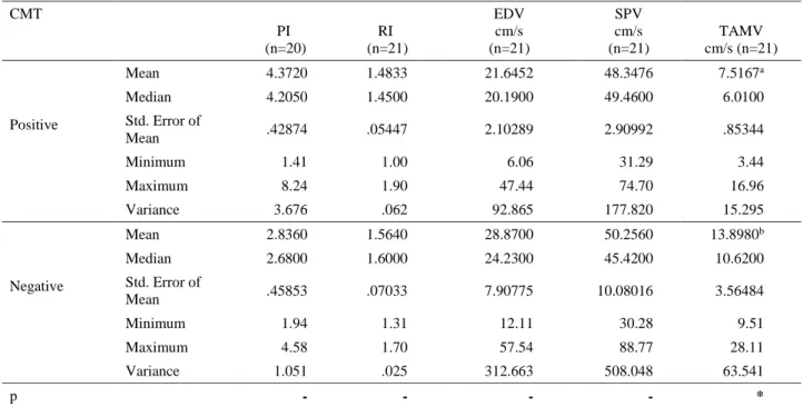
Descriptive values of PI (CMT-positive 4.37±4.21 - CMT-negative 2.84±0.46), RI (CMT-positive 1.48±0.05-CMT-negative 1.56±0.07), EDV (CMT-positive 21.65±2.10- CMT-negative 28.87±7.91 cm/s), SPV (CMT-positive 48.34±2.91- CMT-negative 50.26±10.08 cm/s) and TAMV measured in Simmental breed are presented in Table 1. Only the difference in TAMV was found to be statistically significant between CMT positive and the negative groups. Namely, TAMV was found to be lower in CMT-positive animals (7.52 ± 0.85 cm/sec) compared to CMT-negative animals (13.90 ± 3.56 cm/sec; p=0.021).
Discussion and Conclusion
Although etiology, diagnosis, treatment, and prevention of mastitis have been thoroughly investigated by previous studies, this disease is still present (16). New assessments are required, particularly for subclinical mastitis, considering new technologies. Investigating the dynamics of blood flow to udder using Doppler ultrasonography is a new approach to mastitis (4).
An experimentally induced mastitis study in goats, results of which were determined by CMT, reported increased SPV in udder artery and decreased RI as the disease progressed (i.e., as CMT positivity increased). Same study determined that the increase in SPV was related to increased blood flow to affected region due to increased inflammation, while the decrease in RI was suggested to be related to an increase in vascular
Ankara Üniv Vet Fak Derg, 65, 2018 71
resistance due to the same increased blood flow (18). Braun and Hoegger (5) reported a mean maximum blood flow velocity of 45.4 cm/sec, mean velocity of 33.5 cm/sec, and minimum velocity of 25.8 cm/sec before sedation in 29 healthy Swiss Brown cows, which were sedated. A study investigating milk veins abdominally during lactation reported maximum blood-flow velocity between 23.84 cm/sec and 35.76 cm/sec before birth, increasing to 61.13 cm/sec after birth and gradually regressing to 23.85 cm/sec by day 300 after birth (2). Another study showed a close relationship between milk yield and blood-flow changes in milk veins of cows in dry and lactation periods (3).
Inflammatory changes and metabolic activities in udders are predicted to increase udder blood flow (3, 12, 13, 15). In a transrectal color-Doppler ultrasonographic study investigating blood flow in the pudendoepigastric trunk in experimentally induced Escherichia coli mastitis in cows (14), blood-flow volumes were reported to increase parallel to somatic cell count, demonstrating that transrectal color-Doppler ultrasonography could be used for the early detection of pathological changes in blood flow in early stages of acute mastitis. Similar results were found in the present study. In particular, TAMV values support this view.
In a study conducted in goats (19), PI and SPV were increased in animals with clinical mastitis, resulting from
an increase in blood flow due to infection-related inflammation. In the present study, PI and SPV were found to be higher in CMT-positive animals, although the difference was statistically insignificant. The data presented here reveal that TAMV was lower in CMT-positive animals.
In conclusion, PI, RI, EDV, and SPV values were determined not to differ between positive and CMT-negative animals, while TAMV was found to be lower in CMT-positive animals compared to CMT-negative animals. This result suggests that Doppler data regarding blood flow could change in animals with subclinical mastitis. However, it would be beneficial to conduct this study in various breeds and larger series, as Doppler ultrasonographic measurements are affected by many factors and parameters, including breed, age, udder size, season, temperature, milk yield, body-condition score, sexual-cycle period, and lactation period.
References
1. Berger H, Lietzau M, Tichy A, et al. (2016): Investigations of mammary and uterine blood flow in relation to milk yield, postpartum disease and pregnancy result in dairy cows. Theriogenology, 86, 1906-1912. 2. Braun U, Forster E, Bleul U, et al. (2013): B-mode and
colour Doppler ultrasonography of the milk vein and musculophrenic vein in eight cows during lactation. Res Vet Sci, 94, 138-143.
Table 1. Descriptive values of PI, RI, EDV, SPV, and TAMV.
Tablo 1. PI, RI, EDV, SPV ve TAMV özelliklerine ait tanımlayıcı değerler. CMT PI (n=20) RI (n=21) EDV cm/s (n=21) SPV cm/s (n=21) TAMV cm/s (n=21) Positive Mean 4.3720 1.4833 21.6452 48.3476 7.5167a Median 4.2050 1.4500 20.1900 49.4600 6.0100 Std. Error of Mean .42874 .05447 2.10289 2.90992 .85344 Minimum 1.41 1.00 6.06 31.29 3.44 Maximum 8.24 1.90 47.44 74.70 16.96 Variance 3.676 .062 92.865 177.820 15.295 Negative Mean 2.8360 1.5640 28.8700 50.2560 13.8980b Median 2.6800 1.6000 24.2300 45.4200 10.6200 Std. Error of Mean .45853 .07033 7.90775 10.08016 3.56484 Minimum 1.94 1.31 12.11 30.28 9.51 Maximum 4.58 1.70 57.54 88.77 28.11 Variance 1.051 .025 312.663 508.048 63.541 p - - - - *
a,b: The differences between the groups that had median values with different letters in the same column were significant (*p=0.021). - No significant differences between groups (p>0.05).
a,b: Aynı sütunda farklı harflerle medyan değerleri verilen gruplar arasındaki farklılıklar önemlidir (*p=0.021). - Gruplar arasındaki farklılıklar önemli değil (p>0.05).
Ali Rişvanlı - Halef Doğan - Nevzat Saat - İbrahim Şeker 72
3. Braun U, Forster E (2012): B-mode and colour-Doppler sonographic examination of the milk vein and musculophrenic vein in dry cows and cows with a milk yield of 10 and 20 kg. Acta Vet Scand, 54, 15.
4. Braun U, Hoegger R, Haessig M (2009): Colour-Doppler sonography of the musculophrenic vein in cows. Vet J, 179, 451-454.
5. Braun U, Hoegger R (2008): B-mode and colour Doppler ultrasonography of the milk vein in 29 healthy Swiss braunvieh cows. Vet Rec, 163, 47-49.
6. Christensen K, Nielsen MO, Bauer R, et al. (1989) Evaluation of mammary blood flow measurements in lactating goats using the ultrasound Doppler principle. Comp Biochem Phys A, 92, 385-392.
7. Delamaire E, Guinard-Flament J (2006): Increasing milking intervals decreases the mammary blood flow and mammary uptake of nutrients in dairy cows. J Dairy Sci, 89, 3439-3446.
8. Deng MP, Badri TM, Atta M, et al (2012): Relationship between udder dimensions and milk yield of Kenana × Friesian crossbred cows. Res Opin Anim Vet Sci, 2, 49-54. 9. Erdogan G, Cetin H, Ceylan A, et al. (2016): Comparison
of foetal growth in singleton 1 and twin pregnancies by B-mode and Doppler ultrasonography in Karya ewes. Turk J Vet Anim Sci, 40, 616-621.
10. Götze A, Honnens A, Flachowsky G, et al. (2010): Variability of mammary blood flow in lactating Holstein-Friesian cows during the first twelve weeks of lactation. J Dairy Sci, 93, 38-44.
11. Gračner D, Gilligan G, Garvey N, et al. (2015): Correlation between the milk vein internal diameter surface and milk yield in Simmental cows. Turk J Vet Anim Sci, 39, 741-744.
12. Lough DS, Beede DL, Wilcox CJ (1990): Effects of feed intake and thermal stress on mammary blood flow and other physiological measurements in lactating dairy cows. J Dairy Sci, 73, 325-332.
13. Piccione G, Arcigli A, Fazio F, et al. (2004): Pulsed wave-Doppler ultrasonographic evaluation of mammary blood flow speed in cows during different productive periods. Acta Sci Vet, 32, 171-175.
14. Potapow A, Schmauder S, Petzl W, et al. (2010): Investigation of mammary blood flow changes by transrectal colour Doppler sonography in an Escherichia coli mastitis model. J Dairy Res, 77, 205-212.
15. Potapow A, Schmauder S, Petzl W, et al. (2008): Detection of mammary blood flow changes by transrectal colour-Doppler sonography in an E. coli mastitis model. Reprod Domest Anim, 43, 25.
16. Risvanli A, Kalkan C (2002): The effect of age and breed on somatic cell count and microbiological isolation rates in milk of dairy cows with subclinical mastitis. J Fac Vet Med Yuzuncuyil, 13, 84-87.
17. Rizzo A, Mutinati M, Minoia G, et al. (2012): The impact of oxytocin on the hemodynamic features of the milk vein in dairy cows: A color Doppler investigation. Res Vet Sci, 93, 983-988.
18. Santos VJC, Simplício KMMG, Sanchez DCC, et al. (2015): B-Mode and Doppler sonography of the mammary glands in dairy goats for mastitis diagnosis. Reprod Domest Anim, 50, 251-255.
19. Santos VJC, Simplício KMMG, Sanchez DCC, et al. (2014): Conventional and Doppler ultrasonography on a goat with gangrenous mastitis. Arq Bras Med Vet Zootec, 66, 1931-1935.
20. Schallm OW, Carroll EJ, Jain NC (1971): Bovine Mastitis. Lea-Febiger, London.
21. Seker I, Risvanli A, Yuksel M, et al. (2009): The relationship between CMT scores and ultrasonographic teat measurements in dairy cows. Aust Vet J, 87, 480-483. 22. Svennersten-Sjaunja K, Olsson K (2005): Endocrinology
of milk production. Dom Anim Endocrinol, 29, 241-258. Geliş tarihi:12.08.2016 / Kabul tarihi: 22.05.2017 Address for correspondence:
Prof. Dr. Ali RİŞVANLI
Fırat University, Faculty of Veterinary Medicine, Department of Obstetrics and Gynecology, Elazığ, Turkey, 23159 e- mail: [email protected] Tel: + 90 424 237 00 00/3852 Fax: + 90 424 238 81 73