155 Ankara Üniversitesi Tıp Fakültesi Mecmuası 2008, 61(3) DAHİLİ BİLİMLER / MEDICAL SCIENCES
Olgu Sunumu / Case Report
Renal Invasion Of Hepatocellular Carcinoma: Report Of A Case
Renal İnvazyon Gösteren Hepatosellüler Karsinoma : Olgu SunumuNuray Haliloğlu, Ayşe Erden
Ankara Üniversitesi Tıp Fakültesi, Radyodiagnostik Anabilim Dalı
Received: 05.03.2008 • Accepted: 08.12.2008
Corresponding author
Uzm. Dr. Nuray Haliloğlu
Ankara Üniversitesi Tıp Fakültesi, Radyodiagnostik Anabilim Dalı Phone : + 90 (312) 595 67 81
E-mail address : [email protected]
Local recurrences or metastases may occur after curative treatment of hepatocellular carcinoma. In this report a 47-year-old man with intrahepatic recurrences and extrahepatic growth of hepa-tocellular carcinoma invading inferior vena cava, right adrenal gland and right kidney following surgery is described. Emphasis is placed on the MR imaging findings of renal invasion which is thought to be involved by local route.
Key Words: hepatocellular carcinoma
Hepatosellüler karsinomada küratif tedavilerden sonra lokal nüks ya da uzak metastazlar ortaya çıkabilmektedir. Bu yazıda cerrahi tedaviden sonra intrahepatik rekürrensin yanı sıra inferior vena kava, sağ adrenal bez ve sağ böbreğe invazyona neden olan ekstrahepatik tümör gelişiminin sap-tandığı 47 yaşında bir hasta sunulmakta ve özellikle direk invazyona ikinci olarak değerlendirilen renal tutulumun MR görüntüleme bulguları vurgulanmaktadır.
Anahtar Kelimeler: hepatosellüler karsinoma
Hepatocellular carcinoma (HCC) is the most common primary tumor of the liver. Partial liver resection and orthotopic liver transplanta-tion may be the curative treatment methods for masses limited to the liver. Extrahepatic metastases are usually associated with the ad-vanced disease and there are only palliative treatment choices such as chemotheraphy or transcath-eter chemoembolization for these patients (1,2). Extrahepatic tumor growth with direct invasion of ad-jacent structures is rarely reported in the literature (3-9). Inadequate assessment before surgery, pre-operatively missed occult tumors, insufficient therapy, insufficient tu-mor resection or the aggresive na-ture of the tumor may lead to early recurrence after treatment. In this report we aimed to present a case of recurrent HCC manifesting with paracaval tumor growth and
local-ly invading the right adrenal gland and right kidney and to describe the MR imaging (MRI) findings of extrahepatic involvement.
Case
A 47-year-old man with previously diagnosed and operated recurrent HCC was referred to our MRI sec-tion in order to depict the current status of the tumor. In the retro-spective evaluation of his medical records we found out that he had been diagnosed as moderately dif-ferentiated HCC due to chronic vi-ral hepatitis in May 2001. Because the mass was limited to the right lobe, partial hepatectomy was per-formed in June 2001. He was un-der follow-up in an outside medi-cal center for about 3 years and as hepatic recurrence had occured at
Ankara Üniversitesi Tıp Fakültesi Mecmuası 2008, 61(3)
156 Renal Invasion Of Hepatocellular Carcinoma: Report Of A Case
the end of the first year he under-went transcatheter chemoemboli-sation for 3 times. On the first MRI we performed, besides the multi-ple recurrent intrahepatic masses, an extrahepatic retrocaval tumor tissue which partially invaded the inferior vena cava (IVC) (Figure 1) was depicted. The lesion was ex-tending inferiorly to the medial as-pect of the kidney. Tumor throm-bus in the portal vein branches were also present. As the tumor
was in an advanced stage, despite the palliative treatment the patient continued to detoriate. In a 1-year follow-up period under palliative therapy, both the intrahepatic and extrahepatic masses were pro-gressed by size and renal invasion had occured (Figure 2). The kid-ney masses were not as usually expected in metastatic HCC. The retrocaval tumor originating from the caudate lobe had occupied the place of IVC and invaded the right
kidney from both the parenchyma and the renal sinus (Figures 3,4). The right renal vein was hardly visible as it was displaced by the tumor at the level of the renal hi-lum. We believe that this was a di-rect invasion of the kidney by the hepatic mass following paracaval route.
Discussion
HCC is known as an aggressive tumor. Nevertheless as it frequently devel-ops in cirrhotic liver parenchyma and patients with chronic liver dis-eases are usually under follow-up, at the time of diagnosis metastases are usually not present. As the di-agnostic and the treatment meth-ods of HCC have been improving, the survival rate of the patients has
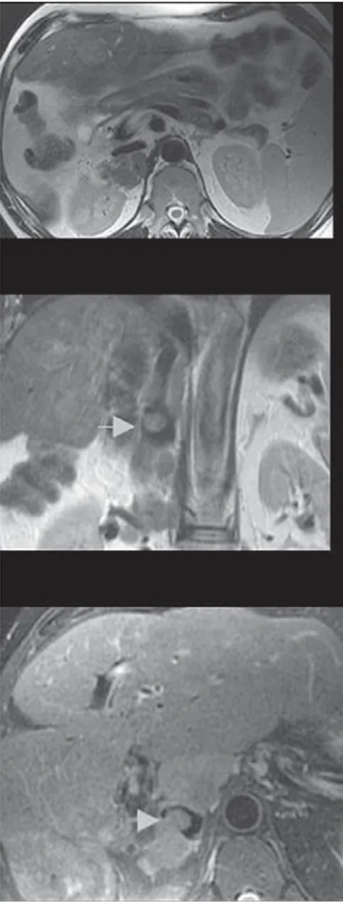
Figure 2. T2-weighted coronal images. The extrahepatic tumor neighbouring the kidney (A) has progressed to invasion (B). Also note the growth in intrahepatic masses (arrows).
Figure 1. Retrocaval tumor tissue is seen on T2 weighted axial image (A). Partial invasion of IVC is visible on T2-weighted coronal (B) and fat suppressed T2-weighted axial (C) ima-ges (arrows).
Figure 3. On T2-weighted coronal image the anatomic region of IVC has been occupied by the extrahepatic tumor. The right adrenal gland is thought to be invaded since it is not visualized on none of the sequences.
Figure 4. Both the parenchyma and the sinus of the right kidney is invaded by HCC. On the portal venous stage of the multiphasic MRI (A) the contrast medium in the masses is washed out (arrows). Also note collateral veins in the abdominal walls (black arrow). The invasion of the right renal hilum is obvious on T2-we-ighted axial image (B).
Journal of Ankara University Faculty of Medicine 2008, 61(3)
157
Nuray Haliloğlu, Ayşe Erden
prolonged and the metastases are seen more commonly. The prog-nosis of extrahepatic metastases is quite poor with a 1-year survival rate of 24.9% (10). Metastases can follow the hematogenous or lym-phatic routes or direct invasion may occur. The most frequent metastatic sites of HCC are known as lung, regional lymph nodes and the skeletal system (1,2,10). Renal metastases are usually bilateral, multiple, small lesions in which tumor cells reach the kidneys by bloodstream. Also retroperitoneal masses neighbouring the kidney may occur due to the dropped tu-mor cells (1). Brain metastases are not frequent and they are usually hemorrhagic deposits manifesting with stroke-like symptoms (11). Rare metastatic sites of HCC, like uterus and psoas muscle have also been described (12,13). Ho et al reported a case of biopsy proven extrahepatic lymph node metas-tases following more than 5 years after liver transplantation which mimicked lymphoproliferative disorder (14). Therefore patients must be carefully examined before and after the therapy. As well as lo-cal recurrences, distant metastases even in unexpected sites should be considered in the follow-up
peri-od. Nevertheless it should be kept in mind that rare metastatic sites of HCC (like brain and gastrointesti-nal tract) are almost never the ini-tial manifestations of extrahepatic metastases (2). The patient we present here had undergone sur-gical resection but recurrence had occured in a year with progression to extrahepatic disease.
Extrahepatic growth of HCC (pe-dunculated HCC) is seen approxi-mately 0.24%-3 % of the cases and the long-term prognosis of this form is quite poor (6). It can be defined as a protruding mass on the surface of the liver with a risk of spontaneous rupture (6,15). Besides peritoneal implantation due to the rupture, direct invasion of the adjacent structures may oc-cur. Okuda et al have described 7 cases with extrahepatic growth of HCC involving the right adrenal gland in 2 of which the tumor was adherent to the right kidney (15). As the right lobe of the liver and right adrenal gland get closer with the increasing age which is more prominent in cirrhotic patients, some of the right sided adrenal metastases may be due to extrahe-patic tumor growth (7).
As far as we are concerned there is no such a case in the literature describing extrahepatic tumor growth invading inferior vena cava, adrenal gland and the kid-ney at the same time. When the patient was referred to us his per-formance status was low and he refused further investigations to detect distant metastases. As the diagnosis and initial therapies were performed in an outside clinic we could not demonstrate the preoperative nature or the op-erability of the tumor and deter-mine the technique of the surgery. Therefore we could not describe the cause of early recurrence and aggressive progression. The ex-tension of the neoplastic tissue in this case is predominately towards to the hilum and the sinus of the right kidney. Although this appear-ance suggested the venous inva-sion, the direction of bloodstream regarding to the site of the primary tumor was inconsistent for throm-botic material to lodge in the renal vein. Inferior vena cava has been reported to be a route for pulmo-nary metastases of HCC via right atrium (10).Thus, we considered local invasion inferiorly into the kidney rather than hematogenous spread.
REFERENCES
1) Hong SS, Kim TK, Sung K, Kim PN, Ha HK, Kim Ay, et al. Extrahepatic spre-ad of hepatocellular carcinoma: a pi-ctorial view. Eur Radiol 2003;13:874-882.
2) Katyal S, Oliver III JH, Peterson MS, Ferris JV, Carr BS, Baron RL. Extra-hepatic metastases of hepatocellular carcinoma. Radiology 2000;216:698-703.
3) Fujii K, Nagino M, Kamiya J, Uesaka K, Sano T, Yuasa N, et al. Complete resection of hepatocellular carcino-ma with direct invasion to the stocarcino-ma- stoma-ch remnant. J Hepatobiliary Pancreat Surg 2004;11:441-444.
4) Cho A, Ryu M, Ochiai T. Succesful resection, using pancreas-sparing duodenectomy, of extrahepatically growing hepatocellular carcinoma associated with direct duodenal in-vasion. J Hepatobiliary Pancreat Surg 2002;9:393-396.
5) Maruyama A, Murabayashi K, Hayashi M, Nakano H, Isaji S, Uehara S, et al. Hepatocellular carcinoma complica-ted by gastrointestinal hemorrhage caused by direct tumor invasion of stomach. J Hepatobiliary Pancreat Surg 1999;1:90-93.
6) Kurachi K, Suzuki S, Yokoi Y, Oku-mura T, Inaba K, Igarashi T, et al. A 5-year survivor after resection of
pe-ritoneal metastases from peduncula-ted-type hepatocellular carcinoma. J Gastroenterol 2002;37:571-574. 7) Mashfigul AS, Tan YM, Thng CH,
Cheow PC, Chung YF, Chow PK, et al. Pedunculated HCC or adrenal metastasis: a diagnostic conundrum. Singapore Med J 2007;48:50-52. 8) Ong JCA, Chow PKH, Chan WH,
Chung AYF, Thng CH, Wong WK. Hepatocellular carcinoma masque-rading as a bleeding gastric ulcer: a case report and a review of the surgi-cal management. World J Gastroente-rol 2007;13:4523-4525.
9) Park MS, Kim KW, Yu JS, Kim MJ, Yoon SW, Chung KW, et al. Radiologic
fin-Ankara Üniversitesi Tıp Fakültesi Mecmuası 2008, 61(3)
158 Renal Invasion Of Hepatocellular Carcinoma: Report Of A Case
dings of gastrointestinal tract invol-vement in hepatocellular carcinoma. J Comput Assist Tomogr 2002;26:95-101.
10) Natsuizaka M, Omura T, Anaike T, Kuwata Y, Yamazaki K, Sato T, et al. Clinical features of hepatocel-lular carcinoma with extrahepatic metastases. J Gastrenterol Hepatol 2005;20:1781-1787.
11) Seinfeld J, Wagner AS, Kleinschmidt BK. Brain metastases from
hepato-cellular carcinoma in US patients. J Neuro Oncol 2006;76:93-98.
12) Ryo E, Sato T, Takeshita S, Ayabe T, Tanaka F. Uterine metastasis from he-patocellular carcinoma: a case report. Int J Gynecol Cancer 2006;16:1894-1896.
13) Wu MH, Wu YM, Lee PH. The psoas muscle as an unusual site for metas-tasis of hepatocellular carcinoma: re-port a case. Surg Today 2006;36:280-282.
14) Ho SGF, Phillips AJ, Browne RFJ, Munk PL, Legiehn GM, Yoshida EM, et al. Recurrent hepatocellular carcinoma after liver transplantati-on mimicking post-transplantatitransplantati-on lymphoproliferative disorder. Austral Radiol 2005;49:315-318.
15) Okuda K, Arakawa M, Kubo Y, Sakata K, Kage M, Iwamoto S, et al. Right-sided pedunculated hepatocellular carcinoma: a form of adrenal metas-tasis. Hepatology 1998;27:81-85.