Ankara Üniversitesi Tıp Fakültesi Mecmuası 2006; 59:7-10
DAHİLİ BİLİMLER / MEDICAL SCIENCES Araştırma Yazısı / Original Article
7 Corresponding author
Dr. Fatih Sinan Ertaş
Bilkent-3 Ufuk Sitesi D1/06800 Ankara, Turkey Tel : +90 (312) 508 25 23 Fax : +90 (312) 312 52 51 E-mail adress : [email protected]
Received: 03.01.2006 • Accepted: 16.01.2006
E
xperimental studies have provided powerful evidence of the dispersion of myocardial recovery times for the occurence ventricular arrhytmias (1-4). Measurement of the variability in QT interval duration among the differ-ent leads of the standard 12-lead electrocardiogram (ECG) (i.e., QT dispersion) has been proposed as a noninvasive method for detecting the inhomogenity of ventricular recovery times (5-9).
Observations have related an increase in QT dispersion on the surface ECG to increased risk of clinically important ventricular arrhythmias (10-14). Useful
Aim: Variability in QT interval duration on the different leads of the 12-lead ECG has been
pro-posed as an indicator of risk for ventricular arrhythmias in different clinical settings, but the value of QTd-V is not clear yet. The aim of this study was to estimate the value of QT dispersion in ven-tricular premature beats (QTd-V) in identifying patients susceptible to reentrant venven-tricular tach-yarrhythmias (VT).
Materials and Methods: We compared the performance of precordial QTd-V, late potentials on
the signal-averaged electrocardiogram and reduced left ventricular ejection fraction for identifi-cation of inducible ventricular tachycardia in 34 patients undergoing electrophysiologic study.
Results: QTd-V in 12 patients with inducible VT (110±50 msec) was found to be greater than that
in 22 patients without inducible VT (65±38 msec, p=0.006). Multivariate analysis including ejec-tion fracejec-tion and presence of late potentials showed that QTd-V was not an independent factor in identifying the susceptible patients to ventricular tachyarrhythmias.
Conclusion: Increased QTd-V is related to susceptibility to reentrant ventricular
tachyarrhyth-mias, however does not appear to provide additional diagnostic information to that provided by late potentials and left ventricular ejection fraction.
Key words: QT dispersion, ventricular premature beat, electrophysiologic study
Amaç: 12 derivasyonlu EKG nin farklı derivasyonları arasında QT interval süresinde değişkenlik
birçok klinik durumda ventriküler aritmiler için bir risk göstergesi olarak öngörülürken, QTd-V nin değeri henüz açık değildir. Çalışmanın amacı reentran ventriküler takiaritmilere (VT) duyarlılığı olan hastaları belirlemede ventriküler premature atımlarda QT dispersiyonu’nun (QTd-V) değerini araştırmaktır.
Gereç ve Yöntem: Elektrofizyolojik çalışma yapılmış 34 hastada indüklenebilir VT nin tahmini için
prekordiyal QTd-V, sinyal ortalamalı EKG de geç potansiyeller ve azalmış sol ventrikül ejeksiyon fraksiyonu karşılaştırıldı.
Bulgular: İndüklenebilir VT li 12 hastadaki QTd-V (110±50 msec) VT nin indüklenemediği 22
has-tadaki QTd-V den (65±38 msec, p=0.006) daha büyük bulundu. Ejeksiyon fraksiyonu ve geç po-tansiyel varlığının dahil edildiği çok değişkenli analiz QTd-V nin ventriküler takiaritmilere duyarlı hastaları belirlemede bağımsız bir faktör olmadığını gösterdi.
Sonuç: Artmış QTd-V tek başına reentran ventriküler takiaritmilere duyarlılıkla ilintili olsa da, geç
potansiyeller ve sol ventrikül ejeksiyon fraksiyonu hesaba katıldığında ilave tanısal bilgi sağlama-dığı gözükmektedir.
Anahtar sözcükler: QT dispersiyon, ventriküler prematüre atım, elektrofizyolojik çalışma
Dispersion of QT interval in premature ventricular
beats is not an independent marker for inducible
sustained ventricular tachycardia
Ventriküler prematüre atımların QT interval dispersiyonu indüklenebilir sürekli ventriküler takikardi için bağımsız bir gösterge değildir
Fatih Sinan Ertaş, Çağdaş Özdöl, Timuçin Altın, Yusuf Atmaca, Ömer Akyürek, Güneş Akgün,
Remzi Karaoğuz, Muharrem Güldal, Çetin Erol
Department of Cardiology, Ankara University School of Medicine
Ankara Üniversitesi Tıp Fakültesi Mecmuası 2006; 59(1)
Dispersion of QT interval in premature ventricular beats is not an independent marker for inducible sustained ventricular tachycardia
8
prognostic value of abnormally increased QT dispersion has been found in patients with acute myocardial infarc-tion (10), cardiomyopathy (11), the long QT syndrome (10,15), drug induced torsades de pointes (16) and sudden cardiac death (17).
The presence of late potentials on the signal-averaged ECG (6,18,19,20,21,22) and abnormal left ventricular ejection fraction ( LVEF) (23) also have been associated with an anatomic substrate for reentrant ventricular ar-rhythmias and inducible ventricular tachycardia (VT) at electrophsiologic study (EPS).
There are only several reports studying the clinical sig-nificance of QT interval dispersion measured in spontane-ous ventricular beats (QTd-V) (24-26) showing significant relationship between QTd-V and the risk of arrhytmic events.
The present study was designed to examine relation of measures of precordial QT dispersion in premature ven-tricular beats and inducible venven-tricular arrhytmias and the independence of these findings from late potentials and ventricular function.
Materials and Methods
Patients who underwent electrophysiologic testing be-cause of symptomatic ventricular arrhythmia at our center were enrolled into the study. The study group consisted of 34 patients (21men, 13 women) who had premature ventricular beats on a 12-lead standard ECG and signal averaged ECG’s before electrophsiologic study. The mean age of the patients was 54 years.
Electrocardiography
Standard ECG’s were recorded with a 12-channel ECG recorder at a paper speed of 25mm/sec. QT interval, QT dispersion, QRS complex duration, QTd-V and duration of the QRS complex of ventricular extrasystole measured manually. The QT intervals were measured from the onset of the QRS complex to the end of the T wave by means of tangential method. When U waves were present, the QT interval was measured to the nadir of the curve between T and U waves, also with the aid of tangential method. The QRS complex duration of normal and ventricular premature beats were measured from the begining of the QRS complex to its end. QTd-V was defined as differ-ence between the maximum and minimum QT interval measured in ventricular premature beats across the 12-lead ECG. A minimum 10-leads was required for QTd-V to be calculated. Index of prematurity of VPBs was obtained by dividing the coupling interval time of ventricular prema-ture beat (VPB) by the QT interval duration of preceeding normal beat.
Signal averaged ECG
After careful preperation of the skin and with the pa-tient lying quietly in the supine position, three orthogonal X,Y and Z leads were acquired with an operator selected template at a frequency of 2000 Hz. Digital filtering was performed on avereged, orthogonal-lead complexes with a fourth-order 40 to 250 Hz bandpass bidirectional digital filter. Late potentials were defined as present when the fil-tered vector QRS duration was >114 msec and either the root-mean square voltage of the terminal 40 msec of the vector QRS was < 20 muV or the low-amplitude signal was > 38 msec.
Electrophysiologic study
Electrophysiologic testing included programmed ven-tricular stimulation using up to three extrastimuli and two basic drive cycle lenghts ( 600 and 400 ms) from the right ventricular apex and outflow tract. Ventricular tachycardia was defined as 1) sustained when its duration was >30s or if defibrillation was required for its termination; and 2) as non-sustained if it lasted >5 beats but <30s.
Statistical analysis
Data are expressed as mean value and standard devia-tion. Paired and unpaired t tests were used for quantitative data where applicable. The Pearson correlation coefficient was used to estimate univariate correlations between the variables. Logistic multible regression analysis was used to evaluate the independent values of the different variables in differentiating the patient groups with and without sus-ceptibility to ventriclar tachyarrhyhtmias. Differences were considered significant when p <0.05.
Results
Group characteristics
Clinical characteristics of the study population groups relevant to the inducibility of VT at EPS are summarized and compared in Table 1. Patients with positive EPS were slightly older than those with negative EPS (57± 15 vs 52±15 years, p=0.37). There were no significant sex differences among the groups. Patients with inducible VT were more likely to have coronary artery disease or cardiomyopathy than those with negative EPS. There were significant difference between the patient groups in terms of the left ventricular ejection frac-tion (%59±12 vs % 45±14 respectively, p=0.005).
QT measurement comparisons:
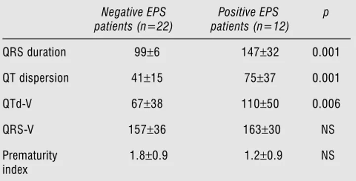
Table 2 summarizes and compares the resting electro-cardiographic findings in two groups. QRS duration of the patients with positive EPS was significantly greater than that of the patients with negative EPS, but the QRS du-ration of ventricular premature beat was not significantly
Journal of Ankara University Faculty of Medicine 2006; 59(1)
F. S. Ertaş, Ç. Özdöl, T. Altın et al. 9
different between the groups. Patients with inducible ven-tricular arrhythmias had a longer QT dispersion of normal QRS complexes and ventricular premature beats (p=0.001 and p=0.006 respectively). The prematurity index did not differ between the groups.
Signal averaged electrocardiogram measurements: Table 3 summarizes and compares the signal averaged ECG findings in the groups. The QRS duration of the pa-tients with inducible ventricular arrythmias was significant-ly longer (p=0.003). The presence of ventricular late po-tentials were much more common in EPS positive patients than EPS negative patients (20% vs 88%, p=0.002).
Regression analysis:
There were no significant correlation between EF and QTd-V in EPS negative patients (r=0.13, p=0.9) or in EPS positive patients (r=0.12, p=0.7). A correlation was found between QTd-V and QTd in EPS positive patients ( r=0.7, p=0.01) but no correlation was found in negative EPS pa-tients (r=0.3, p=0.1). The relationship of QT-V dispersion to the presence of late potentials and an abnormal LVEF who underwent EPS is examined by logistic regression analysis. Among these patients, QTd-V, late potentials (p=0.07) and ejection fraction (p=0.3) were not independently associated with susceptibility to ventricular tachyarrhythmias.
Discussion
The identification of patients who are at high risk for ventricular tachycardia and sudden death is of great im-portance. The strategies such as ventricular ectopic activity and spontaneous arrhythmias can not effectively identify subjects at high risk (27). The newer noninvasive methods such as signal-averaged electrocardiography, heart rate vari-ability and baroreceptor reflex sensitivity offer improved risk stratification (28). However, the positive predictive ac-curacy of each of these methods is still limited with regard to identifying individual patients for therapeutic interven-tions; it is possible that the combination of noninvasive methods may result in better accuracy (29).
The present study demonstrates that prolonged QT dispersion of normal and ventricular premature beats on the standard resting ECG can identify patients with in-ducible VT. These findings confirm the previous report of Dabrowski that demostrate a sigificant relationship be-tween QTd-V and risk of arrhyhmic events (24-26). These observations are consistent with the hypothesis that QT dispersion on the standard ECG is a marker of underlying regional inhomogenity of ventricular repolarization that can be associated with reentrant VT.
Difference from the previous studies from Dabrowski (24-26) showing QT dispersion of ventricular premature beat is
an independent risk for arryhthmic events, the present study demonstrates that QTd-V had a similiar accuracy compared with that of other noninvasive methods used in previous stud-ies for discriminating between patients with different suscep-tibilities to ventricular tachyarrhythmias and does not provide
Table 1. Clinical characteristics of the study patients
Negative EPS patients (n=22)
Positive EPS patients (n=12) Age (yr) 52±15 57±15 Sex (male/female) 14/8 7/5 LVEF 59±12 % 45±14 CAD 2 ( 9.1%) 4 ( 33.3%) HT 6 ( 27.3%) None DCMP 3 (13.6%) 3 (25%) VHD 4 ( 18.2%) None ICMP 1 (4.5%) 5 ( 41.7%) none 6 (27.3%) none
*Data are expressed as mean ± SD
**CAD: coronary ar tery disease; DCMP: dilated cardiomyopathy; ICMP: ischemic
cardiomyopathy; EPS electrophysiologic study; HT: hyper tension; LVEF: left ventricular ejection fraction; VHD: valvular hear t disease;
Table 2. Summary of electrocardiographic measurements
Negative EPS patients (n=22) Positive EPS patients (n=12) p QRS duration 99±6 147±32 0.001 QT dispersion 41±15 75±37 0.001 QTd-V 67±38 110±50 0.006 QRS-V 157±36 163±30 NS Prematurity index 1.8±0.9 1.2±0.9 NS
*QTd-V, QT: dispersion of ventricular premature beat; QRS-V, QRS: complex
duration of ventricular premature beat; NS: non significant
Table 3. Signal avereaged ECG findings in patient groups
Negative EPS patients (n=22) Positive EPS patients (n=12) p LAS 32.7±17.8 65±25 0.006 QRS duration 112±37 164±22 0.003 RMS 43.9±27.8 14.2±13 0.01 Late potentials 20% 88% 0.002
Ankara Üniversitesi Tıp Fakültesi Mecmuası 2006; 59(1)
Dispersion of QT interval in premature ventricular beats is not an independent marker for inducible sustained ventricular tachycardia
10
additional independent diagnostic information. However, measurament of QT dispersion is easy, inexpensive and non-invasive but because measurement of QT interval and its dis-persion are subject to introbserver and interobserver variabil-ity (28,29) and the problems with identification of the end of the T wave and as QTd-V is a dependent risk factor, the measurement of QTd-V should not be included in the non-invasive evaluation of arrhythmic risk.
Limitations
Our major limitations were small size of study popula-tion and the inhomogenity of the study groups.
Prospec-tive studies of larger and homogeneous groups of patients are therefore needed to confirm the present data.
Conclusion
Our findings demonstrate that precordial dispersion of ventricular premature beat on the resting ECG is as-sociated with the presence of inducible reentrant VT in symptomatic patients undergoing EPS, however QT dis-persion of ventricular premature beat does not appear to provide additional independent diagnostic information to that provided by late potentials and left ventricular ejec-tion fracejec-tion.
References
1. Han J, De Jalon PG, Moe GK. Adrenergic effects on ventricular vulnerability. Circ Res 1964; 14:516-24.
2. Han J, Moe GK. Non-uniform recovery of excitability in ventricular muscle. Circ Res 1964; 14:44-60.
3. Merx W, Yoon MS, Han J. The role of local disparity in conduction and recovery time on ventricular vulnerability to fibrillation. Am Heart J 1977; 94:603-10.
4. Kuo CS, Munokata K, Reddy CP et al. Characteristics and possible mechanisms of ventricular arryhythmia dependent on the dispersion of action potential durations. Circulation 1983; 67:1356-7. 5- Day CP, McComb JM, Campell RWF. QT dispersion: an
indication of arryhtmia risk in patients with long QT intervals. Br Heart J 1990; 63:342-4.
6. Day CP, McComb JM, Campell RWF. QT dispersion in sinus beats and ventricular extrasystoles in normal hearts. Br Heart J 1992; 67:39-41.
7. Dritsas A, Gilligan D, Nichoyannopouplos P et al. Amiodarone reduces QT dispersion in patients with hypertrophic
cardiomyopathy. Int J Cardiol 1992; 36:345-9.
8. Cowan JC, Yusoff K, Moore M, et al.Importance of lead selection in QT interval measurement. Am J Cardiol 1988; 61:83-7. 9. Day CP, McComb JM, Matthews J et al. Reduction of QT
dispersion by sotalol following myocardial infarction. Eur Heart J 1991; 12:423-7.
10. Van de Loo A, Arendte W, Hohnloser S.Variability of QT dispersion measurements in the surface electrocardiograms in patients with acute myocardial infarction and in normal subjects. Am Heart J 1994; 74:1113-8.
11. Pye M, Quinn A, Cobbe A. QT interval dispersion: a non-invasive marker of sustained ventricular arrhythmias? Br Heart J 1994; 71:511-4.
12. Sylven J, Horacek M, Spencer C et al. QT interval variability on body surface. J Electrocardiol 1984; 17.179-88.
13. Goldner B, Brandspiegel H, Horwitz L et al. Utility of QT dispersion combined with the signal averaged electrocardiogram in detection patients susceptible to ventricular tachyarrhythmia. Am J Cardiol 1995; 76:1192-4.
14. Perkiomaki J, Koistinen M, Yli-Mayry S et al. Dispersion of QT interval in patients with and without susceptibility to venrtricular tachyarrhythmias after previous infarction J Am Coll Cardiol 1995; 26:174-9.
15. Priori S, Napolitano C, Dielhl L et al. Dispersion of the QT interval: a marker of therapeutic efficacy in the idiopthic long QT syndrome. Circulation 1994; 89:1681-9
16. Hii J, Wyse D, Gillis A et al. Precordial QR interval dispersion as amarker of torsades de pointes: disparete effects of class Ia antiarrhythmic drugs and amiodarone. Circulation 1992; 86:1376-82.
17. Zareba W, Moss A, le Cessie S. Dispersion of ventricular repolarization and arryhthmic cardiac death in coranary artery disease. Am J Cardiol 1994; 74:550-3.
18. Cortina A, Ambrose J, Priet-Granada J et al. Left ventricular function after myocardial infarction: clinical and angiographic correlations. J Am Coll Cardiol 1985; 5:619-24.
19. Merri M, Benhorin J, Alberti M et al. Electrocardiographic quantitation of ventricular replarization. İrculation 1989; 80:1301-8.
20. Garson A. How to measure the QT interval: what is normal? Am J Cardiol 1993; 72:14B-16B.
21. Bazett H. An analysis of time relations of electrocardiograms. Heart 1920; 7:353-70.
22. Lander P, Berari E, Rajagopalan C et al. Critical analysis of the signal-averaged electrocardiogram: improved identification of late potentials. Circulation 1993; 87:105-17.
23. Barr CS, Naas A, Freeman M et al. QT dispersion and sudden death in heart failure. Lancet 1994; 343:327-9.
24. Dabrowski A, Kramarz E, Piotrowicz R. Dispersion of QT interval in premature ventricular beats as a marker of susceptibility to arrhythmic events. Journal of Cardiovascular risk 1998; 5:97-101. 25. Dabrowski A, Kramarz E, Piotrowicz R. Dispersion of QT
interval following ventricular premature beats and mortality after myocardial infarction. Cardiology 1999; 91:75-80.
26. Dabrowski A, Kramarz E, Piotrowicz R et al. Predictive power of increased QT dispersion in ventricular extrasystoles and in sinus beats for risk stratification after myocardial infarction. Circulation 2000; 101:1693-7.
27. Turitto G, El-Sherif N. Complex ventricular arrhytmias and nonsustained ventricular tachycardia: risk stratification and management. In: el-Sherif N, Samet P, editors. Caridac Pacing and Electrophysiology. Philadelphia:Saunders, 1991; 217-33. 28. Farrell TG, Bashir Y, Cripps T et al. Risk stratfication for
arrhythmic events in postinfarction patients based on heart rate variablity, ambulatory elcetrocardiographic variables and the signal averaged electrocaridiogram. J Am Coll Cardiol 1991; 18:687-97. 29. Bigger JT Jr, Steinberg JS. Risk strafication for arrhytmic death after
MI: an overview. In: El-Sherif N, Somet P, eds. Cardiac Pacing and Electrophysiology. Philadelphia: Saunders 1991; 303-23
30. Kautzner J, Yi G, Camm AJ et al. Short and long term
reproductibility of QT, QTc and QT dispersion measurement in healthy subjects. PACE 1994; 17:928-37.
31. Ahnve S. Methodological aspect of QTc interval determiantion. In: Butrous CG, Schwartz PJ, eds. Clinical aspects of Ventricular repolarazation. London: Farrand press, 1989; 1-16.