The Annals of Clinical and Analytical Medicine
Original Research
Mehmet Serdar Dervişoğulları1, Cengiz Akarsu2, Ahmet Ergin3 1Başkent University, Ophthalmology Department, Adana, 2Dünya Göz Hospital, Antalya,
3Balıkesir University, Ophthalmology Department, Balıkesir, Turkey
IOP with tonopen and goldman tonometers
Comparison of the intraocular pressure measurements with the tono-pen and the
goldman applanation tonometer and the effect of central
corneal thickness on measurements
DOI:10.4328/ACAM.5996 Received: 04.08.2018 Accepted: 14.09.2018 Published Online: 17.09.2018 Printed: 01.07.2019 Ann Clin Anal Med 2019;10(4): 479-84 Corresponding Author: Mehmet Serdar Dervisogullari, Ophthalmology Department, Başkent University Medical School, Adana Dr. Turgut Noyan Clinic and Research Center, Adana, Turkey. T.: +90 3223272727 F.: +90 3223271274 Gsm: +905327720616 E-Mail: [email protected]
ORCID ID: https://orcid.org/ 0000-0003-2006-2906
Abstract
Aim: Incorrect values of applanation tonometers according to varying corneal thickness were investigated many times. In this study, we aimed to compare intraocular pressure (IOP) measurements of the Tono-Pen and Goldman applanation tonometer (GAT) in eyes with normal corneas of various thicknesses. Material and Method: IOP was measured with Tono-Pen and GAT respectively in 255 patients’ eyes with normal corneas. Only the right eyes were analyzed for statistical purposes. Central corneal thickness (CCT) was measured using an ultrasound pachymeter after all IOP determinations had been made. Re-sults: With both instruments, IOP varied with CCT. Readings with Tono-Pen showed a mean increase in IOP with increasing CCT of 0,18 mmHg/10μm and an increase of 0,16 mmHg/10 μm with the GAT. The Tono-Pen consistently recorded comparatively higher IOPs than the GAT (p<0,05). Discussion: In our study, the difference between the Tono-Pen and GAT measurements is statistically insignificant and 60% of this difference is within the 1 mmHg range. Although it is thought that Pen may be less affected by corneal thickness due to the measurement from a smaller area we did not obtain data that Tono-Pen is less affected by corneal thickness as a result of linear regression analysis.The Tono-Tono-Pen is more affected by CCT when used to measure IOP in eyes with normal corneas. This is contrary to expectations, based on the theory that Tono-Pen is least affected by the CCT because it needs smaller applanation area.
Keywords
Introduction
Glaucoma is an ocular disease characterized by optic neurop-athy which is caused by mechanisms connected to vascular and mechanical factors and is among the leading causes of preventable blindness in the world. Elevated intraocular pres-sure (IOP) is the most common risk factor. In the pathogen-esis of glaucoma, blood flow in the eye, optic disc structure and ganglion cell degeneration are also important but the most important factor in diagnosis and treatment of the disease is IOP. Many instruments such as Schiotz indentation tonometry, Goldman applanation tonometry (GAT), McKayMarg tonometry, noncontact and contact pneumotonometers have come to the fore with the historical process in IOP measurements made from smooth corneal surfaces. Among them only McKay-Marg tonometry can measure irregular corneal surfaces due to scar-ring, edema or surgery [1] but it is not in production today. GAT developed by Goldman in 1957 according to the Imbert-Fick law has been accepted as the gold standard today. According to the Imbert-Fick law the pressure to flatten the wall of an elastic full of water, for example a balloon, is equal to the applied force divided by the application area. In fact, this law recognizes that cornea is a structure that does not resist other forces than the internal pressure force against fine, applied gentleness in per-fect elasticity. Goldman and Schmidt stated that although the device is calibrated according to the standard corneal thickness (520 μm), it may theoretically be affected by corneal thickness changes [2]. There are many factors that can affect the IOP measurements made with this device. The accuracy of the mea-surement is expected to increase with the application of the ap-propriate measurement technique in large scale. [3] As for the negative aspects of GAT, this device, which is developed with constant corneal thickness and higher values above the value, may cause lower measurements at lower thicknesses, and cor-rections between 1 and 6.8 mmHg are also made for a 0.1 mm change in corneal thickness. [4] Glaucoma prevalence, which is high in myopic patients, and the increase in refractive surgi-cal procedures that change corneal thickness in recent years have made the relationship between IOP and corneal thickness more important [5]. In addition to differences in race, age and gender, central corneal thickness (CCT) and corneal curvature and IOP and curvature associations are also discussed in cor-neal thickness [6]. Medeiros et al. have shown that ocular hy-pertensive cases with early glaucomatous defects with perim-eter have lower CCT values than those without a defect [7]. In this context, the use of devices such as Tono-Pen, which is thought to be less affected by corneal surface irregulari-ties and thickness in recent years, has come to the fore. To-no-Pen works according to the McKay-Marg principle, which expresses the conversion of mechanical energy into electri-cal energy. The measurement area measures from a smaller area (diameter of 1.02 mm) than the GAT (diameter of 3.06 mm) with a transducer movement as small as 10 μm, which may be considered to be less affected by corneal thickness. The positive aspects of the device are that it is easier to mea-sure IOP in situations such as corneal irregularity, lid edema, narrow palpebral aperture, infant, small children, bedridden or wheelchair patients, head tremor or nystagmus patients. CCT can be measured by optic or ultrasonographic methods.
Ultrasonic pachymetry is a more reliable, precise method than optic pachymetry, with repeated measurements of the observer and less variability between different observers [8]. In this study, we aimed to investigate the effect of CCT on the IOP measurements of GAT and Tono-Pen devices. According to our hypothesis corneal thickness does not make a difference in IOP measurements between the Tono-Pen and the GAT. Material and Method
This study was performed in adherence with the tenets of the Declaration of Helsinki and was approved by the Kırıkkale Univer-sity Medical Faculty Ethics Committee (14.01.2004/2004106). Informed consent was obtained from all of the study partici-pants. Participants with less than 2D spherical and 1D astig-matic refraction were enrolled. Two hundred and fifty-five pa-tients who did not have any contact lens and volunteered to participate in the study were prospectively included in the study. Patients with a history of intraocular surgery history, unilateral anophthalmia, any corneal pathology or scarring and evidence of anterior segment inflammation were excluded. Visual acuity, refraction, biomicroscopy and fundoscopy examinations were performed on both eyes of the participants. The age and sex of the participants were recorded. The participants’ ages ranged from 10 to 80 (average 40.86). There were 109 males and 146 females. In all examinations the right eye was first evaluated. All examinations and measurements were made between 09:00 and 14:00 hours. In response to the possibility of the GAT drop-ping the IOP and causing the lower values that can be measured with the Tono-Pen, the GAT and IOP measurement examination was first applied to 128 participants. Measurements with GAT (Zeiss Co, Zurich, Switzerland) were performed on the patients looking at a target at a distance of 6 m, in the primary po-sition, with a drop of 0.25% fluorescein in both eyes subcon-junctivally as a mixture of 0.45% oxybuprocaine hydrochloride (Benoxinate, Alcon Couvreur, Puurs, Belgium) and fluorescein (Fluoroscite 10%, Alcon, Texas, USA). Participants were asked not to move their eyes and to breathe regularly. The blue light was turned on until the end and was brought to an angle of 45 degrees with the eye in the horizontal plane. The arithme-tic averages were obtained by the experienced practitioner by performing 3 measurements within the limit of 1 mmHg (MSD). In the study, the same GAT was used regularly with annual cali-bration. The IOP measurements were then repeated with the Tono-Pen (Tono-Pen XL, Mentor Ophthalmics, CA, USA) instru-ment in a sitting position, looking at the target 6 m away in the primer position, Measurements with Tono-Pen were performed after the drop of 0.45% oxybuprocaine hydrochloride was ap-plied to the lower conjunctival fornix of both eyes. For each patient, a new latex membrane (Mentor OcuFilm Type Covers) was placed on the transducer. After Tono-Pen was activated and the ‘beep’ sound was heard the transducer was contacted gently to the cornea, waiting for at least 1 second until the ‘beep’ sound indicating successful measurement. The procedure was repeated until two measurement results with reliability of 5% were obtained on the liquid display. All the IOP measure-ments with Tono-Pen were performed by the same practitioner (MSD). Tono-Pen was calibrated every morning in line with the manufacturer’s recommendation. After the measurement with
Tono-Pen or GAT, the measurement with the other instrument was made 15 minutes apart. After IOP measurements were completed by both methods, central corneal thickness (CCT) was measured with an ultrasonic pachymeter (Optikon 2000 S.p.A., Rome, Italy). After dropping 0.45% oxybuprocaine hydro-chloride to each lower conjunctival sac, the patient was held in the primary position, the probe was held perpendicular to the cornea and 3 measurements were taken from the undiluted pupil center and averaged. The participant was asked to blink between measurements. Pachymeter measurements were all made by the same person (MSD). Measurements were made in both eyes, and only right eyes were included in the study. Since the measurements are made with corneal contact, the possible complication is keratitis. Therefore antibiotic drops were ap-plied to the eyes after the measurement.
When the alpha value (confidence interval) was 0.05 and the number of participants was 255, the power of the study was determined to be 80% to show a 2% difference between the two instruments’ measurements. IOP measurements of both devices were evaluated by the Student t- test. Simple linear re-gression analysis (dependent on IOP measurement with Tono-Pen or GAT, corneal thickness as an independent variable) was used to determine the measurement averages of both devices in relation to the corneal thickness and regression equations were found. The increase in measured IOP by 10 μm in CCT is calculated from the graphs for each of the Tono Pen and GAT devices. For this statistical evaluation, SPSS Ver. 11.0 (SPSS Inc, Chicago, USA) program was used. The values of p<0.05 were considered statistically significant.
Results
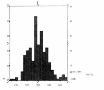
There was no statistically significant difference in the mea-surements of Tono-Pen (p = 0.352), GAT (p = 0,203) and CCT (p = 0,734) (P <0.05) according to the gender. (Table 1) Using Tono-Pen and GAT, a total of 255 values were determined. In the measurements, the mean of the Tono-Pen measurements (± SD) was 16.71 (±3.09) mmHg (8-30 mmHg) and the mean of the GAT measurements was (± SD) 16.10 (±3.07) mmHg (7-26mmhg). The measurement difference between the two devices was statistically significant (p <0.05) and Tono-Pen measurements were higher. Tono-Pen and GAT measurements differed from 0,6 to 1 mmHg (Figure 1). According to the point distribution graph and the regression curve for the compari-son of the thickness of the cornea with Tono-Pen, the regres-sion equation is y = 6,812 + 0,018x (r2: 0.041), where y = IOP measured by Tono-Pen, x = CCT. An increase of 0, 18 mmHg was observed in the IOP measurement made with Tono-Pen in every 10 μm increase in CCT (Figure 2). According to the results of linear regression analysis, GAT is less affected by corneal
thickness (An increase of 0, 16 mmHg was observed in the IOP measurement made with GAT in every 10 μm increase in CCT). In Table 2 and Figures 1, 2 and 3 we can see that the Tono-Pen measurements are higher than the GAT
mea-Table 1. Tono-Pen (mmHg), Goldman applanation tonometry (mmHg) and Central corneal thickness (μm) values.
Gender Tono-Pen
Goldman Applanation
Tonometer
Central Corneal
Thickness Age (years) Male
(n-108)
16,72±3,15 15,87±3,18 550,36±34,16 41,45±15,99 Female
(n-147) 16,72±3,02 16,26±298 545, 98±34,75 40,43±14,33
Figure 1. Measurement differences between Tonopen and Goldman applanation tonometer (x-axis: mmHg, y-axis: n).
Figure 2. Regresyon analysis curve of the Goldman applanation tonometer (y-axis, mmHg) and the central corneal thickness (x-axis, μm).
Figure 3. Regresyon analysis curve of the Tonopen (y-axis, mmHg) and the central corneal thickness (x-axis, μm).
surements. This difference in measurement was statis-tically significant (p<0.05). The mean CCT (± SD) was 547 (± 34.50 μm)( range of 451.60-650 μm) (Figure 4). The regression equation is y = 7,448 + 0,016x (r2 = 0,032), where y = the IOP value measured with GAT and x = the CCT value. According to the point distribution graph for the CCT comparison of GAT and the regression curve, the IOP measure-ment value with GAT increased by 0.16 mmHg at a 10 μm in-crease in thickness (Figure 3). There was no statistically signifi-cant difference between measurements performed with Tono-Pen (p = 0.411) or with GAT (p = 0.579) (Table 2).
Discussion
Glaucoma is one the most important cause of blindness in the world and the only currently accepted treatment is the reduc-tion of IOP. For this reason, correlareduc-tions of IOP measuring de-vices with each other have always been the subject of research. Today GAT is accepted as the gold standard in IOP measure-ment but many factors are thought to affect its measuremeasure-ment results. Opinions have been reported that changes in corneal thickness, which is the determinant of corneal rigidity, will af-fect measurements. [9] In some studies in which invasive IOP measurement methods were used, the values obtained with the applanation method were normal with the corneal thickness increase. [3] Recep et al. have shown that noncontact tonom-eters are also affected by corneal thickness. [10] Feltgen and colleagues found that Tono-Pen and Perkins applanation to-nometry measurements are correlated with manometric mea-surements and did not require any correction.[11] Comparison of GAT with the first sample of Tono-Pen (Tono-Pen-1) was made in 1987 by Minckler et al. and reported that Tono-Pen showed higher values at higher IOP and lower values at lower IOP. [13] Hessemer et al. compared manometric measurements
with TonoPen and reported that Tono-Pen showed lower val-ues at 17 mmHg below and higher at 17 mmHg [14] and Fos-ter et al.observed a deviation of 2 mmHg (between -8 and +4) with Tono-Pen compared to the manometric measurements in their 23 eyes performed phacoemulsification but this deviation could not be correlated with corneal thickness.[15] Similar re-sults were reported in similar studies on rabbits and rats. [16] Eisenberg et al. reported that they measured very well with the Tono-Pen’in laboratory conditions but they found a decrease in the accuracy of the measured values compared with intraocu-lar measurements [17]. Feltgen et al. reported that Tono-Pen and Perkins applanation tonometers were correlated with each other and their measurement results were not affected by cor-neal thickness in their studies comparing applanation tonom-etry measurements with manometric measurements according to corneal thickness [11].
It has been shown that the cornea is thinner in pseudoexfolia-tion glaucoma cases [10]. In order to avoid detecpseudoexfolia-tion of false low IOP values in lower CCT, IOP measuring devices are required to minimize the effect of corneal thickness and make measure-ments close to true IOP values. It is known that changes in IOP measurements after surgery compared to the preoperative pe-riod are determined due to the decrease of corneal thickness after excimer laser photorefractive keratectomy in myopia cas-es. The incidence of increased glaucoma in myopic patients and the increase in refractive surgery applied to myopia today are considered to be significant. In some previous studies, Tono-Pen has been shown to make precise measurements in the eye bank eyes [18]. However in vivo studies have identified some data suggesting that Tono-Pen and GAT measurements are incom-patible with the clinic. For example Tono-Pen reported incon-sistent results on measurements over 30 mmHg [19] did not reflect IOP fluctuation and did not make precise measurements that could be used in glaucoma diagnosis and follow-up [20]. It is thought that the inaccuracies of in vitro studies are related to the absence of precorneal tear film, live corneal epithelium, normal corneal thickness and rigidity, extraocular muscle func-tion, fluctuating blood pressure,respiration and even patient anxiety [21],
In our study the difference between the Tono-Pen and GAT mea-surements is statistically insignificant and 60% of this difference is within the 1 mmHg range. Our sample size and standardized measurement procedures did not allow statistical uncertainty. Although it is thought that Tono-Pen may be less affected by corneal thickness due to the measurement from a smaller area we did not obtain data that Tono-Pen is less affected by cor-neal thickness as a result of linear regression analysis. We also observed slightly higher values with Tono-Pen as the thickness increased and found increases of 0.18 mmHg with TonoPen and 0.16 mmHg with GAT in 10 μm CCT increase. Earlier studies in which Dohadwala et al. evaluated Tono-Pen’s measurements of various corneal thicknesses reported increases in IOP of 0.29 mmHg in males and 0.12 mmHg in females with an increase in corneal thickness of 10 μm [22]. Bhan et al obtained 0,10 mmHg / 10 μm and 0,23 mmHg / 10 μm IOP increments in their studies investigating the relationship between Tono-Pen and GAT corneal thickness [23]. In our study we did not observe a statistically significant difference in Tono-Pen, GAT and CCT Figure 4. Central corneal thickness (μm).
Table 2. There was no statistically significant difference between measure-ments performed first with Tono-Pen (p=0,411) or first with GAT (p=0,579).
group measured first with Tono-Pen (mmHg)
Group measured with GAT (mmHg)
GAT (mmHg) 16,63±3,20 15,56±2,84
values according to sex.
It is known that astigmatism, refraction and visual acuity do not affect corneal thickness. The effect of corneal curvature on IOP measurements has been shown to be negligible. [24] For this reason, we did not evaluate the corneal curvature in our study. In our study we did not divide our values into subgroups, but we performed our measurements within the range of 11-20 mmHg in patients who applied to the eye policlinic for the routine eye examination and observed that the Tono-Pen measurements were higher than the GAT measurements. We have not compared our measurements with manometric measurements but nowa-days it is thought that even manometric measurements have some disadvantages because the applied paracentesis leads to changes in anterior chamber structure and volume, aque-ous production, endothelial function and ocular temperature. The advantages of Tono-Pen compared to GAT are light, por-table, easy to learn, able to measure from a smaller surface easier to measure IOP in corneal irregularities, uncooperative cases and pediatric patients. The measurement made with the Tono-Pen is reflected in the resultant liquid crystal screen and is not open to interpretation. In addition sterile latex tip cov-ers maintain an advantage in postoperative cases and ocular or systemic infections (hepatitis, HIV, prion diseases). The use of the TonoPen in corneal pathology is important in sev-eral ways: It can be used in cases of corneal edema, scarring or band keratopathy. The latex allergy caused by Tono-Pen la-tex tip cover should be questioned before the measurement. Other portable tonometers are also available. Measurements with Schiotz tonometry make the patient more uncomfortable and the values can be influenced by the elastic properties of the eye and the corneal curvature. With Draeger, Kowa and Per-kins tonometric measurements to be more precise, you need to gain experience for quality measurements. The McKay-Marg tonometry can also provide more accurate measurements, but its transport is more difficult and is no longer produced. The ap-pearance of the optic disc and the evaluation of the visual field are much more important than the assessment of the corneal thickness in order to make the decision to start treatment FOR glaucoma. Corneal thickness should be assessed in addition to IOP measurements in cases of ocular hypertension and normo-tensive glaucoma in our findings. Corneal thickness measure-ments are especially important when the clinically determined IOP value is inconsistent and target IOP evaluation needed af-ter the treatment.
Conclusion
In summary, the slightly higher values were seen in the Tono-Pen measurements. This did not appear to cause problems clinically because Tono-Pen also has the above-mentioned characteris-tics. Although GAT is still the gold standard for IOP measure-ment and is used as the most preferred tonometer, Tono-Pen is portable, it can be used in corneal pathologies and animal experiments and its sterility properties are superior.
Scientific Responsibility Statement
The authors declare that they are responsible for the article’s scientific content including study design, data collection, analysis and interpretation, writing, some of the main line, or all of the
preparation and scientific review of the contents and approval of the final version of the article.
Animal and human rights statement
All procedures performed in this study were in accordance with the ethical standards of the institutional and/or national research committee and with the 1964 Helsinki declaration and its later amendments or comparable ethical standards. No animal or hu-man studies were carried out by the authors for this article. Funding: None
Conflict of interest
None of the authors received any type of financial support that could be considered potential conflict of interest regarding the manuscript or its submission.
References
1. Tierney JP, Rubin ML. A clinical evaluation of the electronic applanation tonom-eter. Am J Ophthalmol. 1966; 62: 263-71.
2. Goldman H. Schmidt T. Weiterer Beitrag zur Applantionstonometrie. Ophthal-mologica. 1961; 141: 441-56.
3. Dielemans l, Vingerling JR, Hofman A, Groebe DE, DeJong PTVM. Reliability of intraocular pressure measurement with the Goldman applanation tonometer in epidemiological studies. Graefes Arch Clin Exp Ophthalmol. 1994; 232: 141-4. 4. Argus WA. Ocular hypertension and central corneal thickness. Ophthalmology. 1995; 102: 1810-12.
5. Rashad KM, Bahnassy AA. Changes in intraocular pressure after laser in situ keratomileusis. J Refract Surg. 2001; 17: 420-3.
6. Mark HH. Corneal curvature in applanation tonometry. Am J Ophthalmol. 1973; 74: 223-4.
7. Medeiros FA, Sample PA, Weinreb RN. Corneal thickness measurements and visual function abnormalities in ocular hypertensive patients. Am J Ophthalmol. 2003; 135: 131-7.
8. Giasson C, Forthomme D. Comparison of central corneal thickness measure-ments between optical and ultrasound pachometers. Opt Vis Sci. 1992; 69: 236-41.
9. Whitacre MM, Stein R. Sources of error with use of Goldman-type tonometers. Surv Ophthalmol. 1993; 38: 1-30.
10. Recep ÖF, Hasıripi H, Çağıl N, Sarikatipoğlu H. Relation between corneal thick-ness and intraocular pressure measurement by noncontact and applanation to-nometer. J Cataract Refract Surg. 2001; 27: 1787-91.
11. Feltgen N, Leifert D, Funk J. Correlation between central corneal thickness, ap-planation tonometry and intracameral readings. Br J Ophthalmol. 2001; 85: 85-7. 12. Minckler DS, Baerveldt G, Heuer DK, Quillen-Thomas B, Walonker AF, Weiner J. Clinical evaluation of Oculab Tono-Pen. Am J Ophthalmol. 1987; 104: 168-73. 13. Bordon AF, Katsumi O, Hirose T. Tonometry jm pediatric patients:A compara-tive study among Tono-Pen, Perkins and Schiotz tonometers. J Pediatr Ophthalmol Strabismus. 1995; 32: 373-7.
14. Hessemer V, Rössler R, Jacobi K. Comparison of intraocular measurements with the Oculab Tono-Pen vs manometry in humans shortly after death. Am J Ophthalmol. 1988; 105: 678-82.
15. Foster PJ, Wong JS, Wong E. Acurracy of clinical estimates of intraocular pres-sure in Chinese eyes. Ophthalmology. 2000; 107: 1816-21.
16. Mermoud A, Baerveldt G, Minckler DS, Lee MB, Rao NA. Intraocular pressure in Lewis rats. Invest Ophthalmol Vis Sci. 1994; 35: 2455-60.
17. Mermoud A, Baerveldt G, Minckler DS, Lee MB, Rao NA. Measurement of rab-bit intraocular pressure with the Tono-Pen. Ophthalmologica. 1995; 209: 275-7. 18. Eisenberg DL, Sherman BG, McKeown CA, Scuman JS. Tonometry in adults and children:a manometric evaluation of pneumotonometry, applanation and Tono-Pen in vitro and in vivo. Ophthalmology. 1998; 105: 1173-81.
19. Boothe WA, Lee DA, Panek WC, Pettit TH. The Tono-Pen: a manometric and clinical study. Arch Ophthalmol. 1988; 106: 1214-17.
20. Moses RA, Arnzen RJ,. instanteneous tonometry. Arch Ophthalmol. 1983; 101 :249-52.
21. Lim JI, Blair NP, Higginboham EJ, Farber MD, Shaw WE, Garretson BR. Asses-ment of introcular pressures in gas-containing eyes. Arch Ophthalmol. 1990; 108: 648-88.
22. Khan J, Davis M, Graham CE, Trank J, Whitcre M. Comparison of Oculab Tono-Pen readings obtained from various corneal and scleral locations. Arch Ophthal-mol. 1991; 109: 1444-6.
23. Dohadwala AA, Munger R, Damji KF. Positive correlation btween Tono-Pen intraocular pressure and central corneal thickness. Ophthalmology. 1998; 105: 1849-54.
24. Bhan A, Browninig AC, Shah S, Hamilton R, Dave D, Dua I-IS. Effect of cor-neal thickness on intraocular pressure measurements with the pneumotonometer,
Goldman applanation tonometer and Tono-Pen. Invest Ophthalmol Vis Sci. 2002; 43: 1389-92.
25. Matsumoto T, Makino H, Uozato H, Saishin M, Miyamoto S. The influence of corneal thickness and curvature on the difference between intraocular pressure measurements obtained with a non-contact tonometer and those with Goldman applanation tonometer. J Jpn Ophthalmol Soc. 2000; 104: 317-23.
How to cite this article:
Dervişoğulları MS, Akarsu C, Ergin A. Comparison of the intraocular pressure measurements with the tono-pen and the goldman applanation tonometer and the effect of central corneal thickness on measurements. Ann Clin Anal Med 2019;10(4): 479-84.